The American Heart Association has recently stressed the importance of obesity as a modifiable, independent risk factor for coronary artery disease, ventricular dysfunction, congestive cardiac failure, and cardiac arrhythmias.Reference Klein, Burke and Bray 1 The prevalence of overweight and obesity in children is increasing worldwide at an alarming rate in both developing and developed countries.Reference Mokdad, Serdula, Dietz, Bowman, Marks and Koplan 2 In the United States, approximately 30.9% of adolescents are overweight, of whom 16.1% are obese.Reference Hedley, Ogden, Johnson, Carroll, Curtin and Flegal 3
The relationship between obesity and cardiac structure and function in adolescents is not well studied, and conflicting results have been reported.Reference de Simone, Daniels and Devereux 4 – Reference Daniels, Kimball, Morrison, Khoury, Witt and Meyer 6
Given the marked rise in obesity in youth, an understanding of the pathophysiological implications of these effects early in the lifespan is clearly important. Such information emphasises the urgency of preventive efforts and serves to help defining specific management strategies.
There is little information on how much the excess of body weight impacts left and right ventricle geometry and function in Egyptian adolescents. The aim of this study is to detect any changes in the left and right ventricular structure and function in obese Egyptian adolescents. We also explored the role of tissue Doppler imaging, an emerging technique that measures velocities of the myocardium, as a screening tool for these abnormalities.
Patients and methods
The study was approved by the institutional review board and ethics committee of Suez Canal University. Informed consent was obtained from each patient.
Patients
The study was conducted in the period between May and August 2010. We chose a stratified cluster proportionate systematic random sample from Ismailia Governorate preparatory schools. A total of 4900 adolescents were screened for obesity; obesity prevalence was 5.08%, that is, 249 adolescents. The present study included 28% obese normotensive adolescents, that is, 70 subjects, selected randomly, in the age group of 12–15 from both sexes, with BMI >95th percentile for age and sex. A total of 50 healthy, non-obese adolescents were selected as the control group who were age and sex matched with BMI <95th percentile. Exclusion criteria were a significant concomitant illness, medication known to modify cardiac function, obvious cardiac disease, and patients with syndromes including obesity.
Methods
Clinical assessment
Measurements of height and weight; mid-arm, waist, and hip circumference; and skinfold were recorded. Body mass index values were calculated as weight – in kilograms – divided by height squared – in square metres – and were compared with age standards.Reference Hedley, Ogden, Johnson, Carroll, Curtin and Flegal 3 Waist circumference is defined as the minimal circumference measured at the navel, and hip circumference is the widest circumference measured at the hips and buttocks.Reference Fernandez, Redden, Pietrobelli and Allison 7 , Reference Sardinha, Going, Teixeira and Lohman 8 Fasting blood glucose and lipid profile were also measured.
Echocardiograhic assessment
Echocardiography was performed in the echocardiography laboratory of Suez Canal University Hospital using Vivid-7 Dimension echocardiographic unit (GE Vingmed Ultrasound, Horten, Norway) with a multi-frequency probe ranging between 1.7 and 3.4 megahertz. M-mode, two-dimensional Doppler echocardiography and tissue velocity interrogation were performed for each subject with a simultaneous electrocardiography. Images were taken in the standard views with the patient in the left lateral position.
Left ventricular internal dimensions, septal and posterior wall thickness were measured as per American Society of Echocardiography recommendations.Reference Schiller, Shah and Crawford 9 The ejection fraction, fractional shortening, stroke volume, left ventricular mass,Reference Devereux, Lutas and Casale 10 and left ventricular mass index were calculated. Left ventricular hypertrophy has been underestimated in the obese with the use of left ventricular mass divided by body surface area because this index considers obesity as a physiological variable. A height of 2.7 (in metres) had been validated and recommended for indexing left ventricular massReference de Simone, Daniels and Devereux 4 and was used in this study. Left ventricle hypertrophy is diagnosed when left ventricular mass is over the 95th percentile for healthy children and adolescents (38.6 grams per metre raised to 2.7).Reference Daniels, Kimball, Morrison, Khoury and Meyer 11
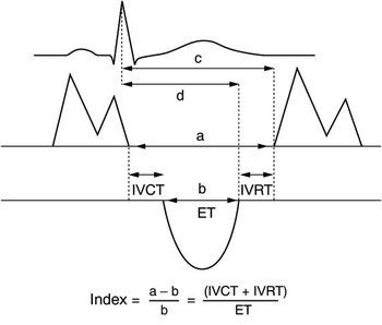
Myocardial performance index (Tei index) was defined as the sum of isovolumetric relaxation time and isovolumetric contraction time divided by ejection time.Reference Tei, Ling and Hodge 12 It has been used to evaluate left ventricular systolic and diastolic performance (global function). It was calculated from the following equation: MPI = (a−b)/b = (IVCT + IVRT)/ET (Fig 1).
Diagram of mitral inflow pattern, aortic outflow pattern, isovolumic time intervals, and Tei index. IVCT = isovolumic contraction time; IVRT = isovolumic relaxation time; ET = ejection time; LV = left ventricle.

Tissue Doppler imaging
From the standard four-chamber and two-chamber view, resting tissue Doppler velocities were derived for the basal segment of the septal and lateral left ventricle wall and for the lateral right ventricle wall. The peak annular velocities of systolic excursion in isovolumic contraction and ejection period (systolic velocity, S), and in early (early diastolic velocity, E′) and late diastole (late diastolic myocardial velocity, A′) were recorded and averaged over three consecutive cardiac cycles. In addition, the ratio of early/late diastolic velocities (E′/A′) was calculated.
Statistical analysis
The Statistical Package for the Social Sciences – SPSS software version 16 was used. All variables are expressed as mean plus or minus standard deviation. Student t tests were performed to compare the continuous variables describing the two groups. Chi-square analysis was used to compare the two groups with regard to the discrete variables. Pearson correlation coefficients were calculated for body mass index, anthropometric and left ventricle structure and function measurements. Stepwise multivariate regression analyses were performed to determine the independent predictors of left ventricular hypertrophy, left ventricular systolic and diastolic function, and right ventricular systolic and diastolic function; independent variables in the multivariate model included body mass index, waist and hip circumference, waist:hip ratio, mid-arm circumference, and age. A p-value <0.05 was considered statistically significant.
Results
Clinical characteristics of the study groups
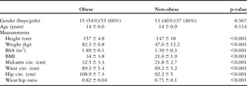
The clinical and anthropometric characteristics of the study groups are shown in Table 1. The anthropometric parameters showed significantly higher values in the obese group. All the study groups have normal fasting blood glucose and lipid profile.
Clinical parameters of the studied population.

BMI = body mass index; BSA = body surface area; Circ. = circumference
p-value significance <0.05
Echocardiographic parameters
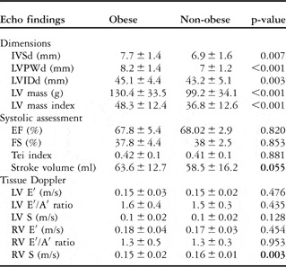
The obese individuals had significantly higher values of left ventricle wall thickness measurements than the non-obese. The left ventricular mass was significantly higher in the obese group when indexed for a height of 2.7 metres (Table 2). However, no statistical difference was found when indexed for the body surface area, (76.7 plus or minus 17.3 for patients, versus 73.5 plus or minus 18.9 for control group, p-value >0.05).
Echocardiographic parameters of the studied population.

A′ = late diastolic myocardial velocity; E′ = early diastolic velocity; EF = ejection fraction; FS = fractional shortening; IVSd = interventricular septum thickness in diastole; LV = left ventricle; LVIDd = left ventricle internal dimension in diastole; LVPWd = Left ventricular posterior wall thickness in diastole; RV = right ventricle; S = systolic velocity
p-value significance <0.05
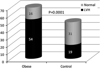
Left ventricular hypertrophy was diagnosed in 77% of obese adolescents compared with 38% in the control group (Fig 2).
Distribution of LVH and normal LVMI in the study groups. LVH = left ventricular hypertrophy; LVMI = left ventricular myocardial infarction.

Left ventricular systolic function had nearly equal values in both groups. The Tei index was almost similar in both groups. The stroke volume showed higher values for the obese group than the non-obese group (63.6 plus or minus 12.7 millilitre versus 58.5 plus or minus 16.2 millilitre respectively, p = 0.05).
Tissue Doppler assessment of the left and right ventricular systolic and diastolic velocities is shown in Table 2. There was no statistical difference between the two groups. Only the value of the S wave of the right ventricle showed significantly lower values in the obese group.
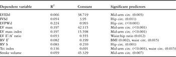
To examine the influence of anthropometric measures on the echocardiographic values, stepwise multiple regression analysis was performed (Table 3). It showed that body mass index was not a predominantly significant parameter in the model, whereas the mid-arm and hip circumference have a significant impact on most of the echocardiographic measurements.
Stepwise multiple regression analysis of the anthropometric measures and the echocardiographic values.

A′ = late diastolic myocardial velocity; E′ = early diastolic velocity; IVSd = interventricular septum thickness in diastole; LV = left ventricle; LVIDd = left ventricle internal dimension in diastole; LVPWd = left ventricular posterior wall thickness in diastole; RV = right ventricle; S = systolic velocity
p-value significance <0.05
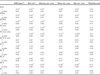
The correlation between anthropometric parameters and echocardiographic findings among the studied groups was shown in Table 4. Body mass index, mid-arm and hip circumference values showed significant correlations with echocardiographic variables. The right ventricle S wave in obese group was inversely correlated with hip, waist, and mid-arm circumference.
Correlation between anthropometric parameters and echocardiographic findings among the studied groups.

A′ = late diastolic myocardial velocity; BMI = body mass index; BSA = body surface area; Circ. = circumference; E′ = early diastolic velocity; IVSd = interventricular septum thickness in diastole; LV = left ventricle; LVIDd = left ventricle internal dimension in diastole; LVPWd = left ventricular posterior wall thickness in diastole; RV = right ventricle; S = systolic velocity; SV = stroke volume
*Significant correlation (p < 0.05)
**Highly significant correlation (p < 0.001)
Discussion
Obesity is an increasingly common condition in industrialised and developing countries, affecting both adultsReference Klein, Burke and Bray 1 and childrenReference Williams, Hayman and Daniels 13 and increasing the social burden due to incident cardiovascular disease.
This study provided evidence that asymptomatic normotensive obese adolescents already exhibit some abnormalities of left and right ventricular structure and possibly function. These changes include: increase in the left ventricle wall dimensions, left ventricular mass index, stroke volume, and minor reduction of systolic right ventricular functions detected by tissue Doppler imaging. However, systolic and diastolic functions of the left ventricle were normal in the study group.
Left ventricle morphology and systolic function
The association of left ventricle remodelling and obesity in the present study is supported by previous studies in adults and children.Reference Li, Li, Ulusoy, Chen, Srinivasan and Berenson 14 , Reference Chinali, de Simone and Roman 15 The Bogalusa Heart Study showed a strong association between left ventricular mass in childhood and the degree of obesity.Reference Li, Li, Ulusoy, Chen, Srinivasan and Berenson 14 In the Strong Heart Study, increased levels of left ventricular mass were found in overweight and obese adolescents. In addition, they found a significant increase in stroke volume in obese adolescents, as in the current study, indicating increased cardiac workload.Reference Chinali, de Simone and Roman 15 In obese subjects, the increase in left ventricular mass is a response not only to substantially increased haemodynamic load, but also to possible neurohormonal effects of clustered metabolic factors influencing left ventricular growth.Reference Chinali, de Simone and Roman 15 In obese children, abnormal left ventricular mass may increase the cardiovascular risk in the future as noticed in the previous studies.Reference Milani, Lavie, Ventura, Kurtz and Messerli 16 Weight control to decrease the severity of obesity should be recommended.Reference Khositseth, Suthutvoravut and Chongviriyaphan 17 Becoming obese is an anabolic event. In addition to the obvious accumulation of excessive body fat, an obese child is characterised by an increase in lean body mass, acceleration of linear growth, and enhanced skeletal maturation.Reference Forbes 18 An expanded circulatory system reflects this somatic growth, with increased plasma volume, hypertrophy of myocardial fibres, and cardiac chamber enlargement.
Enhancement of cardiac mass and chamber size does not actually reflect the amount of body fat, but rather the excess of lean body mass that accompanies the obese state. This relationship is reasonable, as lean body mass, being much more metabolically active than adipose tissue, would be expected to be closely related to the dimensions of its circulatory support.Reference Rowland 19
Most attention has focused on the role of the anabolic effects of hyperinsulinaemia, a reflection of the insulin resistance commonly observed in obese individuals.Reference Wong, O'Moore-Sullivan, Leano, Byrne, Eller and Marwick 20 This model is particularly attractive as insulin is recognised to increase both cardiac and skeletal muscle mass through insulin-like growth factor-1 receptors.Reference Hill and Milner 21
Most echocardiographic studies that assessed systolic function in obese individuals have shown normal results, as demonstrated in this study.Reference Putte-Katier, Rooman and Haas 22 Reduction of indices of systolic function was only found in patients with a considerable degree of obesity, suggesting that left ventricular function is affected late in the course of obesity.Reference Chinali, de Simone and Roman 15 , Reference Pascual, Pascual and Soria 23
Previous studies reported an association of body mass index and resting cardiac output and stroke volume.Reference Chinali, de Simone and Roman 15 , Reference Rowland and Dunbar 24 Despite these indicators of augmented heart size and output, obese individuals often demonstrate evidence of diminished myocardial function, which is directly related to the severity and duration of their adiposity.Reference Chinali, de Simone and Roman 15
Diastolic function
Tissue Doppler imaging is an emerging technique that modifies conventional Doppler to measure the velocity of the myocardium in the systole and diastole. It has proved to be more sensitive to detect subclinical left ventricular dysfunction in several disorders with normal standard parameters of global left ventricular systolic function.Reference Nagueh, Bachinski and Meyer 25
Harada et alReference Harada, Orino and Takada 26 found a reduction in the early diastolic filling in obese children using pulse-wave Doppler. In the present study, we did not find these abnormalities using both conventional and Tissue Doppler imaging. Few studies using Tissue Doppler imagingReference Putte-Katier, Rooman and Haas 22 observed impaired early diastolic function (E′) in the overweight and obese group. These differences may be related to small patient number, patient age, degree, and duration of obesity or racial factors.
Right ventricular function
The most important advantage of Tissue Doppler imaging for assessing right ventricular function is that measurement is independent of geometric assumptions and endocardial border tracing. It also minimises the effects of pre- and afterload on measurements. The right ventricle tissue Doppler parameters showed a significantly lower value of S wave of the right ventricle in the obese group, suggesting minor changes in its systolic function. However, other studies found that obesity is associated with subclinical right ventricular diastolic dysfunction in children.Reference Kinik, Varan, Yildirim and Tokel 27 , Reference Zeybek, Aktuglu-Zeybek, Onal, Altay, Erdem and Celebi 28
Body mass index, mid-arm and hip circumference values showed significant linear correlations with echocardiographic variables similar to previous studies.Reference Chinali, de Simone and Roman 15 , Reference Rowland and Dunbar 24 , Reference Peterson, Waggoner and Kenneth 29
Stepwise multivariate regression analysis showed that the mid-arm and hip circumference followed by body mass index were significant predictors for these early cardiac abnormalities. In other studies, body mass indexReference Rowland and Dunbar 24 or initial skinfold thicknessReference Li, Li, Ulusoy, Chen, Srinivasan and Berenson 14 predicted final left ventricular mass index.
Whether these findings of left ventricular hypertrophy and abnormal left ventricular geometry in younger obese children have any effect on cardiovascular morbidity and mortality when these children grow is interesting and needs to be investigated. However, there was evidence that cardiac adaptation change can occur even in these obese adolescents.
Early intervention during childhood and adolescence to reduce the prevalence of obesity and prevent the transition from overweight to overt obesity might represent a crucial step in averting unfavourable cardiac changes present in the obese adolescents. Owing to the fact that obesity has a variety of causes, a multi-component intervention programme, focusing on more than one strategy, using a variety of settings and involving parents and other adults such as teachers, is likely to be more effective in preventing overweight. The aim of early intervention is to improve diet, increase physical activity, and achieve behavioural change. Social and environmental factors that could have an impact on lifestyle should also be taken into consideration.
Conclusion
Left ventricular hypertrophy, as evidenced by increased left ventricular mass index, was present in obese adolescents. However, both systolic and diastolic functions were essentially normal. Tissue Doppler imaging revealed minor changes in the right ventricular systolic function. The mid-arm and hip circumference has significant predictive value on most of the early cardiac abnormalities, whereas the body mass index was not a predominantly significant parameter in the model. These early abnormalities in cardiac structure and function may have important implications for explaining the myocardial dysfunction that is associated with increased cardiovascular morbidity and mortality caused by obesity.
Acknowledgements
The authors thank Dr Ahmed Tageldien, MD, FRSM, European Board of Cardiology, London, UK, for his assistance in editing this manuscript.