I. INTRODUCTION
Heavy metal pollution in ambient air, soil, water, and even food is a global problem. Reference Guo, Wang, Qu, Shao and Jiang1,Reference Xue, Wang and Liu2 It affects the environment adversely and is a growing threat to human health and the environment. Reference Yang, Gooding, He, Li and Chen3–Reference Aragay, Pons and Merkoçi7 Mercuric ion (Hg2+) – one of the most toxic heavy metal ions – is easily released into the environment through various anthropogenic and natural ways. Reference Aragay, Pons and Merkoçi7–Reference Huang, Wen, Sun, Chou and Fang10 The sources of mercury often include fossil fuel burning, coal and gold mining, solid waste incineration, wood pulping, and chemical manufacturing. Reference Vanatta, Vanatta and Riviello11–Reference Darbha, Ray and Ray14 Hg2+ is not biodegradable and has a strong affinity for many inorganic and organic ligands, especially those containing sulfur atoms. Reference Lawrence, Davis and Compton15 Once introduced into the marine environment, bacteria convert inorganic mercury into methylmercury, Reference Lee, Kim and Kim16–Reference Wang and Chan19 a neurotoxic substance that is readily absorbed by the human gastrointestinal tract, cross the blood-brain barrier, and target the central nervous system. Reference Lee, Cho, Yoon and Kim20–Reference Que, Domaille and Chang23 It will result in prenatal brain damage, various cognitive and motion disorders, and Minamata disease. Reference Chah, Yi and Zare24,Reference Nolan and Lippard25 Therefore, fast and sensitive detection of Hg2+ in aquatic ecosystems plays an important role in environmental protection and has become one of the most important topics in analytical chemistry. Reference Xie, Zheng and Ying26–Reference Knecht and Sethi28 Several traditional analytical approaches have been proposed to achieve this goal, including cold-vapor atomic absorption spectrometry (CV-AAS), cold-vapor atomic fluorescence spectrometry (CV-AFS), inductively coupled plasma-mass spectrometry (ICP-MS), x-ray absorption spectroscopy, ultraviolet–visible (UV–vis) spectrometry, etc. Reference Yuan, Zhang, Zhang and Wang29–Reference Frasco and Chaniotakis33 Although these techniques can offer both wide and low detection limits, they are not only time-consuming, labor-intensive, and laboratory-based but also require certain expensive, sophisticated, and analytical instruments. Reference Sanchez-Rodas, Corns, Chen and Stockwell34,Reference Leopold, Foulkes and Worsfold35 So it is a crucial demand to develop rapid, reliable, and cheap tactics for Hg2+ monitoring.
As it can provide in situ, real-time, and dynamic information of targets, the fluorescence method may be the preferred choice. At present, plenty of organic fluorophore-based fluorescent sensors for Hg2+ have been proposed. Reference Kim, Ren, Kim and Yoon36–Reference Yang, Gou, Blecha and Cao39 Nevertheless, the poor thermal stability in aqueous solution, limited photostability, and low fluorescence quantum yields of organic fluorophore impeded further development of this method. Reference Costa-Fernández, Pereiro and Sanz-Medel40 Because of their remarkable photophysical and photochemical performances, nanomaterials have become one of the most interesting functional materials. Reference Zhang, Wang, Qiao, Tang, Liu and Huo41–Reference Yao, Yang and Duan46 Recently, the increasing availability of nanomaterials has generated great promise in the synthesis, characterization, as well as the design of chemical and biological sensors. Reference Rosi and Mirkin45,Reference Yao, Sun, Yang and Duan47–Reference Hampp, Botah, Odusanya, Anuku, Malatesta and Soboyejo50 Noble-metal nanomaterials, especially gold nanoclusters (Au NCs), offer a great potential for developing applications in potential biochemical analysis owing to their distinct optical, electrical, and chemical properties. Reference Kelly, Coronado, Zhao and Schatz51–Reference Pissuwan, Niidome and Cortie57 A variety of methods have been reported for the preparation of Au NCs using various ligands. Reference Shang, Dong and Nienhaus56,Reference Oh, Susumu, Goswami and Mattoussi58,Reference Daniel and Astruc59 Biomolecules such as amino acids, DNA, small molecule peptides, and proteins were applied to synthesize and stabilize Au NCs owing to their nontoxic, biocompatible nature and the high affinity between gold and sulfur. Reference Kawasaki, Hamaguchi, Osaka and Arakawa60–Reference Liu, Wu, Hsiao, Lai, Shih, Peng, Tang, Chang, Chien, Hsiao, Cheng and Chou62 For example, Au NCs stabilized with histidine or proline were prepared with luminescent property in the visible region. Reference Zheng, Wang, Xu, Xu, Liang and Han63,Reference Mu, Qi, Dong, Qiao, Hou, Nie and Ma64 Modified purine base, 8-mercapto-9-propyladenine, as a stabilizer for Au NCs synthesis was recently reported. Reference Venkatesh, Shukla, Sivakumar and Verma65 Compared with these biomolecules, the bovine serum albumin (BSA)-assisted synthesis of Au NCs is just a greener route and even has a higher quantum yield because the amino, carboxyl, and thiol groups in BSA can serve as effective stabilizing agents. Chen et al. have demonstrated a heating approach for the preparation of Au NCs by using BSA as the template. Later, the synthesis of BSA-stabilized Au NCs via incubated HAuCl4 with BSA at 37 °C for over 12 h was reported. Reference Hemmateenejad, Shakerizadeh-shirazi and Samari66–Reference Zhuang, Ding, Zhu and Tian68 The obtained Au NCs exhibit good biocompatibility and low cytotoxicity; however, most of these recipes are time-consuming. At the same time, they are relatively complicated because both an extrinsic heating device and vigorous stirring are indispensable to provide the reaction system with enough energy and ensure uniform heat. Because of its high efficiency, the ultrasonic technique can significantly reduce the process time and improve the quality of the resultant Au NCs. Reference Liu, Zhang, Wu, Jiang, Burda and Zhu69 So it has been considered a superior technology.
In this contribution, we developed a facile, cost-effective, eco-friendly, well-controlled, and innoxious strategy for the preparation of fluorescence Au NCs using BSA as the template. An ultrasonic cleaner rather than a special ultrasonic equipment was adopted as the energy source to dramatically reduce the cost and enhance the maneuverability. The morphology, size, optical performance, and quantum yield of the synthesized Au NCs were evaluated. They are monodispersed and have large Stokes shifts, strong fluorescence signals, as well as good photostability. Then we applied the visible-emitting Au NCs to optically detect the heavy metal ion Hg2+. This method was sensitive and provided a wide linear range of Hg2+ concentrations under optimal conditions. Finally, the action mechanism between the BSA-Au NCs and Hg2+ has also been investigated.
II. EXPERIMENTAL SECTION
A. Reagents and materials
Chloroauric acid (AuCl3·HCl·4H2O, purity >99.9%) was purchased from Chengdu Chemical Reagent Company (Chengdu, China). BSA was obtained from Sangon Biotech Co. Ltd (Shanghai). Mercuric nitrate was bought from Guizhou Tongren Chemical Reagent Factory (Tongren, China). All the other chemicals were of analytical grade and used without further purification. All aqueous solutions were prepared with ultrapure water (18.25 MΩ·cm).
B. Synthesis of the BSA-stabilized Au NCs
Prior to use, all glassware were thoroughly washed with aqua regia (HCl:HNO3, volume ratio 1:3) and then rinsed with ultrapure water. Typically, 20 mL HAuCl4 solution was added dropwise into the round bottom flask with 20 mL BSA solution under vigorous stirring. The mixture was rapidly stirred for another 3 min, followed by the addition of 1 M NaOH solution to adjust the pH of this solution. Then the mixture was exposed to high intensity ultrasonic irradiation (KQ-100E, Kunshan Ultrasonic Instruments, Jiangshu, China) under ambient air for a certain time. Aliquots of the reaction solution were taken out at regular intervals for further photoluminescence (PL) characterization. After the reaction, brownish red solutions were obtained.
C. Instrumentations
For the preparation of Au NCs, an ultrasonic cleaner (Kunshan Ultrasonic Instruments, Jiangshu, China) was used. The absorption spectrum was recorded on a Lambda 25 UV–vis spectrophotometer (Perkin-Elmer) in the wave length range of 200–700 nm, and a 1.00 cm path length rectangular quartz cell was used for the measurement. LS55 fluorescence spectrophotometer (Perkin-Elmer) was used to collect the fluorescence spectra. Fluorescence images were obtained with a UV lamp. The absolute photoluminescence quantum yield (PLQY) of Au NCs was determined on a Horiba Jobin Yvon-Edison Fluoromax-4 fluorescence spectrometer (Horiba Jobin Yvon, Longjumeau, France) equipped with an integrating sphere (IS80 from Labsphere) and a digital photometer (S370 from UDT) under ambient conditions. The high resolution transmission electron microscope (HRTEM) and selected area electron diffraction (SAED) images of nanoparticles were taken on a Philips Tecnai G2 F20 transmission electron microscopy (Philips, Holland) operated at an accelerating voltage of 200 kV. The samples for HRTEM characterization were prepared by placing a drop of the Au NCs solution on carbon-coated copper grid (400 mesh) and dried at ambient temperature. All the measurements were carried out at room temperature. Measurements of the pH were made with a PHS-25 (China) pH meter.
D. Mercury detection test
The fluorescence analyses were carried out on a fluorescence spectrophotometer (LS55) in the absence and presence of a series of Hg2+ solutions when the spectrophotometer was set in the fluorescence mode. The slit widths of excitation and emission were both 10 nm and the scan speed was set as 400 nm/min. Different concentrations of Hg2+ were added to the calibrated test tube containing 300 μL of Au NCs and 1 mL of 0.01 M phosphate buffer (pH = 7.5). The final volume was further increased to 10 mL with ultrapure water and then mixed thoroughly. The resulting solutions were equilibrated at room temperature for appropriate time and then placed in a quartz cell (10.0 mm width). The fluorescence spectra were recorded at 680 nm with an excitation wave length of 386 nm.
E. Analysis of real samples
We also evaluated the developed method for the detection of Hg2+ in water samples. Tap water and river water were collected from the laboratory and the Jinjiang River (Chengdu, China), respectively. Prior to performing fluorescence measurements, the samples obtained from the Jinjiang River and the local tap water were filtered through a 0.2 mm membrane to remove soil and other particles. For the detection of Hg2+ ions, aliquots of the water samples were spiked with different amounts of standard Hg2+ solutions. The spiked samples were then diluted to 10.0 mL.
III. RESULTS AND DISCUSSION
A. Synthesis of the Au NCs
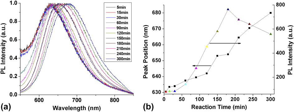
The optical performance of a series of BSA-Au NCs synthesized through a facile sonochemical method under ambient air with different reaction times was explored. As shown in Fig. 1, there was a continuous red shift from 630 to 680 nm in emission with the prolonging of the reaction time. This phenomenon may be caused by the increase in particle size. From Fig. 1(b), we can see that after reacting for 5 min, the solution is able to emit fluorescence at 630 nm, which demonstrates that this strategy is suitable to prepare Au NCs in a very short time. In addition, the PL intensity varies with different reaction time. As the synthesis time is prolonged, the PL intensity first increases, and then declines. It reaches a maximum value when the reaction time is 180 min.
(a) Emission spectra of BSA-Au NCs synthesized with different reaction times. (b) Temporal evolution of emission peak position and intensity.

B. Characterization of the Au NCs
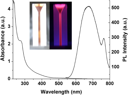
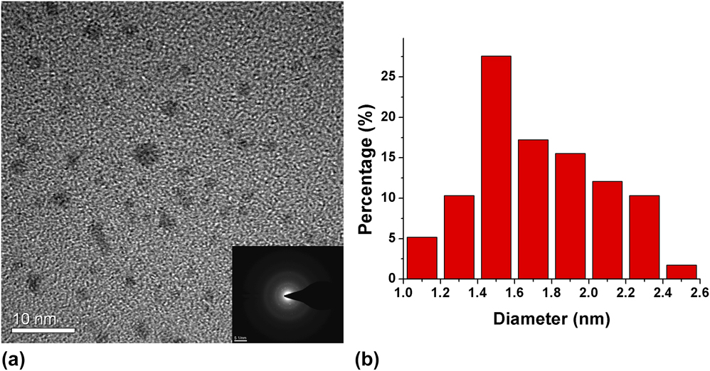
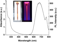
A digital camera was used to observe the solutions of BSA-Au NCs under visible light and ultraviolet light in a darkroom. As presented in the inset of Fig. 2, the obtained BSA-Au NCs exhibit a deep brown color under visible light, whereas after being radiated under a hand-held UV lamp, they emit red fluorescence. Then the as-prepared Au NCs solution was further characterized using a UV–vis scanning spectrophotometer and a fluorescence spectrophotometer, respectively. The UV–vis absorption and room temperature fluorescence spectra of Au NCs are displayed in Fig. 2. It illustrates the typical absorption and emission spectra of Au NCs. The excitonic absorption peak of the Au NCs is about 274 nm and corresponding maximum fluorescence emission is observed at 680 nm. The absolute quantum yield of the obtained Au NCs is 4.5% at the emitting wave length of 386 nm, which tends to be higher than that of Au NCs prepared by other synthetic methods. Reference Mu, Qi, Dong, Qiao, Hou, Nie and Ma64,Reference Venkatesh, Shukla, Sivakumar and Verma65,Reference Lin, Yang, Lee, Huang, Sperling, Zanella, Li, Shen, Wang, Yeh, Parak and Chang70–Reference Chen, Chiang and Chang72 A HRTEM analysis was performed to investigate the morphology of the Au NCs. The HRTEM image and size distribution histogram of the resultant particles are displayed in Fig. 3. As displayed in the figure, spherical particles are obtained with good dispersity. The Au NCs hold a narrow size distribution [Fig. 3(b)] with an average diameter of 1.5 nm. The inset of Fig. 3(a) is the SAED pattern of Au NCs obtained from a selected area of the Au NCs via a convergent beam. It demonstrates that the Au NCs possess a favorable crystal performance.
UV–vis absorption and fluorescence emission spectra of the visible-emitting Au NCs solution. Inset: photographs under visible light (left) and ultraviolet light (right) of the as-prepared visible-emitting Au NCs solution.

HRTEM micrograph (a) and size distribution histogram (b) of visible-emitting Au NCs. Inset: SAED pattern of visible-emitting Au NCs.

C. Optimization of experimental conditions
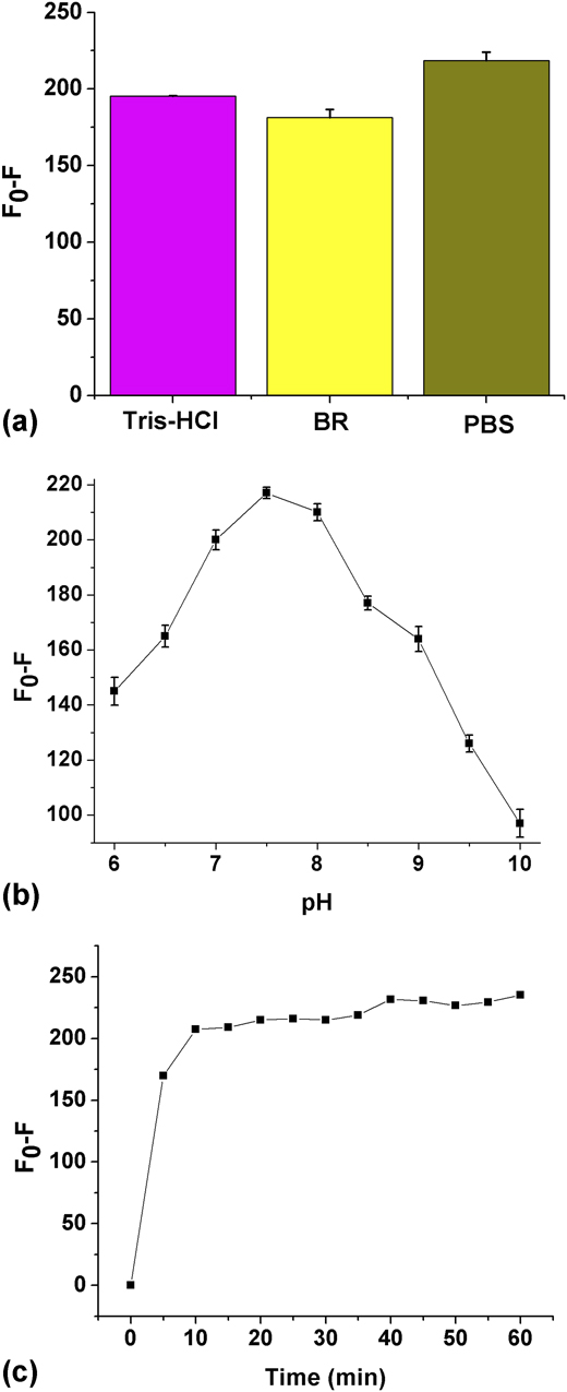
Since the fluorescence quenching of BSA-Au NCs is influenced by the kind of buffer solution and pH, these two important parameters and the response of the sensing material to the quencher were studied. Figure 4(a) shows the relative fluorescence intensity of BSA-Au NCs with Hg2+ in phosphate buffer solution (PBS), Tris-HCl, and Britton–Robinson (BR) buffer, respectively. Clearly, it can be observed that the fluorescence intensity of BSA-Au NCs in the PBS solution is quenched most remarkably. So in this paper, PBS was selected to control the pH of the system. Then the relationship between the decreasing extent of the BSA-Au NCs fluorescence and the pH value was investigated. It can be observed from Fig. 4(b) that the maximum relative fluorescence intensity occurs at pH 7.5. So pH 7.5 was chosen as the optimal pH value in this work. Figure 4(c) shows the effect of the contact time on fluorescence quenching. Clearly, the fluorescence intensity of BSA-Au NCs decreased significantly and the interaction between Hg2+ and BSA-Au NCs was over within 10 min. Therefore, we recorded the fluorescence intensity of the system after a reaction time of 10 min.
Effect of buffer solutions (a) and pH (b) on the interaction between visible-emitting Au NCs and Hg2+. (c) Time-dependent fluorescence intensity of the visible-emitting Au NCs to Hg2+.

D. Analysis of Hg2+ using Au NCs
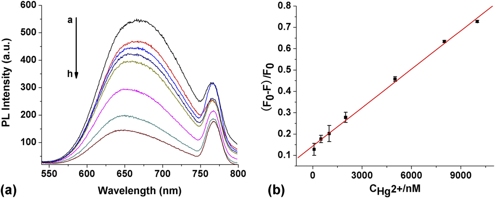
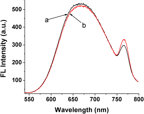
Under the optimum condition, the fluorescence assay was processed directly by mixing BSA-Au NCs and Hg2+ with different concentrations. The fluorescence of the BSA-Au NCs can be quickly quenched by Hg2+. The addition of 1 μM heavy metal solution has resulted in a decrease of the fluorescence intensity and the concomitant blue shift of the emission peak position. By further additions of the Hg2+ solution, the blue shift of the emission peak became more and more significant, moving from 660 down to 640 nm. The analytical potential of the as-prepared BSA-Au NCs was further explored by utilizing the quenching effects of Hg2+ on the BSA-Au NCs fluorescence. Typically, different concentrations of Hg2+ were placed into a quartz cuvette containing 300 μL Au NCs solution. It can be observed in Fig. 5(A) that with the increase of Hg2+, the fluorescence intensity of the BSA-Au NCs is gradually decreased, accompanied by a blue shift of the fluorescence emission peak. The calibration curve was prepared and the correlation of the Hg2+ concentration with the relative fluorescence intensity provided the basis for the quantitative analysis. As displayed in Fig. 5(B), the fitted curve shows a good linear relationship between (F 0–F)/F 0 and Hg2+ concentration in the range 8 × 10⁻⁸ M to 1 × 10⁻⁵ M. It is found that a relatively broad linear range can be obtained. The linear regression equation is described by the following equations: (F 0–F)/F 0 = 6.009 × 10⁻⁵[Q] + 0.1437, where F 0 and F are the fluorescence intensity of the BSA-Au NCs in the absence and presence of Hg2+, respectively. [Q] is the concentration of the quencher (Hg2+). The correlation coefficient R is 0.9982. Moreover, the limit of detection (LOD) was determined to be 1 nM (Fig. 6). Comparing this fluorescent method with electrochemiluminescent, Reference Zhu, Chen, Lin, Qiu and Chen73 colorimetric, Reference Huang and Chang74,Reference Yu and Tseng75 and other similar fluorescence methods based on Au NCs reported by other literature, Reference Xie, Zheng and Ying26,Reference Chen, Chiang and Chang72,Reference Wei, Wang, Yang, Tian, Hou and Lu76–Reference Kawasaki, Yoshimura, Hamaguchi and Arakawa78 the performance of our method is superior. Although the present probe is as sensitive as the electrochemical method, Reference Liu, Nie, Jiang, Shen and Yu79 this method has a broader linear range and is simpler and faster. In addition, the LOD is lower than the maximum level (10 nM) of mercury permitted in drinking water by the US Environmental Protection Agency (EPA). Reference Wang, Huang, Lin, Chen and Chang80 Therefore, the observed quenching effect can be used to monitor the concentration of Hg2+.
(A) Emission spectra of BSA-Au NCs in the presence of different concentrations of Hg2+: (a) 0, (b) 80, (c) 500, (d) 1000, (e) 2000, (f) 5000, (g) 8000, and (h) 10,000 nM Hg2+. (B) Calibration plot of the emission intensity against concentration of Hg2+.

Fluorescence emission spectra of BSA-Au NCs before (a) and after (b) the addition of 1 nM Hg2+.

E. Validation of the assay
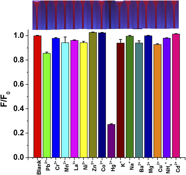
As shown in Fig. 5(A), the fluorescence of the BSA-Au NCs is remarkably quenched upon the addition of Hg2+, which strongly proves that Hg2+ is an effective quencher of BSA-Au NCs fluorescence. Generally, the reported optical sensors for Hg2+ are easily disturbed by other cations. In this sense, to investigate the selectivity of the BSA-Au NCs probe toward Hg2+, 14 common cations, including Pb2+, Cr3+, Mn2+, La3+, Ni2+, Zn2+, Co2+, K+, Na+, Ba2+, Mg2+, Cu2+, NH4 +, and Cd2+ ions (each at a concentration of 10 μM), were added to the solution containing 300 μL BSA-Au NCs. As is evident from Fig. 7, at pH 7.5, only Hg2+ ions induced a significant decrease in the fluorescence, whereas no obvious fluorescence changes are observed in the presence of other cations. Similar results were also observed under UV light (the inset of Fig. 7). The above results indicate that the proposed sensor had a high selectivity toward Hg2+.
Relative fluorescence (F/F 0) of visible-emitting Au NCs solution in the presence of various 10 μM cations. Inset: Photographs of the Au NCs solution in the presence of various 10 μM cations under UV light.

F. Detection of Hg2+ in real samples
To further investigate whether the Au NCs-based fluorescent nanoprobe was potentially applicable to natural systems, real water samples such as tap water and river water were analyzed using our Hg2+-sensing system. The Hg2+ in these water samples was not detectable. The water samples were spiked with Hg2+ at two concentration levels and the experimental results are listed in Table I. It can be seen that the mean recoveries range between 97.00 and 101.89% with relative standard deviations (RSD) under 3.0%. The analytical results are satisfactory. They suggest that this new sensing system is reliable for Hg2+ detection and has great potential for the quantitative analysis of mercury species in environmental samples.
Application of the BSA-Au NCs probe for Hg2+ detection in water samples.

G. Quenching mechanism
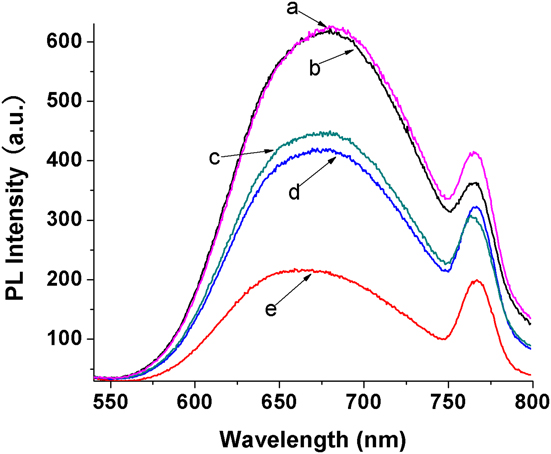
The fluorescence intensity of the BSA-templated Au NCs can be tremendously quenched by a trace amount of Hg2+. Generally, Au+ has strong and specific metallophilic interactions with Hg2+ (Au+(5d10) and Hg2+ (5d10)) Reference Xie, Zheng and Ying26,Reference Shang, Yang, Stockmar, Popescu, Trouillet, Bruns, Gerthsen and Nienhaus81 while thiolates (RS−) have intense interactions with Hg2+ as well. Reference Hu, Sheng, Gong, Zhang and Cai77 To prove these two hypotheses, one or another or both, verified experiments were conducted, as shown in Fig. 8. Firstly, sodium borohydride was added into the mixed solution of the BSA-stabilized Au NCs and Hg2+ and found that the fluorescence intensity of Au NCs had nearly returned to its original level (curve a). This demonstrates that the fluorescence quenching may be attributed to specific metallophilic interactions between Au+ (5d10) of the BSA-templated Au NCs and Hg2+ (5d10). Because, in this process, Hg2+ can be reduced to Hg0 by sodium borohydride, which has a weaker binding energy with Au+ relative to Hg2+. So fluorescence recovers after the addition of sodium borohydride. Secondly, sulfhydryl compounds, glutathione and tiopronin respectively, were added to the mixture of Au NCs-Hg2+ instead of sodium borohydride. The fluorescence intensity of the Au NCs was significantly restored compared to that in the presence of Hg2+ (curves c and d in Fig. 8). However, the fluorescence of the Au NCs was nearly unchanged after adding the glutathione or tiopronin in the absence of Hg2+. So the thiolates of glutathione would first react with Hg2+, which leads to nearly no interactions between Hg2+ and the visible-emitting Au NCs. The results indicate that the nature of the fluorescence quenching may be due to a specific reaction between Hg2+ and the thiolates of visible-emitting Au NCs. On synthesis of the situation, the fluorescence quenching mechanism may seem to include two reasons: one may be the metallophilic interactions between Au+ (5d10) and Hg2+ (5d10) on the surface of the visible-emitting Au NCs and the other may be the strong interactions between the thiolates (RS−) of the BSA-templated Au NCs and Hg2+. The test described above strongly supports our hypotheses, which are in accord with the previous studies. Reference Feng, Huang, Zhou, Cai, Tu and Wang82,Reference Feng, Huang, Chen, Chen, Lin and Wang83
PL spectra of the Au NCs at different conditions: (a) Au NCs + Hg2+ + NaBH4, (b) Au NCs, (c) Au NCs + Hg2+ + GSH, (d) Au NCs + Hg2+ + tiopronin, and (e) Au NCs + Hg2+.

IV. CONCLUSIONS
In summary, we present a facile, economic, and environmentally friendly method to prepare BSA-Au NCs with adjustable emission wave lengths ranging from 630 to 680 nm. Under optimal conditions, the sensing ability of the visible-emitting BSA-Au NCs for the simple, selective, and sensitive detection of Hg2+ in water samples was investigated. The LOD and linear concentration ranges of this method were found to be 1 nM and 8 × 10⁻⁸ M to 1 × 10⁻⁵ M respectively. Common metal ions such as Pb2+, Cr3+, Mn2+, La3+, Ni2+, Zn2+, Co2+, K+, Na+, Ba2+, Mg2+, Cu2+, NH4 +, and Cd2+ ions did not cause interference in the detection of Hg2+ ions when applying this highly sensitive and selective assay. Moreover, a smart method for investigating the interaction of BSA-Au NCs and Hg2+ system was proposed. The results indicated that the quenching of the BSA-Au NCs may be caused by two reasons: one is metallophilic interactions between Au+(5d10) and Hg2+(5d10) and the other is the strong interactions between thiolates (RS−) of the BSA-templated Au NCs and Hg2+.
ACKNOWLEDGMENTS
The authors are grateful for the financial support from the National Major Scientific Instruments and the Equipments Development Special Funds (No. 2011YQ030113), the National Natural Science Foundation of China (No. 21275105), the National Recruitment Program of Global Experts (NRPGE), the Hundred Talents Program of Sichuan Province (HTPSP), and the Startup Funding of Sichuan University for setting up the Research Center of Analytical Instrumentation.