Significant outcomes
• These findings suggest that NBP treatment after ischaemia can extend the time window for subsequent salvage by reperfusion.
Limitations
• Only immunohistochemical analyses of vascular endothelial growth factor (VEGF) were used.
• Only three time points of ischaemia-reperfusion were examined.
Introduction
Ischaemic cerebrovascular diseases pose a severe risk to human health. The standard treatment method involves dissolving the clot to restore blood flow in the blocked vessel, and the more rapidly the blood flow is restored to the brain, the fewer brain cells die Reference Huang, Xia and Zhou1, Reference Srivastava, Taly and Gupta2. However, this treatment strategy is limited by its short time window and potential for reperfusion injury, including haemorrhage development. As there is no really effective therapy for stroke except for thrombolysis in the very early stage, the prevention of stroke has been one of the most concerned issues.
Recent studies have shown that dl-3n-butylphthalide (NBP) can reduce ischaemic cerebral injury and rescue brain tissue after ischaemic stroke via multiple mechanisms including improving blood flow, antithrombotic and anti-inflammatory activity. However, the effect of NBP on prevention of stroke remains unknown Reference Zhang, Wang, Li and Wang3, Reference Liu, Liao and Zeng4. Treatment with NBP within 24-h post-ischaemic stroke may rescue brain tissue by enhancing angiogenesis associated with up-regulation of VEGF and HIF-1 alpha expressions Reference Liao, Lin, Pei, Liu, Zeng and Huang5. VEGF is the most potent and specific angiogenic factor yet identified. After binding to its receptors, VEGF plays important functions in angiogenesis and neuroprotection Reference Schmidt, Koeder and Messing6.
In this study, we investigated the effect of NBP preconditioning on focal cerebral ischaemia-reperfusion injury at three time points (2, 3 and 4 h) after onset of stroke and VEGF expression in a rat model to further define the protective mechanisms of NBP. In addition, we ask whether NBP therapy can extend the thrombolysis treatment time window in an experimental stroke model in rats.
Methods
Animals and materials
Healthy male Sprague-Dawley rats (250 ± 20 g, male) were purchased from the Center of Laboratory Animals, Shanghai Jiaotong University (Shanghai, China). NBP (lot No: 08100111) was purchased from NBP Pharmaceutical (Shijiazhuang, Hebei, China). Immunohistochemical kits were purchased from Boster Bioengineering (Wuhan, Hubei, China). Monoclonal anti-VEGF antibody was purchased from Santa Cruz Biotechnology (Santa Cruz, CA, USA). S-P kits were purchased from Zhongshan Biotechnology (Beijing, China). Nylon sutures (gauge 4, diameter approximately 0.25 mm) were treated with a hot plate to round their tips for subsequent use as intraluminal threads.
Animal model
Cerebral ischaemia-reperfusion was induced in rats by a modified intraluminal thread method Reference Longa, Weinstein, Carlson and Cummins7. Briefly, rats were anaesthetised by intraperitoneal injection of 6% chloral hydrate. An incision was made along the midline of the neck, the right common carotid artery was separated, and an intraluminal thread was inserted via the internal carotid and advanced along the right common carotid artery until feeling a slight resistance, thus obstructing the middle cerebral arterial blood flow to the brain. After maintaining the occlusion for different periods (2, 3 and 4 h), the blood flow was resumed by carefully removing the thread.
Animal treatment groups
Rats were randomly into a sham operation group, an ischaemia-reperfusion group and an NBP group. The rats in the ischaemia-reperfusion group underwent middle cerebral arterial occlusion (MCAO) for different periods (2, 3 or 4 h) followed by reperfusion; the three subgroups (eight rats/subgroup) were defined as ischaemia (2 h) reperfusion, ischaemia (3 h) reperfusion and ischaemia (4 h) reperfusion. The rats in the NBP group first underwent arterial occlusion for different periods (2, 3 or 4 h), and then immediately received intraperitoneal injection of NBP (200 mg/kg, pre-diluted) followed by reperfusion; the three subgroups (eight rats/subgroup) were defined as MCAO2h-NBP-reperfusion, MCAO3h-NBP-reperfusion and MCAO4h-NBP-reperfusion. Rats in the sham operation group and the ischaemia-reperfusion group were treated by intraperitoneal injection of the same volume of physiological saline after the ischaemic period.
Evaluation
Scoring of neurological functions
Rats were evaluated for neurological functions after reperfusion for 7 days using Longa's scoring scale. A score of 0 represents a normal appearance without neurological signs; 1 represents inability to fully extend the left forelimb; 2 represents circling to the left during walking; 3 represents falling to the left during walking and 4 represents no spontaneous walking and a decreased level of consciousness.
Measurement of infarction zone
After evaluation of neurological function, the rats were deeply anesthetised and the chest was opened to expose the heart. A syringe needle was inserted into the ascending aorta via the left ventricle, a small incision was made at the right atrial appendage and physiological saline at 4 °C was injected via the incision until the effluent was clear. The rats were then killed by decapitation, and the brain tissues were collected and sectioned along the coronal plane into five to six thin slices (approximately 2 mm thick). The slices were immersed in a triphenyl tetrazolium chloride solution (2%, 37 °C; Sigma-Aldrich Co. LLC, USA), placed in the dark for 30 min and then fixed with 4% neutral paraformaldehyde solution. The slices were photographed and the infarction areas measured using an image analysis system (Image Pro Plus; Media Cybernetics, Silver Spring, MD, USA). The infarction zone was calculated by: sum of infarction area from all slices × interslice spacing, and was expressed as both absolute volume (mm3) and volumetric fraction (%).
Immunohistochemical analyses of VEGF
As above, rats were deeply anaesthetised and perfused with physiological saline at 4 °C. Next, 250 ml of 4% paraformaldehyde was injected via the incision (initially relatively fast and subsequently slowed down, for approximately 1 h). The rats were killed by decapitation, and the brain tissues were collected and fixed in 4% paraformaldehyde (4 °C for 6 h). The tissues were dehydrated, embedded in paraffin, sectioned and then stained with anti-VEGF antibody (1:150) as follows. The sections were dewaxed and rehydrated, followed by treatment in a trypsin-based antigen retrieval solution (Wuhan Boster Biotech. Co., Wuhan, China) for 30 min. The sections were then transferred into a 3% H2O2 solution and incubated at room temperature for 10 min. The slices were incubated with primary antibody (4 °C overnight), followed by biotin-labelled secondary antibody (room temperature, 30 min) and then visualised with diaminobenzidine (DAB). The sections were cover-slipped and sealed, and examined by light microscopy to identify cells positively stained for VEGF. Cells with brown particles in their cytoplasm or nuclei were interpreted as VEGF positive. Five high magnification fields (×400) were imaged on each slice (HPIAS-2000 imaging system; Wuhan, China), and the number of VEGF-positive cells were counted and averaged.
Statistical analyses
Data were expressed as mean ± SD and analysed by t-tests with SPSS15.0 (SPSS, Chicago, IL, USA). A p-value less than 0.05 was considered statistically significant.
Results
Effect of NBP on ischaemia-reperfusion injury
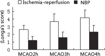
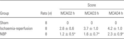
There was a progressive increase in neurological function score with increasing length of the ischaemic period before subsequent reperfusion (Table 1, Fig. 1). Treatment with NBP immediately after ischaemia caused a significant reduction in the neurological function score for all three ischaemic periods (p < 0.01 for all). No significant difference existed in the neurological function score among the subgroups of (2 h), (3 h) and (4 h) NBP-reperfusion groups.
Comparison of neurological function scores between the ischaemia-reperfusion subgroups and the NBP subgroups.

Neurological function scores
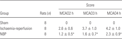
p < 0.01 vs. the corresponding ischaemia-reperfusion subgroup with the same length of ischaemic period.
Effect of NBP on infarction zone
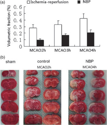
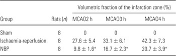
There was a progressive increase in the volumetric fraction of the infarction zone with increasing length of the ischaemic period before reperfusion (Table 2, Fig. 2). Treatment with NBP caused a significant decrease in the volumetric fraction of the infarction zone for all three ischaemic periods compared with the three ischaemia-reperfusion subgroups (p < 0.01 for all). No significant difference existed in the volumetric fraction of the infarction zone among the subgroups of (2 h), (3 h) and (4 h) NBP-reperfusion groups.
(a) Comparison of volumetric fractions of the infarction zones between the ischaemia-reperfusion subgroups and the NBP subgroups. (b) Photomicrographs showing the infarction areas in different layers.

Volumetric fractions of the infarction zones

* p < 0.01 vs. the corresponding ischaemia-reperfusion subgroup with the same length of ischaemic period.
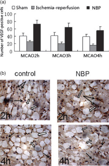
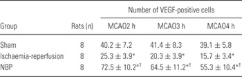
Expression of VEGF also progressively decreased with increasing length of the ischaemic period before reperfusion (Table 3, Fig. 3), to levels significantly less than the corresponding sham operation subgroups (p < 0.01 for all). Treatment with NBP caused a significant increase in VEGF expression in all ischaemia-reperfusion subgroups compared with the corresponding sham operation subgroups (p < 0.01 for all).
(a) Comparison of VEGF expression levels between the different groups. (b) Photomicrographs showing VEGF expression (black arrow) in the border zone of an infarct (blank arrow) in the control and NBP groups at 2 and 4 h of reperfusion (×400).

Expression of VEGF-positive cells

* p < 0.01 vs. the corresponding sham operation subgroup with the same length of sham-ischaemic period.
† p < 0.01 compared with the corresponding ischaemia-reperfusion subgroup with the same length of ischaemic period.
Discussion
In this study, we focused on the potential effect of NBP to reduce ischaemia-reperfusion damage. Our findings indicated that NBP has a protective effect on cerebral ischaemia, probably related to VEGF expression in brain tissues, and results suggest that NBP treatment may extend the time window for subsequent salvage of cerebral ischaemia by reperfusion.
Ischaemic stroke is a serious disease caused by a thrombus (blood clot), and can result in permanent neurological damage and even death Reference Nam, Choi and Jung8. The standard treatment method involves dissolving the clot to restore blood flow in the blocked vessel, and the more rapidly blood flow is restored to the brain, the fewer brain cells die Reference Huang, Xia and Zhou1, Reference Srivastava, Taly and Gupta2. However, this treatment strategy is limited by its short time window and potential for reperfusion injury, including haemorrhage development. Vascular protection (reducing haemorrhage and oedema formation) has emerged as a promising strategy to improve outcome and speed recovery from acute ischaemic stroke.
Numerous basic and clinic studies have shown that NBP has potential for treatment of ischaemic stroke, with multiple actions against various pathophysiological processes including improving microcirculation, inhibition of platelet aggregation, regulation of energy metabolism and inhibition of ischaemia-induced oxidative damage and neuronal apoptosis Reference Chong and Feng9, Reference Dong and Feng10, Reference Graf11. In addition, as angiogenesis can protect the brain from focal cerebral ischaemia, NBP may increase the number of the perfused vessels by stimulating angiogenesis Reference Zhang, Wang, Li and Wang3. Liao et al. showed that treatment with NBP within 24 h post-ischaemic stroke may rescue brain tissue by enhancing angiogenesis associated with up-regulation of VEGF and HIF-1 alpha expressions Reference Liao, Lin, Pei, Liu, Zeng and Huang5. NBP treatment has been shown to rescue brain tissue after focal ischaemia-reperfusion injury in rats Reference Liu, Liao and Zeng4, Reference Graf11. However, the effect of NBP on the time window for salvage of cerebral ischaemia is unknown. In this study, we used a rat model of brain artery occlusion that is pathologically similar to ischaemic stroke encountered clinically, and we quantified the effect of NBP treatment on the time window for salvage of cerebral ischaemia by Longa's scoring scale. We found that neurological function score and infarction volume both increased with the increasing length of ischaemic period, while NBP treatment administered after ischaemia significantly reduced the Longa's score and infarction volume at all ischaemia times. Further, these parameters were similar between the subgroup ischaemia (4 h)-NBP-reperfusion and the ischaemia (2 h)-NBP-reperfusion groups. Thus, these data suggest that NBP can reduce central nervouse system (CNS) ischaemia-reperfusion injury and extend the time window for subsequent salvage by reperfusion.
VEGF is a potent angiogenic, neurotrophic and neuroprotective factor, and is up-regulated by focal cerebral ischaemia in both animal models and in human patients Reference Shi and Liu12, Reference Sims and Muyderman13, 14, Reference Chen, Wu and Chen15. VEGF also plays a vital role during neural and vascular remodelling after stroke 14, Reference Chen, Wu and Chen15, Reference Jin, Mao and Greenberg16, Reference Yasuhara, Shingo and Kobayashi17. VEGF functions via activation of its receptors on the surface of target cells. In cerebral ischaemic lesions, expression of the VEGF receptor Flt-1 is increased in neurons, neuroglial cells and endothelial cells, while expression of another VEGF receptor, Flk-1, is increased predominantly in neuroglial cells and less in endothelial cells. These observations suggest that expression of VEGF after focal cerebral ischaemia may function to limit brain injury Reference Rosenstein and Krum18. In focal cerebral ischaemia, VEGF expression is predominantly increased in the ischaemic penumbra, but rarely in the ischaemic centre, which may relate to impaired VEFG synthesis resulting from extensive cell death and neuronal damage in the infarction zone. Consequently, VEGF expression is higher with increased survival of cells in the penumbra Reference Lennmyr, Ata, Funa, Olsson and Terent19. As such, the levels of VEGF expression reflect the number of surviving cells in the penumbra, helps determine the existence of a penumbra and its size and provides the basis for determining whether a patient should undergo thrombolytic therapy. Nevertheless, VEGF has the potential to increase vascular permeability, leading to increased oedema and haemorrhage in some models Reference Zhang, Wang, Li and Wang3.
In conclusion, we found that NBP significantly reduced infarction volume and improved neurobehavioral deficits in a rat model of MCAO, which may relate to enhanced VEGF expression. Our data also suggest that NBP treatment may extend the therapeutic time window of reperfusion. Further studies are required to completely elucidate the mechanisms that regulate the protective effects of NBP against cerebral ischaemia.
Acknowledgements
This study was supported in part by a grant from the Shanghai Natural Science Foundation (09ZR1424400). The authors report no conflicts of interest. The authors alone are responsible for the content and writing of the article.