Brucellosis is a major bacterial zoonosis with a worldwide distribution. The causative organisms are Gram-negative bacteria that belong to the class α-proteobacteria. These pathogens can affect a wide variety of mammals, including humans. Eight species are known to infect specific animals: sheep and goats (Brucella melitensis), cows (Brucella abortus), pigs (Brucella suis), dogs (Brucella canis), rodents (Brucella neotomae), sheep (Brucella ovis) and marine species (Brucella pinnipediae and Brucella cetacea) (Refs Reference Bricker1, Reference Nielsen2). Among these, three are virulent for humans (B. melitensis, B. abortus and B. suis). Human brucellosis, also known as Malta Fever, remains endemic in the Mediterranean basin, the Middle East, Central Asia, Africa and Latin America (Ref. Reference Corbel3).
Domestic and wild animals acquire brucellosis through ingestion of contaminated milk or from contact with contaminated tissues and fluids associated with birthing or abortion. These animals potentially serve as a source for reintroduction of brucellosis to additional food animals (Ref. Reference Davis and Elzer4). For these reasons, Brucella spp. are of great concern to the agricultural community worldwide (Ref. Reference Boschiroli, Foulongne and O'Callaghan5). In many industrialised countries, brucellosis in food animals has been effectively controlled through the implementation of vaccination, surveillance and quarantine programmes. However, the high cost of these controls can have a significant impact on the agricultural economy.
Transmission of brucellosis to humans occurs through skin abrasions when in contact with infected animals or by the consumption of unpasteurised dairy products. No human-to-human transmission has been identified; however, people may be infected by inhalation of contaminated dust or aerosols. This accounts for Brucella spp. being one of the most common laboratory-acquired pathogens worldwide (Ref. Reference Bouza6), and for their inclusion in the potential biological weapons list.
Pathogenic Brucella spp. cause abortion in female animals by colonisation of placental trophoblasts, and can cause sterility in male animals (Ref. Reference Corbel3). Human infections with B. abortus or B. melitensis may present in varying forms and can become life-threatening. Although the majority of patients present with general symptoms, such as fever, malaise, sweats and lymphadenopathy and/or hepatosplenomegaly, a more severe form of the disease can also occur, usually accompanied with osteoarticular problems or (in males) epididymoorchitis. More serious and hard-to-treat complications include, in descending order of frequency, spondylitis, neurobrucellosis, liver abscesses and endocarditis. The human disease tends to recur, despite adequate antibiotic regimens.
B. melitensis, B. abortus and B. suis have long been considered as potential biological weapons, and are classified as category B pathogens. In fact, one important bioterrorism-related characteristic of B. melitensis is the very small inoculum needed to induce human disease: 10 to 100 microorganisms are sufficient to cause a debilitating infection for several weeks (Ref. Reference Pappas7). Moreover, as Brucella spp. can be easily aerosolised, this pathogen could be used to develop biological weapons. Compared with other potential biological weapons, the inoculation period with Brucella spp. is relatively protracted, ranging from 9 to 60 days. Thus, the bioweapon potential of Brucella spp. may actually be only of historical importance (Ref. Reference Pappas7).
Three animal vaccines exist to prevent brucellosis: two smooth live vaccine strains – S19 for cattle and Rev 1 for small ruminants – and the rough attenuated strain RB51 for cattle. However, these vaccines cannot be used in humans because they offer only moderate protection along with a considerable risk of side-effects, including induction of antibiotic (rifampicin)-resistant cases of certain strains of the disease (Refs Reference Banai8, Reference Blasco and Diaz9, Reference Moriyon10, Reference Nicoletti11). There is presently no safe and effective vaccine that can be used to prevent the human disease and treatment requires a combination of two antibiotics – doxycycline and rifampicin, or doxycycline and streptomycin – for prolonged periods (Ref. Reference Young12).
Brucella cell biology
Invasion of phagocytic and nonphagocytic cells
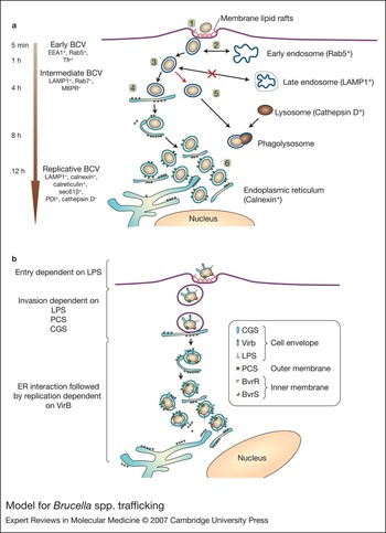
Pathogenic Brucella can invade and multiply intracellularly in both phagocytic cells (J744 and THP1 macrophages) (Refs Reference Celli13, Reference Liautard14) and nonphagocytic cells (HeLa, Vero, trophoblast and 3T3 fibroblasts) (Refs Reference Pizarro-Cerda15, Reference Pizarro-Cerda, Moreno and Gorvel16, Reference Delrue17). Several studies show that once inside the cell, B. abortus is surrounded by a membrane-bound compartment to form the Brucella-containing vacuole (BCV). It is able to escape the endocytic pathway to reach a safe place within the cell where the bacteria are protected from host-defence mechanisms to ensure their replication and thus their virulence (Refs Reference Kohler18, Reference Kohler19) (Fig. 1a).
Model for Brucella spp. trafficking.(Legend; see previous page for figure.) (a) Pathogenic Brucella invades phagocytic and nonphagocytic cells through lipid rafts (1). After lipid-raft-mediated entry, intracellular pathogenic Brucella resides within an early Brucella-containing vacuole (BCV) that interacts with early endosomes (2). These endosomes are marked by positive EEA, Rab5 and TfR expression. Pathogenic Brucella avoids further interaction with the endocytic pathway in an intermediate nonreplicative BCV (3). The bacterium then either interacts and fuses with the endoplasmic reticulum (ER) (4) to generate an ER-derived organelle permissive for Brucella spp. replication, or fails to maintain interaction with the ER and is degraded by fusion with phagolysosomes (5). Once safely in the ER within replicative BCVs (6), Brucella spp. divide rapidly. (b) Model for evasion of lysosomal killing through expression of virulence factors. Brucella spp. entry is dependent on its LPS and the regulatory system BvrR/BvrS at the surface. LPS and other virulence factors, such as the phosphatidylcholine synthase and the cyclic β-1,2 glucan, then play a role in the early stages of intracellular trafficking. Subsequently, interaction and fusion with the ER elements leading to bacterial replication is dependent on the VirB type IV secretion system, since a virB-defective mutant fails to sustain fusion-proficient interactions with the ER and ultimately fuses with lysosomes. Abbreviations: BvrR, Brucella virulence-related regulatory protein; BvrS, Brucella virulence-related sensory protein; CGS, cyclic β-1,2 glucan synthetase; LPS, lipopolysaccharide; PCS, phosphatidylcholine synthase; VirB, VirB type IV secretion system.

Bacterial trafficking within host cells
During the first few minutes after invasion of nonprofessional phagocytes, virulent B. abortus, B. melitensis and the attenuated vaccine strain B. abortus S19 interact with the early endosomal compartment. This is confirmed by the detection of early endosomal markers, such as the transferrin receptor, the small GTP-binding protein Rab5 or the early endosomal antigen (EEA1), in the BCVs (Fig. 1) (Refs Reference Pizarro-Cerda15, Reference Delrue17). This association with early endosomes is transient and starts to decrease significantly 10 min post infection. No early endosome markers are detected 30 min after infection (Refs Reference Pizarro-Cerda15, Reference Delrue17). At this time, BCVs progressively acquire the LAMP1 (lysosome-associated membrane protein 1) marker and remain LAMP1+ up to 4 h post infection. Nevertheless, in contrast to phagosomes containing latex beads or dead bacteria, which transiently interact with late endocytic compartments (characterised by the presence of the small GTP-binding protein Rab7 and mannose-6-phosphate receptors), BCVs do not fuse with late endosomes. Phagosomes containing latex beads or dead bacteria also have a high concentration of cathepsins (nearly 100% of phagosome contents at 2 h post infection). These luminal lysosomal markers are absent from BCVs of virulent strains. BCVs acquire endoplasmic reticulum (ER) markers, such as calreticulin or calnexin. The luminal ER marker glucose-6-phosphatase confirms fusion of BCVs and the ER (Ref. Reference Celli13). At this stage, B. abortus has reached a safe niche that ensures its replication and its virulence. Several virulence factors ensure trafficking of the microorganisms to this safe haven and are discussed below.
Brucella and autophagy
The occurrence of autophagy in Brucella spp. intracellular trafficking remains a matter of debate. In epithelial cells, B. abortus can be found in autophagosomes (Ref. Reference Pizarro-Cerda15); however, autophagic BCVs have not been detected in macrophages (Ref. Reference Celli13). One hypothesis is that autophagy might be a host-defence mechanism that prevails in nonphagocytic cells to fight invading bacteria. Such a method of attack seems limited to nonphagocytic cells. Indeed, macrophages seem to be efficient at killing Brucella and even with wild-type B. abortus infection, 80–90% of bacteria are killed within the first few hours of infection, which is not the case in epithelial cells. However some bacteria resist macrophage killing and are able to survive and replicate (Ref. Reference Celli13).
Virulence factors
Lipopolysaccharides
The lipopolysaccharides (LPSs) of Gram-negative bacteria are composed of a hydrophobic lipid A linked to a charged, densely compact oligosaccharide core, which is associated (smooth-LPS) or not (rough-LPS) with a hydrophilic O-polysaccharide chain (O-chain). In contrast to enterobacteria, such as Escherichia coli, Brucella spp. possess a nonclassical LPS. B. abortus lipid A has a diaminoglucose backbone (rather than glucosamine) and acyl groups are longer (C18–C19 or C28 rather than C12 and C14) and are linked to the core by simple amide bonds (rather than ester and amide bonds) (Ref. Reference Keleti, Feingold and Youngner20). It is proposed that this nonclassical structure confers B. abortus LPS with particular virulence factor characteristics. Indeed, this structure alters LPS–pathogen-associated molecular pattern (PAMP) associations and reduces the endotoxin-related properties typical of LPSs. B. abortus LPS is several hundred times less active and toxic than the classical E. coli LPS (Refs Reference Keleti, Feingold and Youngner20, Reference Moreno, Berman and Boettcher21, Reference Goldstein22) and is a poor inducer of respiratory burst, bactericidal nitrogen intermediates and lysozyme secretion (Refs Reference Rasool23, Reference Riley and Robertson24). Although it has been demonstrated that Brucella spp. LPSs interact with the Toll-like receptor 4 (TLR-4), this interaction has little influence on the production of cytokines, such as tumour necrosis factor α (TNF-α), type I and type II interferons, or antimicrobial proteins, such as the GTPases of the p-47 family (Refs Reference Keleti, Feingold and Youngner20, Reference Goldstein22, Reference Lapaque25).
B. abortus LPS has been shown to impair antimicrobial responses by inhibiting the deposition of complement at the bacterial surface and antibacterial-peptide attacks. In fact, the B. abortus O-chain blocks access of C1Q to the outer membrane protein targets (Refs Reference Moreno, Berman and Boettcher21, Reference Eisenschenk, Houle and Hoffmann26). It has also been shown that Brucella spp. are resistant to a large variety of such proteins, including defensin NP-2, lactoferrin, cecropines, lysozyme, bactenecin-derived peptides and the defensin-like antibiotic polymyxin B, as well as to crude lysosomal extracts from polymorphonuclear leukocytes (Refs Reference Martinez de Tejada27, Reference Freer28, Reference Velasco29). Moreover, the O-chain and lipid A are also implicated in the entry and early stages of B. abortus trafficking (Refs Reference Sola-Landa30, Reference Detilleux, Deyoe and Cheville31). The rough strain of Brucella spp. (which does not possess the O-chain) does not enter by lipid rafts, and fuses rapidly with lysosomes. By contrast, smooth B. suis entry is lipid-raft dependent and impairs phagosome–lysosome fusion during the first few hours of infection (Ref. Reference Porte32).
The LPS of Brucella spp. also acts as a modulator of the immune response. The B. abortus LPS O-chain appears to impair an antimicrobial response by preventing the synthesis of immune mediators. In contrast to that observed with smooth strains of B. suis and B. melitensis, rough strains induce both the secretion of inflammatory cytokines and expression of nitric oxide synthase (iNOS) at a similar level to that reached upon E. coli LPS stimulation (Ref. Reference Jimenez de Bagues33).
Once B. abortus is internalised, it follows the same classical endocytic pathway as protein antigens but with slower kinetics (Ref. Reference Forestier34). B. abortus LPS is next found in a MHC class II compartment (MIIC), the role of which is to load antigenic peptides onto MHC class II molecules. From there, the LPS is subsequently recycled to the cell surface where it forms large stable clusters with MHC class II in LPS macrodomains (Refs Reference Forestier34, Reference Forestier35, Reference Forestier36). In these macrodomains, B. abortus LPS interferes with MHC class II presentation of peptides to specific CD4+ cells but has no effect on MHC class I antigenic presentation (Ref. Reference Forestier36).
The two-component BvrR/BvrS system
The B. abortus BvrR/BvrS (for Brucella virulence regulator) two-component regulatory system is similar to the regulatory and sensory proteins of Sinorhizobium meliloti and Agrobacterium tumefaciens, which are necessary for endosymbiosis and pathogenicity in plants, and very similar to a putative system present in the animal pathogen Bartonella bacilliformis. The two-component BvrR/BvrS system is crucial for the control of polycation resistance, virulence, cell invasion and intracellular replication (Ref. Reference Sola-Landa30). The bvrR and bvrS genes encode proteins of 237 and 601 amino acids, respectively, which regulate the composition of the outer membrane. Two-dimensional gel electrophoresis studies showed differences in various outer membrane proteins (Omps) between the wild type and both B. abortus bvrR and bvrS mutants (Refs Reference Guzman-Verri37, Reference Lamontagne38). Omp25 (also known as Omp3a) is not expressed in bvrR and bvrS mutants and Omp22 (also known as Omp3b) is absent in the bvrS mutant only. Both bvrR and bvrS mutants are less invasive than the wild-type strain and both mutants fail to replicate within phagocytic or nonphagocytic cells and are targeted to lysosomes for degradation (Ref. Reference Sola-Landa30). Dysfunction of BvrR and BvrS also diminishes the characteristic resistance of B. abortus to bactericidal cationic peptides and increases its permeability to surfactants (Ref. Reference Sola-Landa30). BvrR/BvrS-null mutants are incapable of directly activating the small GTPase Cdc42 upon cell contact and consequently these mutants cannot invade cells. They remain bound to the host cell surface, indicating that the two-component system controls the expression of factors necessary for invasion (Ref. Reference Guzman-Verri39). Recent studies showed that the lipid A of the bvrR and bvrS mutants is modified and exhibits altered acylation, acyl-chain fluidity and aggregate structure (Ref. Reference Manterola40). Nevertheless, the BvrR/BvrS system is not implicated in the regulation of genes involved in the biosynthesis of lipid A, because levels of these gene transcripts in wild-type strain and bvrR and bvrS mutants are identical (Ref. Reference Manterola40). This suggests that the overall defect in the bvr null mutants is not restricted to the absence of the BvrR/BvrS proteins, but rather that there is a more complex interaction of molecular events that needs to be dissected.
virB operon
The type IV secretion system, encoded by the virB region, is a key virulence factor for Brucella spp. (Refs Reference Celli13, Reference O'Callaghan41). The virB region is composed of 12 genes that form an operon specifically induced by phagosome acidification in cells after phagocytosis (Ref. Reference Porte, Liautard and Kohler42). In general, type IV secretion systems have been shown to transfer multimolecular complexes across the bacterial membrane to the extracellular space, the host cytosol or into other cells (Ref. Reference Cascales and Christie43); for example, Agrobacterium tumefaciens transfers DNA from bacteria to plant cells (Ref. Reference Christie44), Bordetella pertussis secretes the pertussis toxin through the Ptl system (Ref. Reference Covacci and Rappuoli45) and Legionella pneumophila exports proteins through the Dot/Icm system (Ref. Reference Bardill, Miller and Vogel46). This suggests that Brucella spp. use their type IV secretion system to secrete effector molecules into the host cytosol; however, no effectors have yet been identified. Structure–function studies of the A. tumefaciens VirB system showed that there are three functional groups of proteins: the pilus at the bacterial surface (VirB2 and VirB5), a pore crossing the two membranes (VirB3 and VirB6–VirB10) and two ATPases (VirB4 and VirB11) on the cytoplasmic side of the inner membrane, which energise the assembly of the complex as well as transport of the substrates through the complex (Ref. Reference Ward47). The virB region is essential for intracellular survival and replication in B. suis, B. abortus and B. melitensis because virB mutants are unable to bypass the endocytic pathway to reach their replicative niche in the ER, but rather fuse with lysosomes and are eliminated (Refs Reference Celli13, Reference Comerci48, Reference Kahl-McDonagh49). The VirB apparatus is essential for sustaining interactions and fusion events between the BCV and ER elements (Refs Reference Celli13, Reference Arellano-Reynoso50) (Fig. 1b).
Cyclic β-1,2 glucan
The B. abortus genome encodes a high molecular weight (316.2 kDa) inner-membrane protein encoded by the cyclic β-1,2 glucan synthetase gene (cgs). This protein shares similarities with a similar enzyme in A. tumefaciens (encoded by chvB) and Sinorhizobium meliloti (encoded by ndvB). Moreover, these three genes have highly conserved functions, because Agrobacterium or Sinorhizobium cyclic β-1,2 glucan mutants (cgs–) can be complemented by the Brucella spp. cgs gene (Ref. Reference de Iannino51).
Brucella spp. cgs mutants have reduced virulence in mice and are defective in intracellular replication in HeLa cells (Refs Reference de Iannino51, Reference Briones52). Furthermore, because of its ring appearance, cyclic β-1,2 glucan has been compared to cyclodextrin and, like cyclodextrin, may act as an inclusion agent for hydrophobic molecules (Ref. Reference Bohin53). Some synthetic cyclodextrins are able to act as membrane-damaging agents and can carry and selectively extract cholesterol. Cholesterol, with sphingosine, is the main constituent of lipid rafts present on phagosomes. It has been shown that cgs mutants fail to prevent phagosome–lysosome fusion and they cannot replicate (Ref. Reference Arellano-Reynoso50). However, when HeLa cells are treated with purified cyclic β-1,2 glucan or synthetic methyl-β cyclodextrin, cgs mutants control vacuole maturation by avoiding fusion with lysosomes, and thus allow intracellular Brucella spp. to survive and reach its replication niche (Ref. Reference Arellano-Reynoso50).
Phosphatidylcholine
Phosphatidylcholine (PC) is a typical eukaryotic phospholipid that is absent from most prokaryotes. B. abortus possesses this lipid in its outer membrane, and the mutation of a key enzyme of bacterial PC biosynthetic pathways (PC synthase) results in reduced growth in a rich medium (Ref. Reference Conde-Alvarez54). Moreover, in macrophages, pcs mutants show a reduced ability to avoid lysosome fusion. In vivo, pcs mutants are attenuated (Ref. Reference Conde-Alvarez54), suggesting that the phospholipid contributes to Brucella spp. virulence. It is thought that PC is a bacterial product that is used as a mimicry molecule to avoid recognition by the host.
Clinical implications
Infiltration of Brucella spp. into professional and nonprofessional phagocytes causes the wide spectrum of clinical symptoms of brucellosis presented and sets important therapeutic limitations, both in terms of antibiotic use and in terms of human vaccine development. Human brucellosis can involve practically any body system, and the clinical manifestations are often considered as an equilibrium between expression of pathogen virulence factors and the host immune response. In humans, the immune response is of a predominantly cellular Th1 type, mediated principally by interferon γ; a humoral component may also be implicated in the early stages. The affinity of the pathogen for the reticuloendothelial system means that bone marrow may serve as the ideal niche for isolating the pathogen.
By hiding intracellularly without interrupting the cell life cycle Brucella spp. manage to largely escape the host immune response: thus, bacterial multiplication can continue for prolonged periods, resulting in a relapsing form of the disease, with months or even years of apparently disease-free periods. The recent recognition of positive results for Brucella spp. gene expression in real-time PCR in apparently healthy individuals, years after disease manifestation and successful treatment, suggests that eradication of the pathogen may not even be feasible (Ref. Reference Navarro55).
Importantly, the acidic environment of the BCV serves as a neutraliser of antibiotics. Thus, macrolides are typically inactive against brucellosis. Furthermore, antibiotics with acknowledged in vitro and in vivo activity against Brucella spp. may show an attenuated intracellular activity, necessitating their prolonged and combined administration. Monotherapeutic regimens in the past have been largely disappointing (Ref. Reference Pappas, Akritidis and Tsianos56), and attempts at shortening the duration of therapeutic regimens have shown a statistically significant higher relapse and failure percentage (Ref. Reference Solera57). This phenomenon underlines the dissociation between in vitro susceptibility testing and in vivo antibiotic performance, as exemplified by numerous cases of treatment failure (Ref. Reference Al Dahouk58).
Research in progress and outstanding research questions
The unique and complex nature of Brucella spp. pathophysiology carries significant issues for the development of a human vaccine. Low LPS virulence and immunogenicity (relative to other Gram-negative pathogens) has hampered the development of live attenuated vaccines. This could be because the mutation of a specific single gene has little effect on virulence unless it is a global regulator, or because two mutations result in too great an attenuation that an immune response is not induced. Recent years have seen various attempts to develop a DNA vaccine for Brucella spp. utilising numerous peptides (Refs Reference Al-Mariri59, Reference Cassataro60, Reference Cassataro61, Reference Munoz-Montesino62, Reference Yu, Hu and Cai63). Most of the studies have shown satisfying results in animal models but no clear potential for further experimentation in humans. The main reason behind this failure is that targeting only the Th1 response may be inadequate. In the latter stages, cytotoxicity may be more important in controlling human infection, whereas in the early bacteraemic stages, a humoral response may limit disease spread and the appearance of focal complications. The development of a bivalent (or even trivalent) vaccine encompassing all these aspects is at present stalled by problems with vectors of DNA vaccines, and by an inability to select the optimal combination of peptides. Moreover, the predominant immunogenicity of LPS and outer-membrane peptides in general might minimise the immunogenic response elicited by other vaccine components (for a review, see Ref. Reference Al-Mariri59).
Study of the interaction between Brucella spp. and the host reveals unique properties, which are mainly focused on the low activation profile of the immune response. The two main factors responsible are the Brucella spp. LPS and the residence of the pathogenic bacteria in safe intracellular compartments, such as the ER. The direct consequence is that these properties affect treatment options. In addition, the fact that it is very difficult to predict the clinical manifestation in humans increases the problems of diagnosis, especially in resource-deprived countries. The disease manifests in vague symptoms, tends to be chronic or relapsing, and needs protracted antibiotic combination treatment for eradication, if the latter is feasible. A hope for the future will be to bring basic science and clinical developments in the field of diagnosis and treatment of brucellosis. Indeed, the complex interplay between the pathogen and host immune response further enforces significant problems in human vaccine development because, at the molecular level, pathogenic Brucella spp. have developed a ‘stealth’ strategy by expressing molecules with low stimulatory activity and toxicity for cells, allowing the bacterium to reach its replication niche before activation of antimicrobial mechanisms by the host. In conclusion, brucellosis is a complex disease, which requires not only optimisation of treatment, but also a good relationship between physicians, patients and molecular bacteriologists.
Acknowledgements and funding
This work was supported by a grant from the Ministère de la Recherche et Technologie. We thank the anonymous peer reviewers for their insightful comments.