Introduction
The problem of foreign bodies impacted in the oesophagus is fairly common. Impaction of meat bone in the oesophagus is common in adults and impacted coins are commonly found in children. Unlike bones and coins, which are readily detectable by conventional radiology, swallowed dentures can present a diagnostic challenge. However, few studies have comprehensively discussed the problems associated with managing impacted dentures in the oesophagus.
This study aimed to determine the incidence of denture impaction in the oesophagus; review the clinical features, patient profile and type of dentures swallowed; evaluate the role of plain radiographs in diagnosis; examine the problems encountered in managing such cases; and suggest measures to reduce the incidence.
Materials and methods
The records of all patients admitted with a history of denture ingestion under the care of the first author during the period 1999 to 2010 were reviewed. The types of prostheses and the activities carried out during the occurrence of ingestion were noted. The patients’ symptoms, especially the site of pain felt during deglutition (as indicated by the patient), were compared to the actual site of foreign body lodgement (measured in centimetres from the upper incisors). The radiological investigations included anteroposterior and lateral X-ray views of soft tissue in the neck, chest or abdomen. Computed tomography (CT) was performed in selected cases. Patient morbidity was assessed and the operative notes were reviewed in order to identify the problems encountered in managing such patients.
Results
Out of a total of 262 patients admitted with suspected foreign body ingestion, 47 patients (34 males and 13 females, aged 35 to 84 years) accidentally swallowed dentures. The offending dentures were all partial dentures containing incisors; there were 25 upper and 22 lower dentures. Forty-five of the dentures were successfully removed, and one passed through the gastrointestinal tract naturally. In one case, no foreign body was found, even after extensive search.
The most common activity during ingestion was drinking (39 of 47 patients). The type of drinking associated with this problem is widely prevalent in India: water is continuously poured into the open mouth with the head turned upwards, and is swallowed by movement of the tongue and contraction of the floor of the mouth (Figure 1). In other cases, the dentures were accidentally swallowed during eating (n = 4), sleep (n = 2), an accidental fall (n = 1) and convulsion (n = 1). The patients attended hospital within 30 minutes to 24 hours of the incident, depending upon the distance they had to travel or delays in arranging transport to reach the hospital.
Photograph showing a type of drinking prevalent in India, whereby water is poured continuously into the mouth. Published with patient's permission.

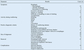
All the patients presented with a definite history of swallowing a foreign body. Difficulty in swallowing was the commonest complaint. Three patients complained of discomfort on swallowing, 33 complained of pain on swallowing, and 9 complained of complete dysphagia and were not even able to swallow saliva. When the dentures were lodged in the cervical or upper thoracic sections of the oesophagus, patients could indicate the site of impaction with remarkable accuracy. Talking was also painful when the prosthesis was lodged in the upper oesophagus, and affected patients preferred to communicate with sign language rather than by talking. When the dentures were impacted lower down the oesophagus, patients complained of retrosternal pain, or back discomfort or pain. Excessive salivation was noted in 13 of the patients. Nausea or vomiting was noted in 12 patients. Three patients complained of difficulty in breathing or a sense of choking. Two patients did not have dysphagia on presentation; one of them subsequently passed the foreign body out through faeces and in the other the foreign body remained undetected. On subsequent questioning, the latter patient said that when he could not find his upper denture, he suspected that he could have swallowed it accidentally. All 47 cases of denture swallowing were reviewed (Table I), but only the data for the 46 confirmed cases were used for statistical calculations in this study.

Clinical evaluation of the oral cavity and pharynx proved unrewarding in most cases. However, there were minor mucosal abrasions in four patients, which could have been inflicted by the foreign body or by attempts to induce vomiting by putting fingers into the throat. Indirect laryngoscopy was difficult because of excessive gag reflex. Patients with dentures lodged in the cervical and upper thoracic sections of the oesophagus complained of severe pain when the trachea was moved laterally against the cervical vertebrae. The tracheal tenderness was universal in these patients; this was the most consistent and reliable clinical finding indicating the presence and site of foreign body lodgement.
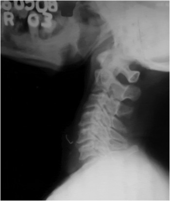
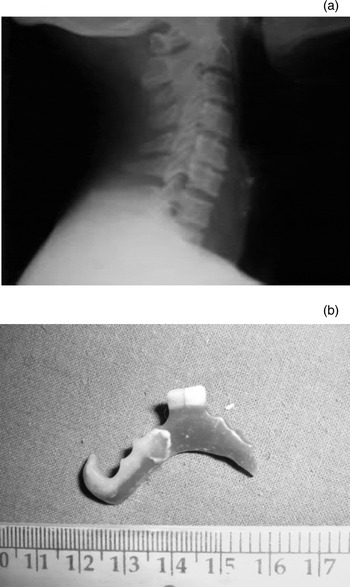
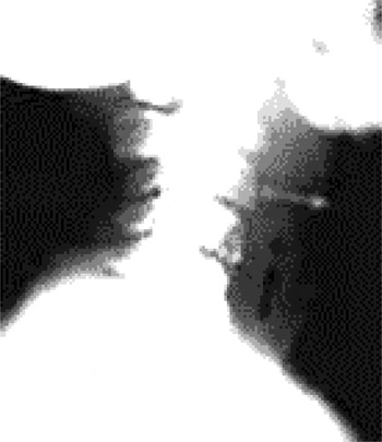
Lateral soft tissue radiography of the neck was carried out on all 47 patients. Only three of the patients showed an impacted foreign body in the neck (Figure 2). Another foreign body was detected on chest radiograph. These four dentures all had metal wires attached. Widening of the prevertebral soft tissue shadow was noted in 23 patients; the foreign body was impacted in the cervical oesophagus in these patients (Figure 3). The presence of an air shadow in the oesophagus on lateral X-ray of neck soft tissue was noted in two cases, associated with prevertebral soft tissue widening (Figure 4). However, this prevertebral soft tissue widening was not apparent in three other patients who had dentures impacted in the same region.
Lateral X-ray showing dentures with metal clasps, readily identifiable on this conventional view.

(a) Lateral X-ray showing widening of the prevertebral soft tissue shadow, and (b) clinical photograph showing the denture that caused the widening.

Lateral X-ray showing widening of the prevertebral soft tissue shadow, together with the presence of gas in the neck.

Contrast studies were not carried out on any of the patients. Computed tomography was performed on only one patient. This patient had negative upper gastrointestinal endoscopy findings but was symptomatic, and suddenly complained of abdominal pain on the second night. The CT scans showed no presence of any foreign body in the abdomen.
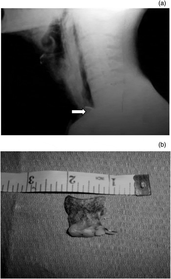
Radiographic findings were suggestive of a foreign body in 30 cases. Foreign bodies per se were only detectable radiographically if they had metal wires attached. All of the visible foreign bodies appeared significantly smaller than the real objects, as the radiolucent parts of the dental prostheses were not visible. The wire of one of the dentures penetrated the oesophageal wall, resulting in surgical emphysema radiologically evident as gas shadows in the soft tissues of the neck (Figure 5).
(a) Lateral X-ray showing the radiopaque shadow of the wire attached to the denture (white arrow), together with surgical emphysema of the neck, and (b) clinical photograph of the offending denture.

Rigid oesophagoscopy was carried out under general anaesthesia in all cases. Flexible upper gastrointestinal endoscopy was performed in the three cases where rigid oesophagoscopy had initially failed to locate the foreign body. Dental prostheses were most commonly found to be impacted in the cervical section of the oesophagus (29 of 46 patients), followed by the mid and lower oesophagus sections (14 and 2 patients respectively). Of the two patients with no radiological or endoscopic evidence of a foreign body, one subsequently passed the foreign body out with faeces. No foreign body whatsoever was found inside the body of the remaining patient.
Of the 45 dentures found impacted in the oesophagus, all but 1 could be removed successfully using a rigid oesophagoscope. Oesophagotomy was needed in one patient, as endoscopic manoeuvres had failed to disimpact the foreign body. The oesophagus was exposed via dissection between the carotid sheath and the thyroid. The foreign body was extracted via a vertical incision in the oesophagus over the denture, and was followed by primary repair.
During oesophagoscopic removal of the dentures, on locating the prostheses, the edge of the plate was grasped with forceps to disimpact and align it along the length of the oesophagus before attempting to pull it out. Locating the denture sometimes proved difficult: the dentures tended to remain hidden because of similarities in the colour of the plate, its curvature and the rugosities on its surface which helped camouflage it within the mucosal folds of the oesophagus. Rotating the oesophagoscope around on its long axis, and frequent gentle sounding of the oesophageal wall with the tip of the cannula on reaching the suspected area, helped in identifying the plate when it was snugly positioned within the oesophageal wall. Ten patients had mucosal abrasion and two patients had mucosal laceration. Surgical emphysema developed in two patients following removal, which improved with conservative measures.
There were no major complications such as oesophageal perforation, neck abscess or mediastinitis. The duration of hospitalisation varied from 2 to 7 days, the majority staying for 3 days or fewer. None of the patients suffered any subsequent long-term complication.
Discussion
The most common impacted oesophageal foreign bodies in adults vary in different parts of the world. Chicken and fish bones are common in Asia,Reference Nandi and Ong1, Reference Abdullah, Kaur and Ng2 while in North America it is meat bolus.Reference Vizearrondo, Brady and Nord3, Reference Webb4 The incidence of dental prosthesis impaction in the oesophagus, though not uncommon, varies from only 0.4 to 0.7 per cent.Reference Nandi and Ong1 The percentage is rather high in our series (17.6 per cent). Abdullah et al. (1998) also reported a rather high incidence of 11.5 per cent.Reference Abdullah, Teong, Mahadevan and Jalaluddin5 We concur with their opinion that small sample size and reviewing cases from tertiary care centres only (where more difficult cases are usually referred) might explain the apparent discrepancy. Lack of patient awareness about the need to change the dentures at regular intervals in India and neighbouring regions is another cause of the high incidence. In the current study, the offending dentures were in use for quite a long time (8 to 18 years without check-up) prior to the incident. Twenty patients in our series were aware that the dentures were loose, but took no remedial measures. Over the years, patients might have had other teeth removed but may have continued to wear the same old denture, resulting in loss of the support of the abutting teeth (Figure 6).
Clinical photograph showing a loose abutting tooth (white arrow), which resulted in loss of support for the denture, evident from the gap visible between the plate of the denture and the gum (black arrow).

All the patients in our series presented with a history of denture ingestion. However, a definite history may not always be available, especially in case of patients with learning and mental health disorders, or those who have suffered from a drug overdose or stroke.Reference Hashmi, Walter, Smith and Latis6 Occasionally, denture impaction is even compatible with near normal swallowing.Reference Mishra, Shukla, Mishra, Bhatia and Agarwal7 Patients may also present with apprehension that he or she might have swallowed the missing denture, as was the case for one patient in the current study. When swallowed, the most likely presenting symptom of an impacted denture is dysphagia, with other complaints related to how far the denture has progressed and time since swallowing.Reference Hashmi, Walter, Smith and Latis6 These other complaints include sore throat, choking sensation, retrosternal pain, sweating, raised temperature, hoarseness and coughing up of blood. A swallowed denture may rarely be lodged in the tracheobronchial tree.Reference Agarwal, Agarwal and Purohit8 Early diagnosis is essential to avoid complications like oedema, infection and perforation. If it is observed that there are some natural teeth missing, especially in neurologically compromised or disadvantaged categories of patients, the possibility of a swallowed denture should be noted.Reference Hashmi, Walter, Smith and Latis6 Tracheal tenderness is a very reliable clinical indicator of an impacted foreign body in the cervical or upper thoracic sections of the oesophagus. In the current study, the presence of tracheal tenderness influenced our decision to perform oesophagoscopy, even in the absence of positive radiological signs.
Poly(methymethacrylate), the plastic used to make dentures, is radiolucent. Wire clasps are radiologically visible, but these are seldom attached to the prostheses. A soft tissue exposure is more likely to suggest the presence of a plastic denture than a standard exposure, but cannot always be relied upon.Reference Hashmi, Walter, Smith and Latis6 Abdullah et al. found the lateral soft tissue neck radiographs helpful in 24 per cent of cases.Reference Abdullah, Teong, Mahadevan and Jalaluddin5 Chest radiographs rarely aid detection of an impacted denture, unless the denture has metal wires attached. All of the four dentures with wires in our series could be detected by conventional radiography. Widening of the prevertebral soft tissue shadow served as indirect evidence of an impacted foreign body in 79 per cent of cases (23 of 29) where the dentures were lodged in the cervical section of the oesophagus. Although a plain radiograph may not identify a swallowed denture, the investigation has been recommended to exclude pneumomediastinum or gas within soft tissue.Reference Nageris and Feinmesser9 Upper gastrointestinal tract radiography using a non-ionic water-soluble contrast medium may be used to rule out perforations (especially when aspiration is a risk).Reference Abdullah, Teong, Mahadevan and Jalaluddin5 Contrast studies prior to endoscopy may also help to establish the level of impaction, the presence of a pharyngeal pouch or the cause of impaction (e.g. strictures).Reference Marais, Mitchell and Wightman10 Barium contrast medium is seldom helpful as it will coat all sides of a radiolucent object and can make subsequent endoscopy more difficult.Reference Hashmi, Walter, Smith and Latis6 In the event of a foreign body seen on the lateral soft tissue neck X-ray but absent on endoscopy, further evaluation by CT should be considered.Reference Abdullah, Teong, Mahadevan and Jalaluddin5 Computed tomography has been advocated by Newton et al. (1987) as an important diagnostic aid because of its substantial advantages over conventional radiography in the detection of acrylic-resin denture base materials or inhaled denture fragments, or in the detection of complications such as perforation with abscess or mediastinitis.Reference Newton, Abel, Lloyd and Yemm11
The radiolucency of acrylic-based denture material continues to impair localisation of a swallowed denture. The radiopacity of dental prostheses on occlusal or intra-oral radiographs does not guarantee similar visibility on lateral soft tissue neck, chest or abdominal films.Reference Davy and Causton12, Reference Goshima, Gettleman, Goshima and Yamamoto13 In addition, Mahadevan et al. (cited in Abdullah et al.) and Abdullah et al. highlighted the significant decrease in the radiographic size of the foreign bodies compared to their actual size.Reference Mahadevan, Abdullah, Kasim and Yunus14, Reference Abdullah, Teong, Mahadevan and Jalaluddin5 This has been corroborated by our study also.
The incorporation of radiopaque material in dental resins should be made mandatory for easy localisation and early removal.Reference Nimmo, Nimmo and Chin15 However, no such product is commercially available that can match the physical properties, appearance and the ease of handling of the currently used radiolucent products. The incorporation of heavy metal salts or glass fillers has been sufficient to weaken the material, thereby increasing the possibility of fracture and the risk of swallowing a denture fragment.Reference Hashmi, Walter, Smith and Latis6 The appearance of such dentures has also been deemed unacceptable. Payne and Henry (1984) suggested that barium fluoride be included in dentures.Reference Payne and Henry16 Abdullah et al. (1998) advocated implanted radiopaque wires to increase their visibility on radiographs.Reference Abdullah, Teong, Mahadevan and Jalaluddin5
The majority of ingested dental prostheses in the current series were 3.5 to 4 cm in width. Nevertheless, a denture does not need to be small to be swallowed, and the swallowing of complete (full) loose dentures has been reported.Reference Hashmi, Walter, Smith and Latis6 The configuration and overall dimensions of the denture are important. A horseshoe-shaped denture swallowed ‘end on’ and vertically may well rotate into the hypopharynx and the oesophagus, though its width would make it difficult to swallow flat and transversely.Reference Hashmi, Walter, Smith and Latis6
• Swallowed dentures often lodge in the oesophagus
• Most dentures are radiolucent and difficult to detect in X-rays
• Cervical oesophagus is the commonest site of impaction
• Dysphagia and tracheal tenderness are the commonest symptom and sign
• Rigid oesophagoscopy generally enables successful removal
The irregular shape of dentures makes them liable to become impacted in the gastrointestinal tract if swallowed. The impaction is more likely to occur at the sites of natural constriction. Once a denture is impacted, it is unlikely to have a spontaneous passage. The impacted denture should be removed under direct vision as soon as the diagnosis is made, as oedema from local trauma tends to grip the object more firmly making later manipulation increasingly difficult. The impacted foreign body may result in partial or complete obstruction, or bleeding or perforation secondary to abrasion. Complications are more likely if the diagnosis is delayed. Recurrent laryngeal nerve palsy and oesophago-aortic fistulas are rare complications. Mucosal necrosis and oesophageal abscess formation can be avoided by early intervention. Perforation of the oesophagus is more common than in other parts of the gastrointestinal tract.Reference Nandi and Ong1, Reference Maleki and Evans17, Reference Dhingra and Bansal18 We did not encounter any large perforation in our series, but two cases of difficult removal were associated with the development of surgical emphysema in the post-operative period, which resolved with conservative measures over a 7-day period. It is safer to have a Ryle's tube inserted under direct vision in cases of difficult removal or if there is bleeding during the endoscopic removal of the denture. The decision to perform endoscopy should be based on clinical judgement. A high index of suspicion and appropriate use of direct endoscopic examination early in the management are the most important factors in preventing serious complications.
Conclusion
After prolonged use, denture fittings may become loose and therefore easier to accidentally swallow. The shape, size and contour of a denture make it a perfect foreign body to get impacted in the gastrointestinal tract. In the current study, drinking was the most common activity associated with denture ingestion. The cervical section of the oesophagus was the commonest site of impaction. Although dysphagia or odynophagia are common symptoms, and a definite history is usually forthcoming, their absence does not preclude the presence of an impacted denture.
Dentures are often radiolucent (unless there are wire attachments). As it is difficult to confirm the presence of dentures radiologically, a high degree of suspicion is vital to early detection of an impacted denture. This applies in cases where the patient is asymptomatic or when no definite history is available, or when the patient is unconscious. Tracheal tenderness was found to be the most consistent and reliable sign of a foreign body in the cervical or upper thoracic sections of the oesophagus. It may sometimes be difficult to detect a denture hiding within the mucosal folds because of the similarities in colour, surface rugosities and contour of the plate. Rotating the oesophagoscope around on its axis, and gentle sounding with the tip of the suction cannula, can aid localisation of a difficult-to-detect denture.
Some form of radiopaque marker incorporated into the denture may help in localisation in the event of accidental swallowing. Educating individuals about the proper use of dentures would help to reduce the incidence of accidental swallowing. Dentures should come with a warning tag, and users should be advised about proper use and care, and the need to visit the doctor for regular maintenance of the denture.