Introduction
The new mineral proposal for ‘marshallsussmanite’ (IMA2013-067) was approved in October 2013 and the abstract was published in the Mineralogical Magazine (Origlieri et al., Reference Origlieri, Downs and Yang2013). In the proposal, the authors made no comparison between ‘marshallsussmanite’ and schizolite yet they knew schizolite existed as they referred to the crystal structure reported by Ohashi and Finger (Reference Ohashi and Finger1978). The crystal structure of ‘marshallsussmanite’ has not been officially published, but it is recorded in IMA proposal and can be found as a ‘preproof’ on the Mineralogical Magazine website (Origlieri et al., Reference Origlieri, Downs, Yang, Hoffman, Ducea and Post2017). Since then, Nagashima et al. (Reference Nagashima, Imaoka, Fukuda and Pettke2018) refined the structure of a ‘marshallsussmanite’.
In this brief account, we have re-characterised the cotype material from Tutop Agtakôrfia, Greenland in order to show that ‘marshallsussmanite’ and schizolite are the same mineral species. This validates the authenticity of schizolite and re-establishes the type material. For historical reasons the name schizolite must be given priority. The discreditation of ‘marshallsussmanite’, with a reinstatement of the name schizolite, has been proposed and it has been approved by the IMA-CNMNC (Voting proposal 18-B, February 19, 2018, Hålenius et al., Reference Hålenius, Hatert, Pasero and Mills2018). Two samples of schizolite are examined here in detail (labelled NHMD 1899.856 and NHMD 1899.8), both originating from the original type locality.
History of schizolite characterisation
The type locality of schizolite is Tutop Agtakôrfia (Petersen and Johnsen, Reference Petersen and Johnsen2005), in the Ilímaussaq alkaline complex, Julianehåb district, South Greenland. The type material is stored in the Geological Museum, now a part of the Natural History Museum of Denmark. In 1901 schizolite was described as a new mineral species by Winther based on material collected by Flink in 1897: “The new mineral, on account of its properties, is allied to the pectolite group.” He goes on to say: “Both in composition and crystallographic properties the schizolite differs distinctly from pectolite, so it is undoubtedly correct to classify it as a special mineral.” Winther (Reference Winther1901) named the mineral schizolite based on its ‘schizo’ or ‘split’ cleavage. Shortly afterwards, Bøggild (Reference Bøggild1903) reported that the mineral is triclinic and not monoclinic, working on material that he himself had collected at the type locality in 1899.
These first studies of schizolite from Ilímaussaq showed a difference in appearance and composition, with the Tutop Agtakôrfia material typically being pinker than material from the other localities where the colour is often more grey-to-brownish (Flink, Reference Flink1898; Winther, Reference Winther1901; Bøggild, Reference Bøggild1903). Bøggild (Reference Bøggild1903) compared the composition of schizolite from different localities within Ilímaussaq and found that the pink material has higher Mn2+ content than the other coloured schizolite, even when the two types occur at the same locality.
Schaller (Reference Schaller1955) effectively discredited this species as a manganoan pectolite, stating that “The term schizolite can be discarded as an unnecessary species name.” The simplest formula was established as Na(Ca,Mn)2Si3O8(OH). In this study, there was only powder diffraction data and chemical analysis, no crystal-structure analysis, and the differentiation of species in the series was not possible at this time without the structure. Further, this was done prior to the establishment of the Commission of New Minerals, Names and Classification, International Mineralogical Association (IMA-CNMNC) in 1959 and cannot be considered as an official discreditation.
Ohashi and Finger (Reference Ohashi and Finger1978) established the validity of the schizolite species with a crystal-structure analysis that defined the ordering of the cations Ca and Mn2+ between the two octahedrally-coordinated sites, M1 and M2, making a distinction between the three isostructural species; pectolite (NaM1CaM2CaSi3O9H), schizolite (NaM1CaM2MnSi3O9H) and serandite (NaM1MnM2MnSi3O9H). However, these authors did not emphasise that this made schizolite a different species. In fact, Petersen and Johnsen (Reference Petersen and Johnsen2005) list schizolite as a doubtful species, but this can now be rectified.
Study material
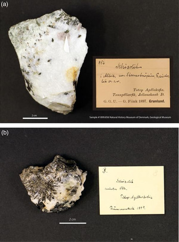
Two samples of cotype material are investigated here, both originating from the type locality: (1) Sample NHMD 1899.856: This material was studied by Winther (Reference Winther1901) after being collected by Gustaf Flink in 1897 (Fig. 1a). This specimen is part of a suite of 14 specimens studied by Winther, with an additional seven that were also probably part of his research. Winther named the new mineral schizolite, and as this is the first description of the mineral, it is undoubtedly the type material. (2) Sample NHMD 1899.8: This material was collected by Bøggild and Brumnurstedt, in 1899 and studied in greater detail by Bøggild (Reference Bøggild1903) (Fig. 1b). It is part of a suite of 11 specimens. Both specimens were graciously provided by the Natural History Museum of Denmark in Copenhagen. All material used for optical measurements, crystal structure analysis and microprobe data collections has been returned; none has been retained at the Canadian Museum of Nature.
(a) Schizolite collected by Gustaf Flink in 1897 and used in the original description by Christian Winther (Reference Winther1901); catalogue NHMD 1899.856. (b) Schizolite collected by Ove Balthasar Bøggild and Brumnurstedt in NHMD 1899; catalogue NHMD 1899.8

Methods and results
Physical and optical properties
At the type locality, schizolite occurs as stacked prismatic columns, up to 2 cm long. These occur as either individual crystals or aggregates of crystals. The mineral is pale pink to brownish with a white streak and vitreous lustre. It is translucent to opaque, non-fluorescent and has a hardness (Mohs) ≈ 5. The mineral is brittle with two perfect cleavages making it splintery or somewhat fibrous. The calculated density is 3.09 g cm–3.
Pale pink schizolite (NHMD 1899.856) is biaxial (+) with α = 1.626 ± 0.003, β = 1.630 ± 0.002, γ = 1.661 ± 0.002, 2Vmeas = 72(4)° and 2Vcalc = 40°. Pale brown schizolite (NHMD 1899.8) is biaxial (+) with α = 1.640 ± 0.003, β = 1.643 ± 0.002, γ = 1.658 ± 0.002, 2Vmeas = 52(2)°, 2Vcalc = 49°. There is no pleochroism observed in either specimen. Optical parameters were measured on a crystal fragment using a spindle stage with Na light, λ = 590 nm, and the measured 2V was determined from extinction curves. It was particularly difficult to measure α in this mineral, as the observation direction is down a crystal fragment edge. Comparative optical properties for current and other members of the series are given in Table 1.
Comparison of schizolite, ‘marshallsussmanite’, serandite and pectolite.

Chemical analysis
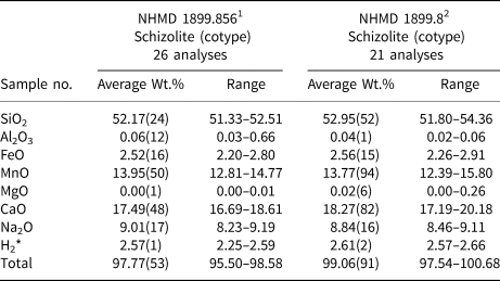
The chemical analyses were performed on a JEOL 8230 electron microprobe operating in wavelength-dispersion (WD) mode using Probe for EPMA software (https://www.probesoftware.com). The operating voltage was 20 kV, the beam current was 20 nA and the beam diameter was 10 µm. At these operating conditions, no intensity change due to sample damage was observed. Peak intensities were counted for 20 s and backgrounds were calculated using a mean atomic number correction (Donovan et al., Reference Donovan, Lowers and Rusk2011). The following lines and standards were used for quantification: albite for NaKα; diopside for MgKα and CaKα; sanidine for KKα and AlKα; hematite for FeKα; rutile for TiKα; tephroite for SiKα and MnKα; sanbornite for BaLα; and chromite for CrKα. Raw intensities were converted to concentrations using the default φρZ corrections of the Probe for EPMA software (c.f. Armstrong, Reference Armstrong1988). Results of microprobe analyses are presented in Table 2. The final chemical formulae (Table 1) are calculated for each analyses on the basis of 9 anions per formula unit (apfu); H2O (wt.% oxide) was calculated on the basis of stoichiometry, with (OH) = 1 apfu.
Chemical analyses: schizolite and pectolite from Tutop Agtakôrfia.

1Na1.02Ca1.09Mn0.69Fe0.12Si3.04H1O9; 2Na0.98Ca1.12Mn0.67Fe0.12Si3.04H1O9; atomic fractions calculated of the basis of 9 oxygen atoms.
* H2O calculated on the basis of 1 H apfu
Powder X-ray diffraction
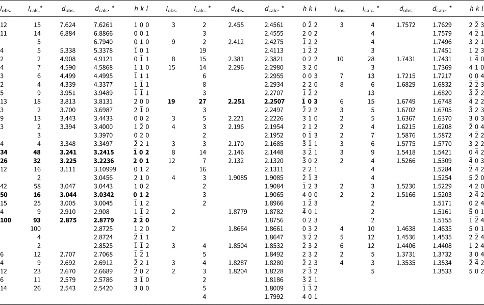
X-ray diffraction data (Table 3) for schizolite NHMD 1899.856 were acquired with a Bruker D8 discover equipped with CuKα radiation and a Deskris Eiger2R_500K detector, calibrated at a distance of 17.562 cm using a statistical calibration (Rowe, Reference Rowe2009). Schizolite, as expected, is characterised by an intermediate diffraction pattern between end-members of the pectolite–serandite series. The unit cell was refined in the triclinic system, based on the powder data, thanks to the increased detector resolution and the flexibility the Rietveld software for cell-refinement purposes. The recent improvement in data resolution and area detection technology facilitates the characterisation of lower symmetry structure, for which accurate pattern indexing was historically difficult and often impossible.
Powder X-ray diffraction data for schizolite (Kα1) (sample# NHMD 1899.856)

* Calculated from structure data modified with Rietveld software.
Refined cell a = 7.85062(14) b = 6.97283(8) c = 6.91131(11) Å, α = 90.695(2), β = 94.5174(18), γ = 102.8861(12)°, V = 367.487(2) Å3
The strongest lines are given in bold
The discrepancy between the measured and calculated intensities in Table 3, largely result from the inability of the diffraction experiment to resolve the high-intensity peaks at d = 2.8779 and d = 2.8725 Å, corresponding to hkl indices (120) and (2 ${\bar 2}$0), respectively. The (120) is the maximum (100%) peak in the calculated powder pattern, whereas (2
 ${\bar 2}$0) is calculated to be 93% of this intensity. As the 100% reference in the experimental pattern consists of both calculated peaks, the observed peak intensities are systematically, proportionally lower than predicted. Accounting for this would result in a better match between observed and calculated data.
Single-crystal X-ray diffraction data and structure refinement
Two crystal-structure analyses on schizolite were carried out. In both cases, single crystals were fixed to glass fibres and a Bruker single-crystal diffractometer equipped with an APEX II area detector and a microfocus sealed X-ray tube operating at 50 kV and 0.99 mA, located at the X-ray Crystallography Laboratory at the University of British Columbia (Vancouver, Canada), was used for the collection of intensity data. In all cases, data were collected with MoKα radiation at 293(2) K and measured φ and ω scans of 0.5° per frame. The APEX III software package was used to determine a data collection strategy that ensured complete coverage of the Ewald Sphere with reasonable data redundancy. The program SAINT (V3.38, Bruker, 2013) was used to index and refine the final unit-cell parameters, as well as reduce, scale and apply an adsorption correction.
Non-merohedral twinning was observed in sample NHMD 1899.856. Using CELL_NOW (V. 2008/4; Sheldrick, Reference Sheldrick2008), more than two domains were found; these were verified visually using the RLATT routine available in the APEX III software package. Only the two most prominent domains (rotated 2.9° about the b axis) were used during the integration routine. The structure was refined on a merged data set of the two domains. TWINABS-2012/1 (Bruker, 2012) was used to calculate a multi-scan absorption correction. The twinned structure was solved against an HKLF4 file, containing reflection intensities averaged over the two twin domains, and was refined again with a HKLF5 file, containing domain-separated data. No twinning was observed in the data for sample NHMD 1899.8.
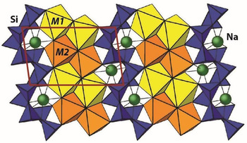
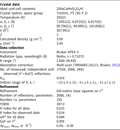
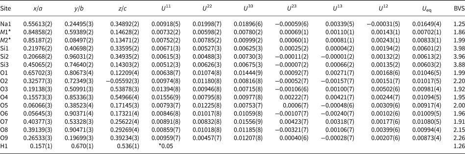
For the crystal taken from NHMD 1899.856, the structure was solved with Direct Methods using SHELXS-97 (Sheldrick, Reference Sheldrick1997) and refined by the Least Squares Method using version 2014 of SHELXL (Sheldrick, Reference Sheldrick2015). For the crystal taken from NHMD 1899.8, the structure was refined against the structure presented in Ohashi and Finger (Reference Ohashi and Finger1978). The transformation matrix from Ohashi and Finger (Reference Ohashi and Finger1978) to our cell is [−½−½0 / 00−1 / −½−½0]. The M1 and M2 sites were refined with split-occupancies (with the constraint Mn + Ca = 1). All positions were refined anisotropically, with the exception of hydrogen (U iso = 0.05Å2), which, in all cases was readily observed in the difference-Fourier prior to being included in the refined model. The O–H distance was allowed to refine freely to O3–H1 = 1.22(1), O4–H1 = 1.28(1) and O3–H1····O4 = 2.4726(4) Å. Scattering factors of neutral atoms were taken from the International Tables for X-ray Crystallography (Ibers and Hamilton, Reference Ibers and Hamilton1974). For all crystals, assigning phases to a set of normalised structure-factors gave mean values of |E 2 – 1| in excess of 0.95, in accord with a centrosymmetric space-group. Weighting parameters were manually updated during final Least Square cycles, and this process was repeated until convergence. For sample NHMD 1899.856, details of collection and refinement are given in Table 4, and final atomic coordinates are given in Table 5; structural formulae, calculated by site-refinement are given in Table 1. Compatible structure data for sample NHMD 1899.8 have been deposited as supplementary material (see below). The final structure is entirely consistent with previously reported intermediate compositions of the serandite–pectolite solid-solution series (Fig. 2). It shows the tri-periodic [Si3O10] chains, dreierinfach chains that zigzag parallel to c between double-wide ribbons of octahedra are made up of two crystallographically-unique octahedrally-coordinated sites, M1 and M2 (Fig. 2). The final structures for NHMD 1899.856 and NHMD 1899.8 are very similar, but not identical i.e. small differences in Mn2+ and Ca content but consistent in the ordering of the M1 and M2 sites. Summary data for four minerals are given in Table 1. Consistent with previous authors, (Nagashima et al., Reference Nagashima, Imaoka, Fukuda and Pettke2018; Ohashi and Finger, Reference Ohashi and Finger1978; Prewitt, Reference Prewitt1967; Rozhdestvenskaya and Vasilieva, Reference Rozhdestvenskaya and Vasilieva2014) Ca and Mn preferentially order at M1 and M2, respectively, resulting in the ideal structural formula for schizolite being written as Na(M1CaM2Mn)Si3O8(OH).
The schizolite crystal structure. It shows the tri-periodic [Si3O10] chains that zigzag parallel to c between double-wide ribbons of edge-sharing octahedra. The ribbon is formed by dimers of M1 (yellow) and M2 (orange) octahedra.

Data and experimental details for schizolite, Tutop Agtakôrfia NHMD 1899.856 (cotype), Greenland.

 $R_1 = \sum \vert {\vert {F_{obs}} \vert - \vert {F_{calc}} \vert } \vert / \sum \vert {F_{obs}} \vert $;
 $wR_2 = \left[ {\sum \left \lsqb {w{ \left \lpar {F_{obs}^2 -F_{calc}^2} \right \rpar }^2} \right \rsqb / \sum w{\left \lpar {F_{obs}^2} \right \rpar }^2} \right]^{1/2} $ G
 $oF = \left[ {\sum \left \lsqb {w{\left \lpar {F_{obs}^2 -F_{calc}^2} \right \rpar }^2} \right \rsqb /\lpar {n - p} \rpar } \right]^{1/2}$w = 1/[δ2(F 02) + (0.0269 x P)2 + 0.20 x P] where P = (Max (F 02, 0) + F c2) / 3
Final refined atomic coordinates, site-occupancy factors (s.o.f.) and thermal parameters for schizolite Tutop Agtakôrfia, Greenland (NHMD 1899.856).

M1* s.o.f. Ca 0.792(1) and Mn 0.208(1); M2* s.o.f. Ca 0.792(1) and Mn 0.208(1)
U iso parameter fixed during refinement; BVS – bond-valence sum.
Discussion
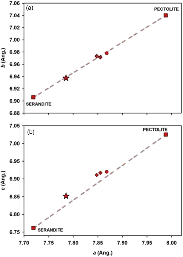
The unit cell and optical parameters, as well as calculated empirical chemical formulae for all phases of interest are listed in Table 1. Close inspection of these confirm the equivalence of schizolite and ‘marshallsussmanite’. In all cases, the values of these parameters fall between those of the series end-members, pectolite and serandite. Parameters for ‘marshallsussmanite’ are reasonably similar to those of the two schizolite samples presented here. The variation in unit-cell parameters for the phases presented in Table 1 is shown in Fig. 3. A clear, linear trend relates the variation in parameters for pectolite–schizolite–serandite. Data corresponding to schizolite (NHMD 1899.8 and NHMD 1899.856) and ‘marshallsussmanite’ all plot along the trend, centrally located between the end-member values. These observations are in-line with general expectations, as the composition of schizolite can be expressed as being equal parts of the two end-members.
Variation in unit-cell parameters for serandite (Jacobson et al., Reference Jacobsen, Smyth, Swope and Sheldon2000) and pectolite (Prewitt, Reference Prewitt1967). The data of Ohashi and Finger (Reference Ohashi and Finger1978) (circle), this study (diamond) and ‘marshallsussmanite’ (star) are plotted for comparison: (a) a vs. b cell parameter; (b) a vs. c parameter.

Conclusions
Schizolite and ‘marshallsussmanite’ are the same mineral species (Table 1). As the mineral name schizolite was published first, it is this name that must be used. The name ‘marshallsussmanite’ has now been discredited (Voting proposal 18-B, Hålenius et al., Reference Hålenius, Hatert, Pasero and Mills2018). It is unfortunate that the name ‘marshallsussmanite’ is now in the literature and widely spread throughout mineral trade, having been sold under that name. Circumstances such as this exemplify why withholding the approved name of new minerals in advance of the official publication may be a useful consideration. The present authors greatly appreciate data of new minerals being published quickly to prevent overlap of research efforts.
Author ORCIDs
Ralph Rowe, 0000-0002-8735-7592.
Acknowledgements
Theresa Smith, Mineralogical & Geological Museum at Harvard University, kindly provided information on the specimen used by Ohashi and Finger (Reference Ohashi and Finger1978) in the first structure determination of schizolite. We would like to thank the following individuals for the use of their single-crystal diffractometers: Anita Lam and Brian Patrick, X-ray Crystallography Facility – University of British Columbia. We further thank two reviewers and for their thoughtful reviews and comments on the manuscript.
Supplementary material
To view supplementary material for this article, please visit https://doi.org/10.1180/mgm.2019.21.