Introduction
Cellular mechanisms leading to noise-induced hearing loss remain unclear. Noise leads to mechanical damage and metabolic changes in cochlear structures. Mechanical damage starts with the loss of stereocilia of hair cells in the organ of Corti in the cochlea. Hair cells die (apoptosis) as the noise continues; damage is also observed in the supporting cells. Nerve fibres and nerve endings degenerate, and these effects are irreversible. The damage and loss of outer hair cells are greater than those of inner hair cells.Reference Lim, Choi, Kang, Ahn and Chung1,Reference Bohne and Harding2
Metabolic changes occur in the cells, and some of these changes can be prevented. In recent years, several studies have been conducted to explain these metabolic changes, and protective measures and treatment methods have been developed for each of these changes.Reference Mukherjea, Ghosh, Bhatta, Steth, Tupal and Borse3 Many metabolic and biochemical changes have been identified.Reference Ohlemiller, McFadden, Ding, Lear and Ho4–Reference Ohinata, Miller, Altschuler and Schacht7 Reactive oxygen species initiate the apoptotic signal in outer hair cells. Accumulation of reactive oxygen species in the cochlea and concomitant inflammation play an essential role in acoustic trauma. Therefore, reactive oxygen species reducing antioxidant and anti-inflammatory agents can be used in noise-induced hearing loss cases.
Oxytocin has anti-inflammatory and antioxidant effects.Reference Tuğtepe, Sener, Biyikli, Yuksel, Cetinel and Gedik8,Reference Dusunceli, Iseri, Ercan, Gedik, Yegen and Yegen9 Oxytocin decreases glutathione and superoxide dismutase consumption, inhibits nicotinamide adenine dinucleotide phosphate hydrogen oxidase and myeloperoxidase activity, increases nitric oxide levels, and prevents apoptosis.Reference Elberry, Refaie, Kamel, Ali, Darwish and Ashourg10–Reference Tsuruya, Tokumoto, Ninomiya, Hirakawa, Masutani and Taniguchi12 Many studies have demonstrated the anti-inflammatory activity of oxytocin in different organ injuries.Reference Tuğtepe, Sener, Biyikli, Yuksel, Cetinel and Gedik8,Reference Bıyıklı, Tuğtepe, Şener, Velioğlu-Öğünç, Çetinel and Gedik13–Reference Rashed, Hashem and Soliman15
Although the protective effects of intraperitoneal and intratympanic oxytocin against high-dose cisplatin ototoxicity have been demonstrated, there are no publications in the literature concerning its activity on acoustic trauma.Reference Bekmez Bilmez, Aydin, Şanli, Altintoprak, Demir and Atalay Erdoğan16 This study aimed to investigate whether oxytocin prevents acoustic trauma induced internal ear injury. We hypothesised that intratympanic oxytocin would have a protective effect on rats exposed to acoustic trauma.
Materials and methods
This study was approved by the local ethics committee (on 25 April 2016, decision number 16/31). Regulations concerning animal care and handling, which are stated in the International Declaration of Helsinki, were observed.
Test animals
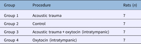
The study was conducted on 28 healthy, 11-month-old, male Sprague Dawley rats, with an average weight of 300–350 g. The rats were kept in light for 12 hours and in darkness for 12 hours, at 25°C, with ad libitum access to food and water, and a background noise level of less than 50 dBA. Rats with middle-ear or tympanic membrane pathology were excluded from the study. Twenty-eight rats were randomly assigned to four groups (Table 1).
Intervention groups

Study procedure
Distortion product otoacoustic emission (DPOAE) and auditory brainstem response (ABR) tests were performed in all rats under general anaesthesia. The rats were anaesthetised with 60 mg/kg of ketamine hydrochloride (Ketalar ampoule; Pfizer Medical, Istanbul, Turkey) and 6 mg/kg of xylazine hydrochloride (Rhompun ampoule; Bayer Medical, Istanbul, Turkey), administered intraperitoneally.
Auditory threshold values were obtained. A total of 14 ears were evaluated from each group. The DPOAE signal-to-noise ratios at each frequency (1191, 2001, 3359, 4755, 6728 and 9511 kHz) and ABR threshold values obtained before acoustic trauma (baseline measurements) for the 14 ears were compared with those obtained on days 1, 7 and 21 post-intervention.
Finally, all animals were sacrificed, and cochlear tissues were isolated for immunohistochemical assessments.
Acoustic trauma group
After baseline measurements, this group (group 1) was exposed to white noise, emitted from two free-field loudspeakers located 50 cm away. This was conducted in an audiometry cabin using a clinical audiometer (AC40; Interacoustics, Middelfart, Denmark), with noise presented at 107 dB SPL for 15 hours. No additional procedure was performed in this group. The DPOAE and ABR measurements were performed on days 1, 7 and 21 after acoustic trauma.
Control group
No procedure was performed in this group (group 2). The measurements were performed on the same days as for the other groups.
Acoustic trauma plus intratympanic oxytocin group
This group (group 3) received 0.1–0.3 ml intratympanic oxytocin (Synpitan Fort, 5 IU/5 ml ampoule; Deva İlaç, Çerkezköy, Turkey) on days 1 and 2 after baseline measurements. The rats were then exposed to noise. Oxytocin was administered intratympanically to the postero-inferior quadrant of the tympanic membrane using a dental injector, under general anaesthesia, on days 1, 3, 5 and 7 after acoustic trauma. The measurements were performed on days 1, 7 and 21 after acoustic trauma.
Intratympanic oxytocin group
This group (group 4) was administered 0.1–0.3 ml of intratympanic oxytocin on days 1, 2, 4, 6, 8 and 10 after the baseline measurements. The post-intervention measurements were performed on the same days as for the other groups.
Distortion product otoacoustic emission measurement
The Madsen Capella DPOAE instrument (Otometrics, Taastrup, Denmark) was used in this study. The ratio between frequencies f2 and f1 (f2/f1) was set at 1.22. The intensity of f1 and f2 stimuli were L1 and L2, respectively; L1 was kept at 55 dB and L2 was kept at 55 dB. The DPOAEs test was performed using two different loudspeakers placed in the external ear canal for the two stimuli (f1 and f2). The DPOAEs were recorded as signal-to-noise ratios, at frequencies of 1191, 2001, 3359, 4755, 6728 and 9511 kHz, using a microphone placed in the external ear canal. The selected frequencies are those automatically determined by the device.
Auditory brainstem response measurement
The ICS Chartr EP 200 instrument (Otometrics) was used for the ABR test. The ABR signals were recorded using subcutaneous needle electrodes. The active electrode was placed on the vertex, reference electrodes were placed on the bilateral mastoid and the ground electrode was placed in the dorsal region. Click stimuli were presented via ‘insert’ headphones. Stimuli were delivered in the rarefaction polarity. The filter was set to 100–3000 Hz, the repetition rate was 21.1 per second and the timeframe was set to 15 ms. A total of 2000 samples were taken to obtain the average signal. Impedance was confirmed to be between 0 kΩ and 3 kΩ before the measurement.
The threshold level was defined as the lowest reproducible and visually detectable intensity level. The stimulus was initially delivered at 80 dB nHL, and the intensity level was reduced incrementally by 20 dB until the threshold was reached. As the threshold was approached, intensity increases of 10 dB were used and the threshold was detected. A minimum of two traces were created for each measurement to ensure reliability of the wave. The ABR threshold was defined as the lowest intensity level at which the wave II of ABR could be observed. The ABR waves were evaluated by two audiologists and their threshold levels were determined.
Immunohistochemical method
The tissues removed were fixed in neutral formalin for 24 hours. They were then placed in 0.1 mol/l ethylenediamine tetra-acetic acid solution (Sigma-Aldrich, St Louis, Missouri, USA) for three weeks for the decalcification of osseous tissues. This was followed by an overnight washing under running water. Afterwards, the tissues were dehydrated through a graded ethanol series, cleared in xylene and processed for embedding in paraffin wax, according to routine protocols.
The slides were stained with caspase 3, 8 and 9 primary antibodies (all at 1:100 dilution; Thermo Scientific, Waltham, Massachusetts, USA). The terminal deoxynucleotidyl transferase dUTP (2'-deoxyuridine 5'-triphosphate) nick-end labelling (‘TUNEL’) technique was also used. Caspases 3, 8 and 9 are involved in the execution of apoptosis. The terminal deoxynucleotidyl transferase dUTP nick-end labelling method reveals the apoptotic cells undergoing DNA fragmentation. The measurements for caspase 3, 8 and 9 staining techniques were obtained from the basal turn of the cochlea. The number of all terminal deoxynucleotidyl transferase dUTP nick-end labelling positive immunoreactive cells for all subjects was counted from the apical to basal turns, and analysed. Two observers blinded to the experimental information evaluated the immunolabelling scores independently.
Statistical method
Data were analysed using the SPSS version 16 statistical software package (IBM, Chicago, Illinois, USA). Homogeneity of variances was verified by Levene's test and conformity of normal distribution was checked by the Kolmogorov–Smirnov test. Non-parametric tests were used for the analysis of DPOAE signal-to-noise ratios. The Kruskal–Wallis test was used for comparisons between the groups and the Friedman test was used for intra-group comparison of the measurements for different time-points.
In cases where differences between groups were significant using the Kruskal–Wallis test, the Bonferroni correction was applied to the p-value obtained, using the two-sample Mann–Whitney U test to determine which groups were different (p < 0.0083). The Bonferroni correction was also applied to the p-value obtained using the two-sample Wilcoxon signed-rank test, to demonstrate which measurements differed significantly when there was a significant difference between the intra-group measurements in the analyses performed using the Friedman test (p < 0.0083).
Descriptive statistics included the mean, standard deviation (SD), minimum and maximum values in the parametric tests, and the mean, SD, median, minimum and maximum values, and 25th and 75th quartiles in the non-parametric tests.
Results
Histopathological findings
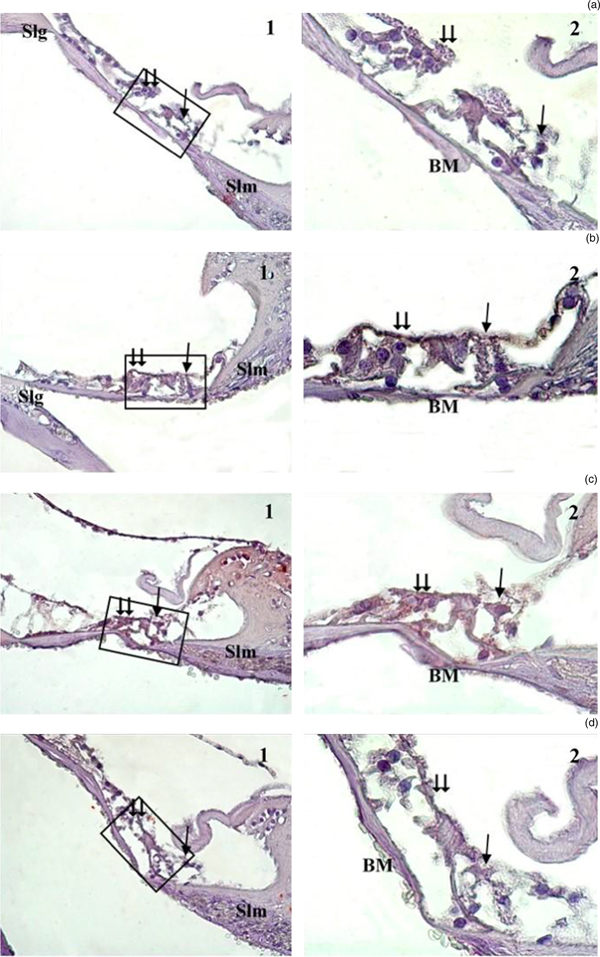
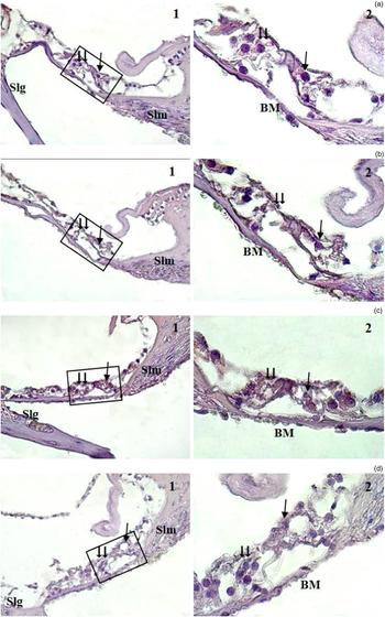
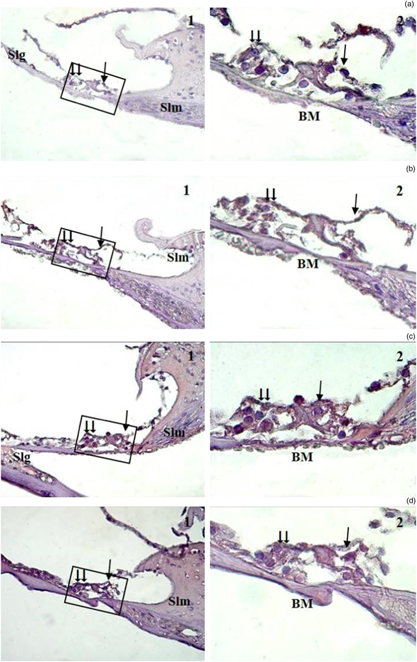
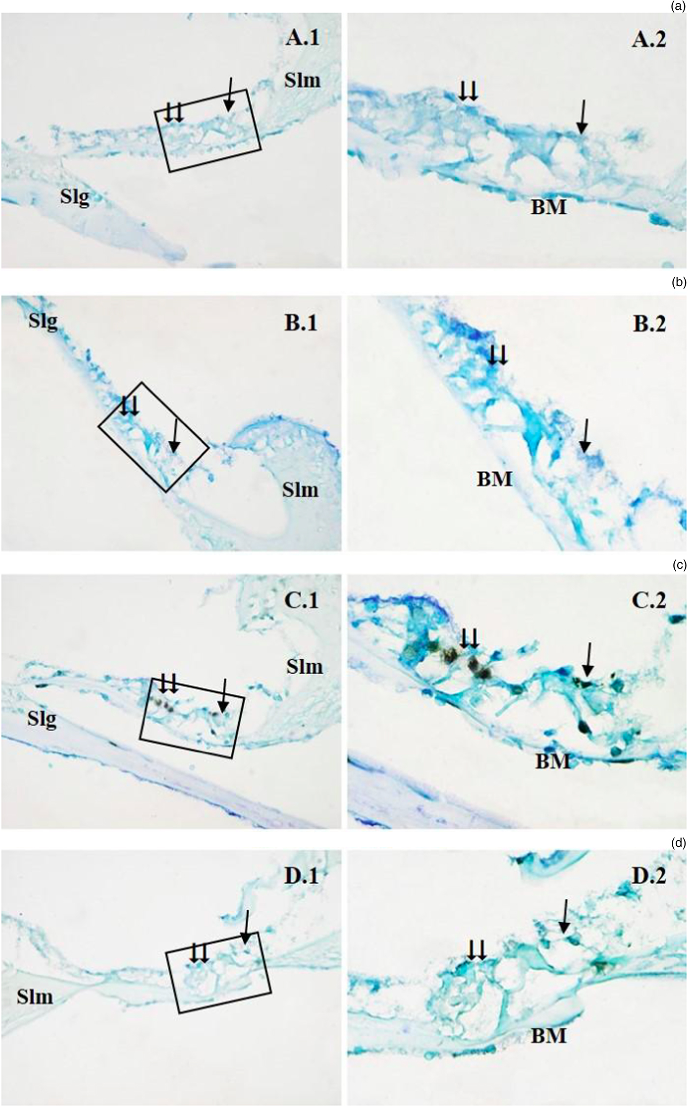
On caspase 3 immunostaining, a weak immunoreaction was observed in the rats’ organ of Corti in groups 2 and 4, whereas there was strong immunoreaction in group 1 and weak-to-moderate immunoreaction in group 3 (Figure 1). On caspase 8 staining, weak immunostaining was observed in groups 2, 3 and 4, whereas in group 1 a moderate-to-strong reaction was detected in the outer and inner hair cells (Figure 2). Caspase 9 staining was similar to, but slightly stronger than, caspase 3 staining (Figure 3). There was a significant difference between groups 1 and 3 in caspase 3, 8 and 9 staining.
Corti organ caspase 3 immunostaining, with Mayer's haematoxylin solution, in the (a) control, (b) oxytocin, (c) acoustic trauma, and (d) acoustic trauma plus oxytocin groups. 1 = Corti organ ×40 magnification; 2 = Corti organ ×100 magnification; Slg = spiral ligament; ⇉ = outer hair cell; → = inner hair cell; Slm = spiral limbus; BM = basilar membrane

Corti organ caspase 8 immunostaining, with Mayer's haematoxylin solution, in the (a) control, (b) oxytocin, (c) acoustic trauma, and (d) acoustic trauma plus oxytocin groups. 1 = Corti organ ×40 magnification; 2 = Corti organ ×100 magnification; Slg = spiral ligament; ⇉ = outer hair cell; → = inner hair cell; Slm = spiral limbus; BM = basilar membrane

Corti organ caspase 9 immunostaining, with Mayer's haematoxylin solution, in the (a) control, (b) oxytocin, (c) acoustic trauma, and (d) acoustic trauma plus oxytocin groups. 1 = Corti organ ×40 magnification; 2 = Corti organ ×100 magnification; Slg = spiral ligament; ⇉ = outer hair cell; → = inner hair cell; Slm = spiral limbus; BM = basilar membrane

The terminal deoxynucleotidyl transferase dUTP nick-end labelling positive cells were almost absent in the organ of Corti cochlear tissues of groups 2 and 4 (Figure 4). In group 1, strong terminal deoxynucleotidyl transferase dUTP nick-end labelling positive cells were visible in outer and inner hair cells, and in the internal pillar and phalangeal cells in the organ of Corti. In group 3, the organ of Corti was preserved; the terminal deoxynucleotidyl transferase dUTP nick-end labelling positive reaction was significantly reduced, particularly in outer and inner hair cells, and a positive reaction was found only in the internal pillar cells. However, the difference between groups 1 and 3 was not statistically significant.
Corti organ terminal deoxynucleotidyl transferase dUTP nick-end labelling (‘TUNEL’) method, stained with Methyl green solution, in the (a) control, (b) oxytocin, (c) acoustic trauma, and (d) acoustic trauma plus oxytocin groups. 1 = Corti organ ×40 magnification; 2 = Corti organ ×100 magnification; Slg = spiral ligament; ⇉ = outer hair cell; → = inner hair cell; Slm = spiral limbus; BM = basilar membrane

Auditory brainstem response thresholds
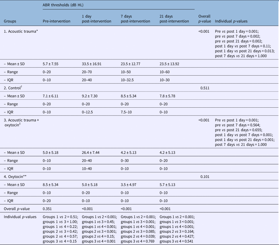
There was no statistically significant difference in ABR thresholds between any study groups for the baseline measurements (p = 0.351). There was a significant difference between the groups in ABR thresholds measured on days 1, 7 and 21 post-intervention (p < 0.0001). There were significant differences in ABR thresholds at day 1 post-intervention between groups 1 and 2, groups 1 and 4, groups 2 and 3, and groups 3 and 4. There were significant differences in ABR thresholds on day 7 post-intervention between groups 1 and 2, groups 1 and 3, and groups 1 and 4. There were significant differences in ABR thresholds on day 21 post-intervention between groups 1 and 2, groups 1 and 3, and groups 1 and 4.
In group 1, there was a significant difference in ABR thresholds measured prior to acoustic trauma and those measured on days 1, 7 and 21 after acoustic trauma. There was no statistical difference in ABR thresholds between the four measurements performed in group 2 (p = 0.511). In group 3, there was a significant difference between ABR thresholds measured before acoustic trauma and those measured on day 1 after acoustic trauma (p = 0.001), whereas no statistically significant differences were observed between the ABR thresholds before acoustic trauma and those on days 7 and 21 after acoustic trauma (p = 0.564 and 0.655, respectively). There were no statistically significant differences in ABR thresholds between the four measurements performed in group 4 (p = 0.101) (Table 2).
Comparison of ABR thresholds

*n = 14; †n = 14; ‡n = 14; **n = 14. ABR = auditory brainstem response; SD = standard deviation; IQR = interquartile range
Signal-to-noise ratios
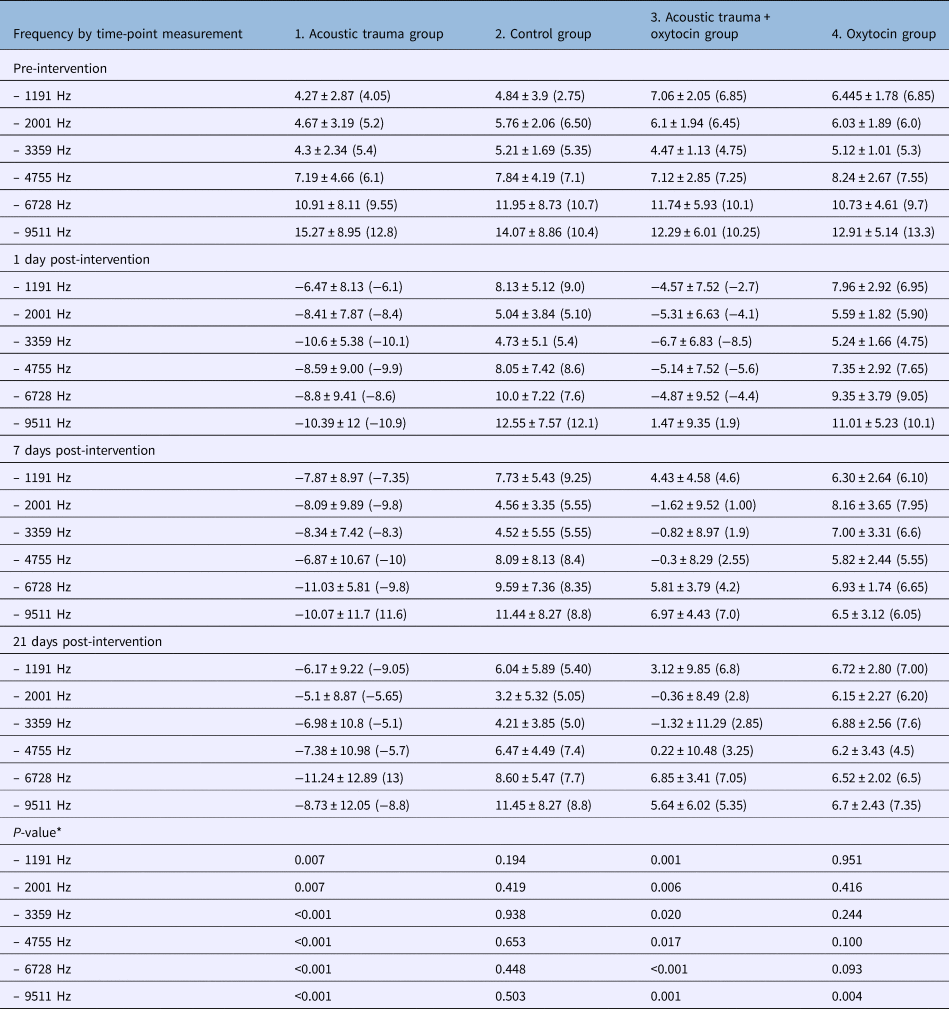
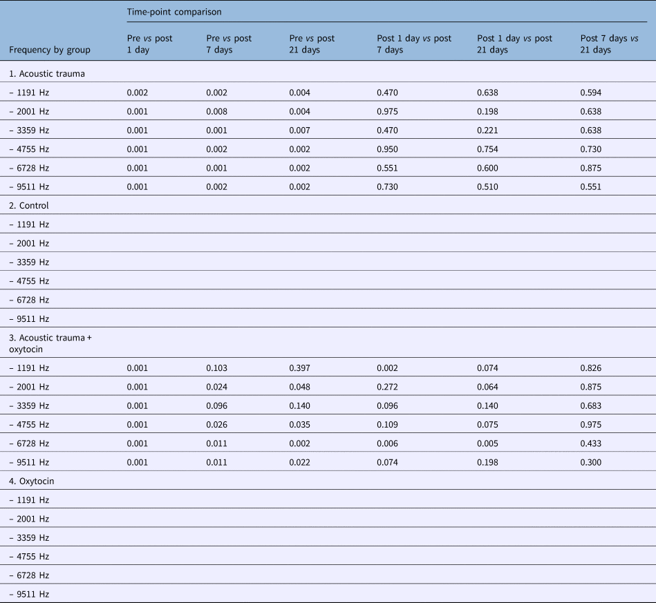
In the group 1, significant differences were observed between distortion product otoacoustic emission signal-to-noise ratios measured: before and on day 1 after acoustic trauma, before and on day 7 after acoustic trauma, and before and on day 21 after acoustic trauma, at all frequencies.
In group 2, no significant differences were observed in signal-to-noise ratios at the four different time-points, for any frequency.
In group 3, significant differences were observed in signal-to-noise ratios measured: before and on day 1 after acoustic trauma at frequencies 2001, 3359, 4755 and 9511 kHz; before and on day 1 after acoustic trauma, and on day 1 and day 7 after acoustic trauma, at the frequency of 1191 kHz; and before and on day 1 after acoustic trauma, before and on day 21 after acoustic trauma, on day 1 and on day 7 after acoustic trauma, and on day 1 and day 21 after acoustic trauma, at the frequency of 6728 kHz.
Although there were no statistically significant differences in signal-to-noise ratios between the four measurements at frequencies 1191, 2001, 3359, 4755 and 6728 kHz in group 4, there was a significant difference in signal-to-noise ratios measured before and on day 7 post-intervention, and before and on day 21 post-intervention, at the frequency of 9511 kHz (Tables 3 and 4).
Comparison of DPOAE signal-to-noise ratios

Data represent mean ± standard deviation (median) distortion product otoacoustic emission signal-to-noise ratios, unless indicated otherwise. *Friedman test. DPOAE = distortion product otoacoustic emission
DPOAE signal-to-noise ratio statistical values for different time-point comparisons

Data represent p-values. Wilcoxon signed-rank test was used to compare the intra-group measurements with each other (p < 0.0083 with Bonferroni correction for level of significance). DPOAE = distortion product otoacoustic emission
Discussion
The increase of reactive oxygen species and concomitant inflammation in the cochlea play essential roles in acoustic trauma. Oxytocin has been the subject of several studies in recent years because of its antioxidant and anti-inflammatory effects. However, no studies have been identified in the literature regarding its use in an acoustic trauma model. The results of this study indicate that oxytocin may be useful in preventing noise-induced inner-ear damage.
Acoustic trauma leads to the apoptosis of outer hair cells.Reference Hu, Henderson and Nicotera17 In acoustic trauma, reactive oxygen species damage outer hair cells by altering the structure of their cellular DNA, damaging the cell membrane through lipid peroxidation and reducing cochlear blood flow.Reference Ohinata, Miller, Altschuler and Schacht7,Reference Van Campen, Murphy, Franks, Mathias and Toraason18,Reference Miller, Brown and Schacht19 Therefore, antioxidant and anti-inflammatory agents that reduce reactive oxygen species can potentially be employed as rescue therapies or used in the prophylaxis of acoustic trauma induced hearing loss.Reference Mukherjea, Ghosh, Bhatta, Steth, Tupal and Borse3 This study aimed to evaluate whether intratympanic oxytocin (with proven links to antioxidant and anti-inflammatory activity), administered before and after acoustic trauma, prevents inner-ear injury caused by acoustic trauma. The study was conducted on rats in an acoustic trauma model, using distortion product otoacoustic emission (DPOAE), ABR and histopathological assessment methods.
A literature review revealed that the acoustic trauma models used in previous studies differed significantly in terms of duration, intensity and method.Reference Kurioka, Matsunobu, Niwa, Tamura, Satoh and Shiotani20 Aksoy et al.Reference Aksoy, Dogan, Yenigun, Veyseller, Ozturan and Ozturk21 investigated the efficacy of thymoquinone in an acoustic trauma model in rats, using 105 dB SPL white noise for 4 hours. They reported that more localised damage occurred with narrow-band noise, with most energy in a one-octave band and in pure sounds, and found that homogeneous damage occurred over the entire cochlea with broadband noise (white noise) which has equal sound intensity at all frequencies. In this study, rats were exposed to acoustic trauma using white noise at a level of 107 dB SPL for 15 hours in a free-field environment.
• Accumulation of oxidant agents in the cochlea and concomitant inflammation play essential roles in acoustic trauma
• Antioxidant and anti-inflammatory agents have been used for acoustic trauma in experimental models
• Oxytocin has anti-inflammatory and antioxidant effects
• This is the first study to investigate the efficacy of intratympanic oxytocin against acoustic trauma in rats
• It showed that oxytocin can repair damage caused by acoustic trauma in laboratory rats
The ABR and DPOAE measurements obtained before and after acoustic trauma were compared. The increase in ABR thresholds after acoustic trauma and the decrease in DPOAE signal-to-noise ratios suggested that investigators had created a successful acoustic trauma model.Reference Aksoy, Dogan, Yenigun, Veyseller, Ozturan and Ozturk21 However, the lack of a standard trauma model in the literature is a critical problem. Because of the inability to develop an ideal acoustic trauma model, the standardisation and comparison of study results are difficult. In this study, two (groups 1 and 3) of the four groups were exposed to noise; the ABR thresholds and DPOAE signal-to-noise ratios measured on day 1 after acoustic trauma were significantly worse than the baseline measurements. This shows that the acoustic trauma model we created was effective.
Rashed et al.Reference Rashed, Hashem and Soliman15 biochemically and histologically demonstrated the potent antioxidant and protective role of systemic 1 mg/kg oxytocin against cisplatin nephrotoxicity in rats. Tuğtepe et al.Reference Tuğtepe, Sener, Biyikli, Yuksel, Cetinel and Gedik8 showed that intraperitoneal oxytocin administration improved renal functions and the microscopic damage caused by renal ischaemia and reperfusion-dependent oxidative damage, in rats. Elberry et al.Reference Elberry, Refaie, Kamel, Ali, Darwish and Ashourg10 reported that pre-cisplatin oxytocin administration was effective against cisplatin-induced renal injury. In light of this information, we investigated the protective effect of intratympanic oxytocin administration in an acoustic trauma model. To the best of our knowledge, no studies on this subject have been published previously.
The administration of local drugs has gained importance in inner-ear disease treatment.Reference Salt and Plontke22 These techniques allow maximum drug concentration in the inner ear, while minimising systemic effects.Reference Wang, Dellamary, Fernandez, Harrop, Keithley and Harris23 We thought that oxytocin would not be sufficiently effective because of its short half-life after systemic administration, and that intratympanic administration would result in higher concentrations in the inner ear. Bekmez Bilmez et al.Reference Bekmez Bilmez, Aydin, Şanli, Altintoprak, Demir and Atalay Erdoğan16 have administered intratympanic and intraperitoneal oxytocin for cisplatin ototoxicity; they reported that both routes have protective effects, but the intratympanic route is more effective. In that study, an intratympanic oxytocin dose of 1–3 ml was used. The same effective and safe dose was also used in this study. Group 3 received intratympanic oxytocin on days 1 and 2 after baseline measurements. Acoustic trauma was then provided, followed by the administration of the same dose of oxytocin on days 1, 3, 5 and 7. As the intratympanic administrations were performed under general anaesthesia in this study, oxytocin was administered on day 2, before acoustic trauma, and on days 1, 3, 5 and 7 after acoustic trauma, to minimise complications due to anaesthesia. Although some studies indicate the need for using treatments before acoustic trauma, other studies suggest that they should be used immediately after acoustic trauma or for 5 days after acoustic trauma.Reference Yamane, Nakai, Takayama, Iqucgi, Nakaqawa and Kojima24,Reference Henderson, Bielefeld, Harris and Hu25
In this study, there was a statistically significant difference in ABR thresholds between groups 1 and 3 on days 7 and 21 after acoustic trauma. In group 1, the ABR thresholds on days 1, 7 and 21 after acoustic trauma were impaired compared with baseline measurements. In group 3, the ABR thresholds on day 1 after acoustic trauma were also impaired compared with the baseline measurements; however, thresholds were statistically improved on days 7 and 21 after acoustic trauma. This result indicates that oxytocin has therapeutic activity against noise toxicity.
There was no significant difference between the ABR thresholds in groups 1 and 3 on day 1 after acoustic trauma, suggesting that intratympanic oxytocin administration does not have a protective effect against noise when administered 2 days before acoustic trauma. There were no significant differences in ABR thresholds between the four measurements in group 4, suggesting that oxytocin was not ototoxic in the ear.
The DPOAE test is an objective method for measuring damage to or loss of outer hair cells. The main pathology in noise-induced hearing loss cases is found in the outer hair cells. Thus, it can be concluded that DPOAEs measurement is an effective auditory parameter for the detection of acoustic trauma. Although ABR thresholds may be normal, DPOAE signal-to-noise ratios may show changes related to outer hair cell damage.Reference Davis, Qiu and Hamernik26 This means that the ABR and DPOAE results may not be always correlated.
The absence of significant differences between baseline measurements and DPOAE signal-to-noise ratios on days 7 and 21 after acoustic trauma in group 3 suggests that oxytocin eliminates the effect of acoustic trauma. These findings suggest that intratympanic oxytocin treatment is helpful for repairing acoustic trauma induced internal ear damage, primarily in outer hair cells. Immunohistochemical staining results also correlated with the ABR and DPOAE measurements.
Conclusion
This experimental study demonstrates that oxytocin can repair inner-ear damage caused by acoustic trauma in laboratory rats. To our knowledge, this is the first study of its kind. Further clinical studies are required to evaluate whether there are similar effects in humans.
Competing interests
None declared