INTRODUCTION
Sperm morphology can be used in order to contribute to understanding of the reproductive biology and elucidating taxonomic and phylogenetic relationships in Caenogastropoda (Ponder et al., Reference Ponder, Colgan, Healy, Nützel, Simone, Strong, Ponder and Lindberg2007).
The family Olividae includes carnivorous or detritivorous semi-infaunal marine gastropods of moderate size, they are usually found in shallow waters from tropical and temperate seas (Smith, Reference Smith, Beesley, Ross and Wells1998). Ten species of Olividae are reported off the Argentine coast, within the genera Olivella, Amalda and Olivancillaria (Pastorino, Reference Pastorino2003, Reference Pastorino2009; Teso & Pastorino, Reference Teso and Pastorino2011). Olivancillaria d'Orbigny 1840, was revised recently by Teso & Pastorino (Reference Teso and Pastorino2011) and restricted to eight living species that inhabit shallow waters to about 70 m depth in the Argentine Malacological Province, from Bahia State, Brazil (12°15′S 37°47′W) to Punta Pardelas (42°37′S 64°15′W), Chubut Province, Argentina. Some of them are the target of subsistence fisheries (Scelzo et al., Reference Scelzo, Martinez Arca and Lucero2002; Narvarte, Reference Narvarte2006).
Little is known about reproductive biology and ecology of Olivancillaria species. Borzone (Reference Borzone1995) described embryonic development and egg capsules of Olivancillaria deshayesiana (Ducros de Saint Germain, 1857) and Olivancillaria carcellesi (Klappenbach, 1965). Teso & Penchaszadeh (Reference Teso and Penchaszadeh2009) reported the phenomenon of imposex in O. deshayesiana from the Mar del Plata area, and Teso et al. (Reference Teso, Signorelli and Pastorino2011) studied the phenotypic variation of shell size and shape of O. carcellesi from four south-western localities using geometric morphometric methods. Recently, Teso et al. (Reference Teso, Arrighetti and Penchaszadeh2012) studied the reproductive cycle of the same population over two years.
Previous sperm works on Olividae were done in Olivella fulgurata (Adams & Reeve, 1850) by light microscopy (Tochimoto, Reference Tochimoto1967) and Koike (Reference Koike1985) with a few transmission electron microscopy micrographs. In contrast to many other neogastropod families, a detailed description of spermatozoa at ultrastructural level for species of Olivancillaria in the south-western Atlantic Ocean has not been intensively examined except for some preliminary results on euspermatozoa of O. deshayesiana (Giménez et al., Reference Giménez, Arrighetti, Teso, Hermida, Zabala and Penchaszadeh2009).
The high diversity in Olividae could be related to a high diversity of sperm morphology. The primary goals of the present study were to describe the comparative sperm ultrastructure of O. carcellesi and O. deshayesiana and to compare the results obtained with available information on other neogastropods.
MATERIALS AND METHODS
Reproductively mature males of Olivancillaria deshayesiana (N = 10, MACN-In 37505) and O. carcellesi (N = 8, MACN-In 37506) were collected by bottom trawling in 4–12 m depth from Mar del Plata (38°02′S 57°31′W), Buenos Aires Province of Argentina, and by SCUBA diving in 6–8 m depth from Punta Pardelas (42°37′S 64°15′O), Chubut Province of Argentina, respectively. Gonads of eight mature males of O. carcellesi and ten mature males of O. deshayesiana were fixed in Bouin's solution, dehydrated in a graded ethanol series (70%, 80%, 96% and 100%), embedded in paraffin wax and resin, cut at 5 µm thickness and stained with Harris haematoxylin and eosin for light microscopy. Other small pieces of the testis were fixed in 2.5% glutaraldehyde in phosphate buffer (0.1 M, pH 7.0) for 4 hours at 4°C. Subsequently, the tissue pieces were placed in a 1% solution of osmium tetroxide (in 0.1M phosphate buffer) for 1.5 hours and washed in buffer. Tissues were dehydrated using an ascending series of ethanol concentrations (20% to absolute ethanol), placed in a 1:1 ethanol: propylene oxide solution for 15 minutes and embedded in Spurr's epoxy resin. Ultrathin sections were cut using either a Reichert or an LKB IV ultramicrotome and stained with uranyl acetate and lead citrate (Reynolds, Reference Reynolds1963). All sections were examined and photographed using Zeiss (Oberkochen, Germany) EM 109T, Hitachi 300 and Jeol 1010 transmission electron microscopes (TEMs) operated at 75–80 kV.
The length of acrosomal complex, apical bleb, accessory membrane and basal invagination of the nucleus were measured from TEM pictures with N= number of structures. The diameter of glycogen piece and endpiece were measured from TEM pictures.
RESULTS
The euspermatozoa of Olivancillaria carcellesi and O. deshayesiana share the same general morphology, being filiform with an acrosomal complex, nucleus, midpiece, glycogen piece, and end piece.
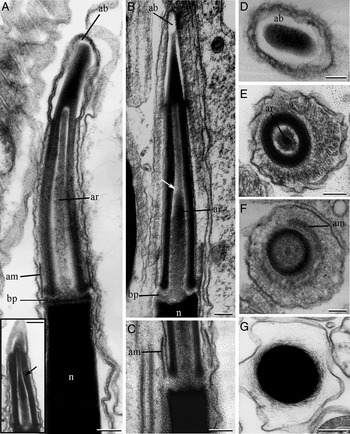
The acrosomal complex consists of a tall, conical, membrane-bound acrosomal vesicle, an axial rod and a basal plate, and is similar in both species. The acrosomal vesicle is 0.99 ± 0.2 µm long (N = 14) in O. carcellesi (Figure 1A) and 1.1 ± 0.2 µm long (N = 19) in O. deshayesiana (Figure 1B). Apically, the plasma membrane of the acrosomal vesicles separated from the vesicle contents, forming an apical bleb that measured in length 0.35 ± 0.01 µm (N = 14) in O. carcellesi (Figure 1A, D) and 0.40 ± 0.03 µm (N = 19) in O. deshayesiana (Figure 1B). The acrosomal vesicle is invaginated posteriorly, containing the axial rod, and is a slight narrowing of the acrosomal vesicle about half way along the length of the invagination part of the acrosome. In O. carcellesi (Figure 1A, inset, E) this narrowing is situated at 0.7 in relation to acrosomal vesicle length and in O. deshayesiana (Figure 1B, F) the narrowing is situated to 0.5 in relation to acrosomal vesicle length. A basal plate is situated between the base of the acrosomal vesicle and the nucleus. An accessory membrane is situated between the basal plate and the acrosomal vesicle, in the posterior region of the acrosomal complex, measured in length 0.12 ± 0.02 µm (N = 8) in O. carcellesi (Figure 1A) and 0.18 ± 0.02 µm (N = 10) in O. deshayesiana (Figure 1C, F).
Euspermatozoa of Olivancillaria carcellesi (A, D, E, G) and Olivancillaria deshayesiana (B, C, F): (A) longitudinal section (LS) through the acrosomal complex showing the apical bleb, axial rod, accessory membrane, basal plate and the anterior part of the nucleus of O. carcellesi. Inset: note the slight narrowing (arrowhead) from acrosomal complex in oblique section (B, C) LS through the acrosomal complex showing the slight narrowing (arrowhead), apical bleb, axial rod, accessory membrane, basal plate and the anterior part of the nucleus of O. deshayesiana; (D) transverse section (TS) of the apical bleb region of the acrosomal vesicle; (E) TS of the anterior region of the acrosomal complex showing the invagination and the axial rod material of O. carcellesi; (F) TS of the posterior region of the acrosomal complex showing the accessory membrane of O. deshayesiana; (G) TS of the nucleus of O. carcellesi; (E–G) some residual cytoplasm indicating slight immaturity was observed. ab, apical bleb; am, accessory membrane; ar, axial rod; bp, basal plate; n, nucleus. Scale bars: A, A inset-B, D–E, G = 0.1 µm; C = 0.05 µm; F = 0.2 µm.

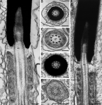
The mature nuclei are filiform, highly electron-dense, and were measured at 17.81 ± 2.07 µm (N = 30) in O. carcellesi (Figure 1A, G) and 12.82 ± 1.62 µm (N = 30) in O. deshayesiana in length (Figure 1B, C). The nucleus contains a basal invagination that includes a centriolar derivate and is continuous with the initial portion of a 9 + 2 microtubular pattern axoneme (Figure 2A, B, D, F). The length of this basal invagination is 0.68 ± 0.09 µm (N = 10) and 0.39 ± 0.05 µm (N = 12) in O. carcellesi and O. deshayesiana, respectively. Posterior to the nucleus, the axoneme is enclosed in a mitochondrial sheath to form the midpiece (Figure 2A, C, E, F). The midpiece of O. deshayesiana measured 3.26 µm in length (figure 19 in Giménez et al., Reference Giménez, Arrighetti, Teso, Hermida, Zabala and Penchaszadeh2009).
Euspermatozoa of Olivancillaria carcellesi (A–C) and Olivancillaria deshayesiana (D–F): (A) longitudinal section (LS) through the nucleus and anterior portion of midpiece with mitochondria spiralling around the axoneme of O. carcellesi; (B) transverse section (TS) through the nucleus with the axoneme of O. carcellesi; (C) TS through the midpiece of O. carcellesi; (D) TS through the nucleus with the axoneme of O. deshayesiana; (E) TS through the midpiece of O. deshayesiana; (F) LS through the nucleus and anterior portion of midpiece with mitochondria spiraling around the axoneme of O. deshayesiana; (B, D, E) some residual cytoplasm indicating slight immaturity was observed. ax, axoneme; mp, midpiece; m, mitochondria. Scale bar: A–F = 0.1 µm.

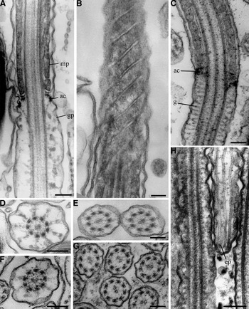
An annular complex, which consists of a double electron-dense ring attached to the inner surface of the plasma membrane, is located at the immediate junction of the midpiece and glycogen piece in both species (Figure 3A, C). The midpiece has mitochondrial elements arranged helically (Figure 3B). Posterior to the annulus complex, the axoneme is associated with nine longitudinal and nine radiating tracts of dense granules (Figure 3D, F). The diameter of the glycogen piece in both O. carcellesi and O. deshayesiana decreases towards the posterior region, the endpiece. The diameter of the endpiece is 0.27 ± 0.04 µm (N = 30) in O. carcellesi and 0.16 ± 0.02 µm (N = 30) in O. deshayesiana (Figure 3E, G). The end piece succeeds the glycogen piece, consisting of the continuing 9 + 2 microtubular axoneme and surrounding plasma membrane (Figure 3H).
Euspermatozoa of Olivancillaria carcellesi (A, B, D, E) and Olivancillaria deshayesiana (C, F–H): (A) longitudinal section (LS) through the junction of the midpiece, annular complex and glycogen piece of O. carcellesi; (B) transverse section (TS) showing helical midpiece elements of O. carcellesi; (C) LS through the junction of the midpiece, annular complex and glycogen piece of O. deshayesiana; (D) TS showing the glycogen piece of O. carcellesi; (E) TS through the endpiece of O. carcellesi; (F) TS of the glycogen piece of O. deshayesiana; (G) TS through the endpiece of O. deshayesiana; (H) LS of the endpiece of O. deshayesiana; (A, D, F) some residual cytoplasm indicating slight immaturity was observed. ac, annular complex; gp, glycogen piece; mp, midpiece. Scale bars: A, C–H = 0.1 µm; B = 0.2 µm.

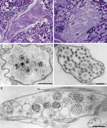
Vermiform cells are observed in O. carcellesi and O. deshayesiana by light microscopy (Figure 4A, B). In transverse sections of TEM these structures show vesicles with granular material, vesicles with less electro-dense material, and small, rounded mitochondrial elements (Figure 4C–E). In addition, 3–6 peripheral axonemes in O. carcellesi and 4–7 peripheral axonemes in O. deshayesiana were found in contact with plasma membrane.
Vermiform cells: (A) Olivancillaria carcellesi; (B) Olivancillaria deshayesiana; (C) transverse section (TS) of vermiform cells of O. deshayesiana showing peripheral axonemes and vesicular content; (D) TS of O. deshayesiana showing only axonemes in contact with plasma membrane; (E) longitudinal section of the vermiform cell of O. deshayesiana. Note peripheral axonemes in contact with plasma membrane. ax, axoneme. Scale bars: A, B = 10 µm; C, E = 0.5 µm; D = 0.1 µm.

DISCUSSION
The euspermatozoa of Olivancillaria carcellesi and O. deshayesiana shared characters include an acrosomal complex with an apical bleb and an accessory membrane, a solid electron-dense nucleus, and a midpiece with mitochondrial elements helically coiled around the axoneme. All these characters are described by Healy (Reference Healy and Taylor1996) for all Caenogastropoda except for the families Ampullaroidea, Cyclophoroidea and Cerithioidea.
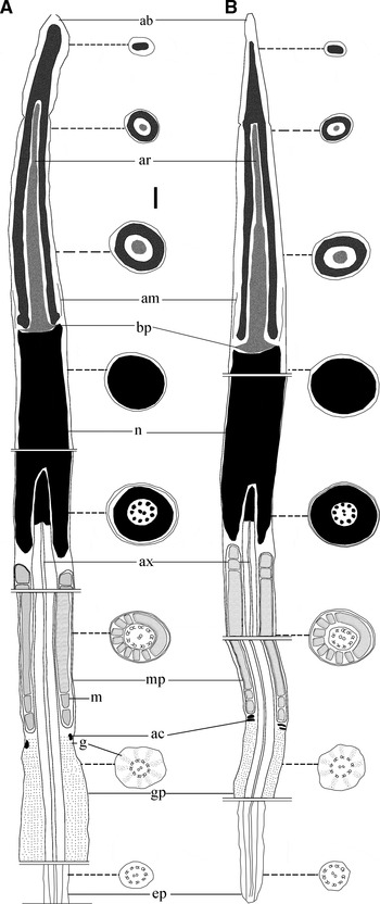
The structure of the acrosomal complex is similar in both species, like other caenogastropods. In O. carcellesi and O. deshayesiana the acrosomal vesicle decreased in diameter from the base to the apical bleb (Figures 1 & 5). The slight narrowing of the acrosomal vesicle are different within O. carcellesi and O. deshayesiana and also with other species of the family Volutidae, e.g. Zidona dufresnei (Donovan, 1823), Provacator mirabilis (Finlay, 1926), Adelomelon ancilla (Lightfoot, 1786), Adelomelon beckii (Broderip, 1836) and Odontocymbiola magellanica (Gmelin, 1791); Mitridae, e.g. Mitra (Strigatella) fastigium Reeve, 1845; and Cypraeidae, e.g. Cypraea tigris Linnaeus, 1758 (Koike, Reference Koike1985; Healy, Reference Healy1986; Giménez et al., Reference Giménez, Healy, Hermida, Lo Nostro and Penchaszadeh2008, Reference Giménez, Arrighetti, Teso, Hermida, Zabala and Penchaszadeh2009; Zabala et al., Reference Zabala, Hermida and Giménez2009; Arrighetti & Giménez, Reference Arrighetti and Giménez2010; Giménez, Reference Giménez2011).
Comparative eusperm morphology of Olivancillaria carcellesi (A) and Olivancillaria deshayesiana (B). Note internal view of eusperm observed by transmission electron microscopy in longitudinal section and transverse section. ab, apical bleb; ac, annular complex; am, accessory membrane; ar, axial rod; ax, axoneme; bp, basal plate; ep, endpiece; g, glycogen; gp, glycogen piece; mp, midpiece; m, mitochondria; n, nucleus. Scale bar = 0.1 µm.

The nucleus is highly electron-dense and has an axoneme inserted in the basal invagination like those of other caenogastropods (Kohnert & Storch, Reference Kohnert and Storch1984; Koike, Reference Koike1985). This invagination is different in size in both species (Figures 2A, F & 5A–B) and short as in many members of Caenogastropoda (Healy, Reference Healy1986, Reference Healy and Ponder1988, Reference Healy and Taylor1996). The length of the nucleus in O. carcellesi and O. deshayesiana is shorter than other neogastropods Nucella lapillus (Linnaeus, 1758) 39 µm and Nucella crassilabrum (Lamarck, 1815) 36 µm Gallardo & Garrido (Reference Gallardo and Garrido1989); Z. dufresnei 25 ± 3 µm and P. mirabilis 20 ± 2 µm Giménez et al. (Reference Giménez, Healy, Hermida, Lo Nostro and Penchaszadeh2008); A. beckii 26.67 ± 3.21 µm Arrighetti & Giménez, Reference Arrighetti and Giménez2010; O. magellanica 32 ± 3 µm Giménez (Reference Giménez2011).
In O. carcellesi and O. deshayesiana, the midpiece has mitochondrial elements with external membranes fused and helically arranged around the axonemes (Figure 3B), as other Caenogastropoda families (West, Reference West1978; Jaramillo et al., Reference Jaramillo, Garrido and Jorquera1986; Gallardo & Garrido, Reference Gallardo and Garrido1989; Hodgson, Reference Hodgson1993; Giménez et al., Reference Giménez, Healy, Hermida, Lo Nostro and Penchaszadeh2008, Reference Giménez, Arrighetti, Teso, Hermida, Zabala and Penchaszadeh2009; Zabala et al., Reference Zabala, Hermida and Giménez2009; Arrighetti & Giménez, Reference Arrighetti and Giménez2010; Giménez, Reference Giménez2011).
The glycogen piece, annular complex and endpiece of O. carcellesi and O. deshayesiana do not differ from the configurations shown in other caenogastropods (Healy, Reference Healy and Ponder1988, Reference Healy, Jamieson, Adiyodi and Adiyodi2000). The double-ring complex of both species of Olivancillaria as well as other Caenogastropoda (Buckland-Nicks et al., Reference Buckland-Nicks, Williams, Chia and Fontaine1982a, Reference Buckland-Nicks, Williams, Chia and Fontaineb; Healy, Reference Healy1986, Reference Healy and Ponder1988; Giménez et al., Reference Giménez, Healy, Hermida, Lo Nostro and Penchaszadeh2008; Zabala et al., Reference Zabala, Hermida and Giménez2009; Arrighetti & Giménez, Reference Arrighetti and Giménez2010; Giménez, Reference Giménez2011) differs from the annulus of other groups such as the Cerithioidea, which have a single ring (Healy, Reference Healy1982).
To our knowledge, the other structure found in the testis in both species (Figure 5A, B) does not to resemble any other type discussed until now (Nishiwaki, Reference Nishiwaki1964; Melone et al., Reference Melone, Donin and Cotelli1980; Healy & Jamieson, Reference Healy and Jamieson1981, 1982, 1986, Reference Healy1993, 1996, 2000; Buckland-Nicks, Reference Buckland-Nicks1998; Giménez et al., Reference Giménez, Healy, Hermida, Lo Nostro and Penchaszadeh2008; Zabala et al., Reference Zabala, Hermida and Giménez2009; Arrighetti & Giménez, Reference Arrighetti and Giménez2010; Giménez, Reference Giménez2011). In addition, Teso et al. (Reference Teso, Arrighetti and Penchaszadeh2012) did not find this structure in seminal vesicles of O. deshayesiana during their two-year study. Although these structures could be paraspermatozoa based in the presence of peripheral axonemes and vesicles, further studies on other members of the family are necessary to confirm our hypothesis.
The slight narrowing in the acrosomal vesicle invagination is situated in different levels between O. deshayesiana and O. carcellesi (Figure 1A, B). This morphology could be considered as a specific character. The length of the nucleus in both species is shorter than other neogastropods. We suggest that these characteristics could have taxonomic importance because they have not been observed in other caenogastropods. The study on these two species is the first step in the sperm morphology analysis within the Olividae family. Further studies in other members of Olividae, could be necessary in order to complete the sperm morphology map associated with molecular phylogeny.
ACKNOWLEDGEMENTS
Special thanks to Jennifer Antonides for her help in the English version and improvement of this manuscript, and Guido Pastorino, Diego Urteaga, Javier Signorelli and Maria Bagur for capturing most of the samples. The MS greatly benefits from the comments of two anoymous referees. This work was partially supported by grants from PADI Foundation in 2007, the Lerner Gray Memorial Fund (AMNH) to V.T. in 2007, the National Agency for Scientific and Technical Promotion, Argentina (PICTR 1869 and PICT 942), UBACyT X 117, and the Consejo Nacional de Investigaciones Científicas y Técnicas, Argentina (CONICET, PIP 2788).