Introduction
The developmental competence of oocytes is recognized as the ability to undergo maturation, fertilization, as well as proper zygote formation (Duranthon & Renard, Reference Duranthon and Renard2001; Krisher, Reference Krisher2004; Sirard et al., Reference Sirard, Richard, Blondin and Robert2006). In several mammalian species, including bovines, pigs and goats, in vitro maturation (IVM) and fertilization (IVF) conditions are well defined. Moreover, there are increased numbers of oocytes that reach MII stage as well those that are able to be successfully fertilized by a single spermatozoon (Rodrigues Bde et al., Reference Rodrigues Bde, dos Santos and Rodrigues2004; Hirao, Reference Hirao2011; Yuan & Krisher, Reference Yuan and Krisher2012). In the bitch, the influence of the oestrous cycle stage and of several media supplements, e.g. progesterone and estradiol, on the meiotic competence of canine oocytes was determined. However, decreased numbers of canine oocytes have been found to reach the MII stage, as compared with other species such as bovine, (Luvoni et al., Reference Luvoni, Luciano, Modina and Gandolfi2001; de Avila Rodrigues & Rodrigues, Reference de Avila Rodrigues and Rodrigues2003; Rodrigues Bde et al., Reference Rodrigues Bde, dos Santos and Rodrigues2004; Songsasen & Wildt, Reference Songsasen and Wildt2007), and only 20–25% of canine oocytes have been shown to be meiotically competent after IVM (Willingham-Rocky et al., Reference Willingham-Rocky, Hinrichs, Westhusin and Kraemer2003; Songsasen & Wildt, Reference Songsasen and Wildt2007; Rodrigues & Rodrigues, Reference Rodrigues and Rodrigues2010; Kempisty et al., Reference Kempisty, Woźna, Piotrowska, Bukowska, Jackowska, Antosik, Jaśkowski and Brüssow2012). Until now, only one morula and one blastocyst have been produced after IVF in dogs (Otoi et al., Reference Otoi, Murakami, Fujii, Tanaka, Ooka, Une and Suzuki2000, Reference Otoi, Shin, Kraemer and Westhusin2004). Therefore, the determination of factors influencing the maturation ability of canine oocytes is of high significance. As developmental competence of oocytes is also recognized as the ability of gametes to undergo fertilization and maturation, the role of zona pellucida glycoprotein (ZP) expression in competent and incompetent cells may be used as a ‘molecular marker’ of fertilization and developmental ability. Until now, only few data have been published related to zona pellucida structure and its modification during folliculogenesis, oogenesis or in vitro maturation in mammalian species (Schwartz et al., Reference Schwartz, Magerkurth and Michelmann1996; von Witzendorff et al., Reference von Witzendorff, Ekhlasi-Hundrieser, Dostalova, Resch, Rath, Michelmann and Töpfer-Petersen2005; Rath et al., Reference Rath, Töpfer-Petersen, Michelmann, Schwartz, von Witzendorff, Ebeling, Ekhlasi-Hundrieser, Piehler, Petrunkina and Romar2006). Michelmann et al. (Reference Michelmann, Rath, Töpfer-Petersen and Schwartz2007) analysed the structure of porcine zona pellucida and sperm–egg binding patterns by scanning electron microscopy. They suggested that the structure of zona pellucida is of heterogeneous morphology.
ZPs are described as the molecules that form protein structure of the zona pellucida and reside in the extracellular glycoprotein matrix surrounding the oocyte (Martinova et al., Reference Martinova, Petrov, Mollova, Rashev and Ivanova2008; Gupta et al., Reference Gupta, Bhandari, Shrestha, Biswal, Palaniappan, Malhotra and Gupta2012). These proteins are encoded by ZP genes, ZP1, ZP2, ZP3 and ZP4 (Gupta et al., Reference Gupta, Bhandari, Shrestha, Biswal, Palaniappan, Malhotra and Gupta2012). The ZP3 proteins are recognized to represent the primary sperm receptor. ZP2 functions as the secondary receptor and plays, compared to ZP3, a subordinate role during the initial steps of fertilization (Jackowska et al., Reference Jackowska, Kempisty, Antosik, Bukowska, Budna, Lianeri, Rosińska, Woźna, Jagodziński and Jaśkowski2009; Antosik et al., Reference Antosik, Kempisty, Jackowska, Bukowska, Lianeri, Brüssow, Woźna and Jaśkowski2010; Ikawa et al., Reference Ikawa, Inoue, Benham and Okabe2010; Kempisty et al., Reference Kempisty, Jackowska, Piotrowska, Antosik, Woźna, Bukowska, Brüssow and Jaśkowski2011; Kempisty et al., Reference Kempisty, Woźna, Piotrowska, Bukowska, Jackowska, Antosik, Jaśkowski and Brüssow2012). As shown in our previous study (Kempisty et al., Reference Kempisty, Jackowska, Piotrowska, Antosik, Woźna, Bukowska, Brüssow and Jaśkowski2011), the porcine pZP3 protein is differentially located and distributed in the oocyte. The canine ZP4 protein is defined as a factor that induces the acrosome reaction and inhibits binding of spermatozoa to zona pellucida in a time- and dose-dependent manner. It was found that protein kinase A, but not G, proteins are responsible for activation of the ZP4-induced acrosome reaction (Chiu et al., Reference Chiu, Wong, Chung, Lam, Pang, Lee, Sumitro, Gupta and Yeung2008). Presumably, correlation between ZP4 protein expression and developmental competence of canine oocytes may be responsible for the induction of acrosome reaction of spermatozoa.
As the efficiency of IVF in dogs is low and the reasons are insufficiently known, the study of ZP genes and ZP protein expressions in relation to the maturation ability of gametes could be of benefit. Therefore, we aimed to investigate the expression pattern and the cellular distribution of zona pellucida glycoproteins in canine oocytes before and after IVM.
Material and methods
Collection of oocytes
Ovaries and reproductive tracts were recovered from 27 normal healthy females, aged 3–5 years, after routine ovariohysterectomy at the Institute of Veterinary Sciences, Poznan University of Life Sciences, and immediately transported to the laboratory. Only bitches at the anoestrus stage of the cycle were used. Therefore, vaginal cytology swab specimen and venous blood samples were collected from each female after induction of anaesthesia for the surgery. Stage of the oestrus cycle was assessed by vaginal cytology, blood serum P4 concentration was determined using P4-kit (Ovucheck Premate®; Camelot Farms, College Station, TX, USA) and the reproductive tissues were evaluated morphologically (Hidalgo et al., Reference Hidalgo, Gómez, Prieto, Duque, Goyache, Fernández, Fernández, Facal and Díez2004). The females were considered to be in anoestrus when the vaginal cytology revealed parabasal cells on vaginal swabs and progesterone concentrations were lower than that in the kit standard for low progesterone. Furthermore, the morphological criteria for the oestrus stage of bitches were evaluated using the classification described previously (Otoi et al., Reference Otoi, Willingham, Shin, Kraemer and Westhusin2002).
Ovaries were placed in physiological saline solution (PSS) at 39°C and, then washed three times with PSS. After washing, the ovaries were sliced with a scalpel blade at room temperature to collect cumulus–oocyte complexes (COCs).
IVM of canine cumulus–oocyte complexes
Cumulus–oocyte complexes were washed three times in a tissue culture medium (TCM-199-HEPES, Gibco®, Carlsbad, CA, USA). Cumulus–oocyte complexes were selected according to the homogeneity of the cytoplasm and the number of cumulus oophorus cells, based on the classification described previously by Hewitt & England (Reference Hewitt and England1997). Only grade I COCs (n = 50, in 500 μl of IVM medium), (Otoi et al., Reference Otoi, Willingham, Shin, Kraemer and Westhusin2002), i.e. those with more than five layers of compact cumulus cells and a homogeneous dark cytoplasm, were matured in culture dishes containing 500 μl of the maturation medium (TCM199, supplemented with 50 UI/ml of gentamycin, Sigma-Aldrich; penicillin/streptomycin, Sigma-Aldrich). The COCs were matured in the maturation medium (TCM199) supplemented with 17β-estradiol (2.0 μg/ml), follicle-stimulating hormone (FSH; 0.5 μg/ml), luteinising hormone (LH; 5.0 μg/ml), Na-Pyr (25 mM), progesterone (1 μg/ml), epidermal growth factor (EGF;100 ng/ml), and bovine serum albumin (BSA; 0.3%) in an atmosphere of 5% CO2 in humidified air at 39°C for 72 h. The COCs that had compact cumulus cell layers were first incubated with bovine testicular hyaluronidase (Sigma-Aldrich, St. Louis, MO, USA) for 2 min at 38°C to separate cumulus and granulosa cells. Thereafter, cells were removed by vortexing the oocytes in 1% sodium citrate buffer and by mechanical displacement using a small-diameter glass micropipette. The granulose cell-free oocytes were used for RQ-PCR, and confocal microscopic analysis.
Assessment of nuclear maturation
After maturation of COCs, the cumulus cells were removed by vortexing. The oocytes were then fixed for 15 min in 2.5% (w/v) glutaraldehyde (Merck, Darmstadt, Germany) in phosphate-buffered saline (PBS), followed by washing twice with PBS, staining with 0.1 mg/ml 4′,6-diamino-2-phenylindole (DAPI; Sigma, St. Louis, MO, USA) in PBS, and mounting on slides. Evaluation of nuclear status was conducted using confocal microscopy (Fluoview 10i, Olympus, Tokyo, Japan). The oocytes were classified as either MII-oocytes with a polar body or two shiny chromatin spots or as oocytes with configuration other than described. Additionally, germinal vesicle (GV) chromatin configuration was also observed, which is characterized by initially decondensed chromatin with nucleolus not surrounded by heterochromatin (named as NSN configuration). During the maturation of oocytes, the chromatin configuration of the GV state is changed into the GVBD state. The oocytes may also undergo GVBD at the advanced stage of atresia.
Transcript analysis
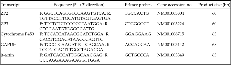
Total RNA was isolated from oocytes before and after IVM (each of 100 oocytes) using an RNeasy mini column (Qiagen GmbH, Hilden, Germany). RNA samples were repurified and their quality was examined by microchannel electrophoresis, using an Agilent Bioanalyzer (Agilent Technologies Inc., Palo Alto, CA, USA). The concentrations of RNA samples were calculated using the NanoDrop system (Thermo Scientific, Wilmington, USA) and samples were then resuspended in RNase-free water and reverse-transcribed into cDNA. Reverse transcription reaction were performed using 1 μg/μl of RNA sample. Briefly, first-strand complementary DNA synthesis was performed by priming with oligo-dT16. The samples were redissolved in an 8.5 μl solution consisting of 1 μl 50 mol/l oligo-dT16, 0.2 μl 0.1 mol/l dithiothreitol (DTT), 0.05 μl RNase inhibitor (20 IU/μl) (RNasin; Promega, Madison, WI, USA), 7.25 μl sterile nuclease-free water, 1 μg/μl of RNA sample and incubated at 37°C for 60 min. The primers were annealed by incubating the samples at 70°C for 6 min and then immediately placed on icy water for 1 min. Real-time quantitative PCR amplification reactions were carried out in an AB7500 Real–Time PCR System (Applied Biosystems) using Universal PCR Master Mix (Roche, Mannheim, Germany). All reactions were performed in a 20-μl volume with 6 pmol forward and reverse primers, 2 μl of cDNA (1 μg/μl of RNA concentration used for reverse transcription) and 4 pmol of probe (Table 1). ZP2 and ZP3 mRNA expression were studied using primer probes (cat. no. 59–04688562001, cat. no. 45–04688058001, respectively, Roche, Mannheim, Germany) according to the manufacturer's instructions. All reactions were conducted in triplicate that represented three different mRNA templates classified into the same group of oocytes. In case of the negative control, cDNA was not added.
Oligonucleotide sequences and primer probes used for RQ-PCR analysis

The relative expression levels of ZP2 and ZP3 mRNA in each sample were standardized employing internal standards of the two housekeeping genes; GAPDH and β-actin (cat. no. 119–04693531001 and cat. no. 44–04688040001, respectively), (Roche, Mannheim, Germany). In order to ensure that granulosa cells did not contaminate the oocytes, we demonstrated the absence of cytochrome P450 aromatase (cat. no. 69–04688686001, Roche, Mannheim, Germany) transcript by reverse transcription (RT) and RQ-PCR.
Confocal microscopic observation of ZP3 and ZP4 protein distribution in canine oocytes
In total, 200 canine oocytes before and after IVM (each n = 100 oocytes per group) were incubated with 300 μg/ml bovine testicular hyaluronidase Sigma-Aldrich Co. (St. Louis, MO, USA) for 2 min at 38°C to remove cumulus cells. Oocytes were fixed in 2.5% paraformaldehyde in PBS and 0.2% Triton-X 100 for 30 min at room temperature and washed three times in PBS/PVP (0.2%). In order to block non-specific binding, the samples were incubated in 3% BSA in PBS with 0.1% Tween 20 for 30 min at RT. Oocytes were incubated for 12 h at 4°C with goat polyclonal anti-ZP3 (C-20), (Santa Cruz Biotechnology, Santa Cruz, CA, USA; sc-23717) antibody (Ab) or with goat polyclonal anti-ZP4 (I-14), (Santa Cruz Biotechnology, Santa Cruz, CA, USA; sc-49587), diluted 1:500 in PBS/ 1.5% BSA/0.1% Tween 20. After repeated washes with PBS/0.1% Tween 20, the samples were incubated for 1 h at RT with fluorescein isothiocyanate (FITC)-conjugated anti-goat IgG Ab produced in rabbits, diluted 1:200 in PBS/0.1% Tween 20. Following this wash in PBS/0.1% Tween 20, the oocytes were mounted on glass slides in an antifade drop and observed under the LSN 510 confocal system (Carl ZEISS microscope, Axiovert 200M) preserving 10 sections of each observed oocyte. FITC was excited at 488 nm by an argon laser, and emissions were imaged through a 505–530 nm filter.
Statistical analysis
Two-way analysis of variance (ANOVA) followed by the Tukey post-hoc test was used to compare the results of the real-time PCR reaction as well as fluorescence intensity of protein expression. The differences were considered to be significant at *P < 0.05, **P < 0.01 and ***P < 0.001. GraphPad Prism version 4.0 software (GraphPad Software, San Diego, CA, USA) was used for statistical calculations. All confocal microscopic images were analysed using Imaris 7.2 software (BitPlane, Zurich, Switzerland).
The experiments were approved by the local ethical committee.
Results
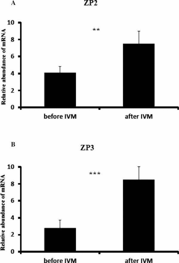
RQ-PCR analysis revealed an increased expression of ZP2 after IVM as compared with oocytes analysed before IVM (P < 0.01), (Fig. 1 A). Similarly, ZP3 mRNA was significantly higher expressed in oocytes after IVM (P < 0.001), (Fig. 1 B).
Relative abundance of ZP2 and ZP3 transcripts in canine oocytes. Canine oocytes, before and after in vitro maturation were immediately used to isolate RNA, which was reverse-transcribed into cDNA. Presence of ZP2 (A) and ZP3 (B) transcripts was evaluated by RQ-PCR analysis. ZP2 and ZP3 mRNA expression was evaluated before and after IVM. Results are presented as means ± standard error of the mean (SEM) with the level of significance of *P < 0.05, **P < 0.01, ***P < 0.001.

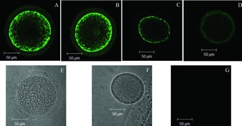
Based on confocal microscopic observations, we found significant differences in ZP3 protein expression before and after IVM (Fig. 2 A–D). In canine oocytes analysed before IVM, the ZP3 protein was expressed at higher level. In both, before and after IVM, the ZP3 protein was distributed in the peripheral area of cytoplasm. An increased ZP4 protein expression was also detected before, as compared with after, IVM.
Confocal microscopic observation of ZP3 glycoprotein expression and distribution in canine oocytes. Canine oocytes were stained before (A, B) and/or after IVM (C, D) with goat polyclonal anti-ZP3 antibodies. The negative controls (only primary antibody), canine oocytes with intact zona pellucida (E); before IVM (F); after IVM, (G). Bars correspond to 50 μm.

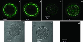
Before IVM, the ZP4 protein was distributed in the peripheral area of cytoplasm and zona pellucida, whereas the localization was changed into the entire cytoplasm when analysed after IVM (Fig. 3 A–D).
Confocal microscopic observation of ZP4 glycoprotein expression and distribution in canine oocytes. Canine oocytes were stained before (A, B) and/or after IVM (C, D) antibodies. The negative controls (only primary antibody) canine oocytes with intact zona pellucida (E); before IVM (F); after IVM (G). Bars correspond to 50 μm.

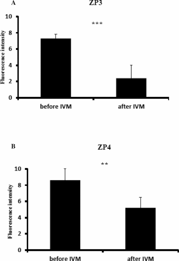
After using a quantitative method to analyse the expression of ZP3 and ZP4 proteins, we found higher expression of ZP3 before IVM as compared with after IVM (P < 0.001) (Fig. 4 A). Similarly, when analysing ZP4 expression, we observed also higher expression before IVM (P < 0.01) (Fig. 4 B).
Fluorescence intensity of canine oocytes stained with anti-ZP3 and anti-ZP4 antibodies. The quantitative expression of ZP3 and ZP4 proteins in canine oocytes was analysed before or after IVM (A, B), respectively. The fluorescence intensity was measured using Imaris software. The differences were considered to be significant at *P<0.05, **P<0.01 and ***P<0.001 (mean ± SEM). The software of GraphPad Prism version 4.0 (GraphPad Software, San Diego, CA) was used for statistical calculations. All confocal microscopic images were analyzed using Imaris 7.2 (BitPlane, Zurich, Switzerland) software.

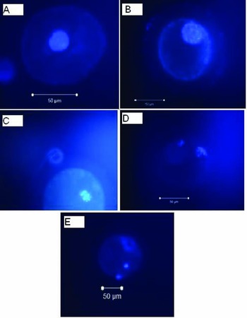
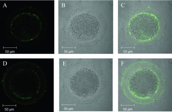
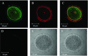
The different stages of canine oocytes maturation are shown in Fig. 5. The confocal microscopic observation of anti-ZP3, anti-ZP4, and Nomarski and merged images are presented in Fig. 6. To confirm the confocal microscopic observation of canine oocytes, antibody binding specificity, as well as the expression and distribution of α-tubulin and f-actin, were applied (Fig. 7). We observed 2% (n = 8) of GV oocytes, 14% (n = 56) of GVBD oocytes, 12% (n = 48) of metaphase I (MI) oocytes and 19% (n = 76) of MII stage oocytes after 72 h of IVM. The oocytes with other chromatin organization structures were identified as abnormal and degenerative.
Confocal microscopic observations and determination of oocyte maturation. Oocytes from bitches were stained with DAPI (A–E). Bars correspond to 50 μm. (A) Oocyte in GV stage; (B) oocyte in GVBD stage; (C) oocyte in MI stage; (D) oocyte in MII stage; and (E) oocyte with I polar body.

Confocal microscopic observations of ZP3 and ZP4 expression. Canine oocytes were stained with goat polyclonal anti-ZP3 antibodies (A–C). (A) Correspond to anti-ZP3 staining; (B) correspond to Nomarski; (C) correspond to merged image of (A) and (B). (D–F) Represents oocytes stained with goat polyclonal anti-ZP4 antibody, Nomarski and merged image, respectively. Bars correspond to 50 μm.

Confocal microscopic observations α–tubulin and f-actin expression in canine oocytes. Canine oocytes were stained with anti- α-tubulin and f-actin antibodies (A–C). (A) Correspond to α–tubulin staining with FITC; (B) correspond to f-actin staining with TRITC; (C) correspond to merged image of (A) and (B). (D–F) represents negative control for ZP3 and ZP4 (D); Nomarski (E); and merged (F). Bars correspond to 50 μm.

Discussion
Previous studies in the pig have suggested that developmental potential and fertilization ability may be strongly related to briliant crezyl blue test (BCB) staining, oocyte morphology and follicular size (Kölle et al., Reference Kölle, Dubois, Caillaud, Lahuec, Sinowatz and Goudet2007; Antosik et al., Reference Antosik, Kempisty, Jackowska, Bukowska, Lianeri, Brüssow, Woźna and Jaśkowski2010; Kempisty et al., Reference Kempisty, Jackowska, Piotrowska, Antosik, Woźna, Bukowska, Brüssow and Jaśkowski2011). The experiments examining the expression and/or distribution of zona pellucida proteins are currently of high significance due to the low number of fully matured canine oocytes recovered in vitro as well as the decreased fertilization rate.
Dogs ovulate at prophase in the first meiotic division, a fundamental point as normal embryo development and pregnancy after IVF is rarely achieved in this species. The extremely specific oviductal conditions during final oocyte maturation in vivo are hard to mimic under in vitro conditions and IVM media may not be suitable for oocyte maturation compared with other species. The conditions of the final stages of oocytes development in dogs are extremely difficult to imitate (Reynaud et al., Reference Reynaud, Fontbonne, Marseloo, Viaris de Lesegno, Saint-Dizier and Chastant-Maillard2006; Concannon, Reference Concannon2011).
It has been demonstrated previously that the zona pellucida structure becomes permanently modified during folliculogenesis and oogenesis (Blackmore et al., Reference Blackmore, Baillie, Holt, Dierkx, Aitken and McLaughlin2004; Parillo et al., Reference Parillo, Zelli, Verini Supplizi, Fagioli and Gargiulo2005; Martinova et al., Reference Martinova, Petrov, Mollova, Rashev and Ivanova2008; Diederich et al., Reference Diederich, Hansmann, Heinzmann, Barg-Kues, Herrmann, Aldag, Baulain, Reinhard, Kues, Weissgerber, Haaf and Niemann2012). Only few data have been published on zona pellucida modification in mature and immature canine oocytes. Blackmore et al. (Reference Blackmore, Baillie, Holt, Dierkx, Aitken and McLaughlin2004), using paraformaldehyde-fixed prepubertal canine ovarian tissue sections and in situ hybridization of mRNA in canine ovaries, suggested that ZP1 mRNA was distributed in the cytoplasm of oocytes of primordial, primary and early secondary follicles. They also demonstrated the localization of ZP2 and ZP3 gene transcripts in granulosa cells of growing follicles, but not in the granulosa cells or oocyte cytoplasm. Finally, they concluded that ZP1 synthesis was restricted to the canine oocyte while synthesis of ZP2 and ZP3 was restricted to granulosa cells. These data partially support our results obtained from the IVM experiment, in which the expression of the ZP4 gene and/or distribution of encoded proteins were related to canine oocyte maturity, which reflects previous processes occurring during folliculogenesis. Our analysis has shown that the expression of ZP2 and ZP3 mRNA or ZP3 and ZP4 proteins was associated with the canine oocyte maturation stage as higher levels of investigated transcripts were observed in oocytes post IVM as compared with before IVM. This situation may be explained by the accumulation of ZP mRNA during folliculogenesis and oogenesis. Moreover, increased levels of zona-encoding mRNAs, used as template for respective proteins synthesis, in MII-competent gametes may suggests that these oocytes are characterized by an increased fertilization ability.
The role of zona pellucida protein expression has been investigated in only a few studies. In an ultrastructural study, Parillo et al. (Reference Parillo, Zelli, Verini Supplizi, Fagioli and Gargiulo2005) characterized and localized the oligosaccharide sequences of zona pellucida glycoproteins at different stages of follicular development in the canine ovary. They found the regionalization of oligosaccharide chains within three concentric bands of the zona matrix: an inner surface close to the oocyte plasma membrane, an intermediate portion and an outer layer in contact with the follicular cells. They showed variations in the carbohydrate residues in canine zona pellucida that were related to follicular growth. Their previous study (Parillo & Verini Supplizi, Reference Parillo and Verini Supplizi1999) indicated also that zona structure depends on species and on follicle developmental stage. Moreover, the similarity in lectin affinity between ooplasm and zona pellucida of oocytes present in follicles at different stages of development confirms the involvement of oocytes in zona pellucida formation. Our results have shown that the level of ZPs mRNAs and proteins may be associated with the maturation stage as the expression of these molecules differs significantly in pre-IVM and/or post-IVM canine oocytes.
Rankin et al. (Reference Rankin, O’Brien, Lee, Wigglesworth, Eppig and Dean2001) have shown that ZP2-null mice (ZP−/−) are able to ovulate. We demonstrated that ZP3 mRNA and protein expression fail to correlate in pre-IVM and/or post-IVM canine oocytes. This failure is due lower expression and protein level of ZP3 after IVM. However, an increased level of ZP3 mRNA after IVM, was determined. This unique process may be associated with induction of alternative splicing mechanisms and/or significant reduction of protein synthesis after IVM. This finding contradicts previously obtained results that expression of mRNA and proteins in COCs and/or single oocytes differ significantly before and after IVM. It may be further suggested that only mature oocytes are able to induce the acrosome reaction following activation of ZP4 protein up-regulation of adenylyl cyclase/protein kinase-A (cAMP/PKA) activity and down-regulation of signal-regulated kinase (ERK) activity (Chiu et al., Reference Chiu, Wong, Lee, Lam, Chung, Lee, Koistinen, Koistinen, Gupta, Seppälä and Yeung2010). The highest expression levels of ZP2 and ZP3 mRNA found in canine oocytes after 72 h of IVM led us to assume that after this maturation time canine oocytes are ready to induce the acrosome reaction (acrosome competent). This may be supported by our observation of ZP4 protein distribution using confocal microscopy, in which maturated oocyte ZP4 protein was distributed over the entire cytoplasm, whereas prior to IVM the ZP4 protein was localized the peripheral areas of the cytoplasm and zona pellucida. We have failed to observe differences in ZP3 protein distribution within canine oocytes before and after IVM; this indicates that the developmental capacity and/or maturation stage of canine oocytes is not linked to ZP3 localization. However, we found that ZP4 protein may be translocated between cytoplasm and zona of oocytes and is related to the maturation stage. ZP4 protein localization in the zona pellucida after IVM may be associated with gamete preparation for fertilization, which may be serve as a marker of gamete competence for fusion (El-Mestrah, Reference El-Mestrah, Castle, Borossa and Kan2002).
The differential expression profile of canine ZPs genes and semi-quantitative analysis of protein distribution in relation to maturation stage has been demonstrated in the present study. Variable expression of canine ZP3 mRNA has been also observed by Nickson et al. (Reference Nickson, Boyd, Eckersall, Ferguson, Harvey and Renton1993). They found that the highest expression of ZP3 was after 48 h of in vitro culture (IVC). After in vitro fertilization at 12 h of culture, both male and female pronuclei were found, but no further development of zygotes was detected during the 72 h in vitro maturation period. These results may suggest that differential expression of canine ZPs genes and proteins may be related to the time of IVC and the maturation stage. This led us also to assume that canine zona pellucida is permanently modified during IVM with special relation to differential expression of genes and encoded proteins as well as distinct protein distribution within the oocytes.
In the present study we found an increased number of 50% of abnormal/degenerative canine oocytes. Van Soom et al. (Reference Van Soom, Vandaele, Goossens, de Kruif and Peelman2007) observed that COCs cultured for 48 h showed enhanced rates of CC apoptosis leading to chromatin degeneration or arrest. This process was associated with progesterone profile of the ovary donor. It was suggested that apoptosis in CCs may be one of the factors responsible for the inhibition of oocyte meiotic competence. Moreover, adenosine triphosphate (ATP) depletion, which is associated with reductions in energy levels in COCs cultured in vitro, may serve as a possible reason for COCs degeneration, as ATP is responsible for maintenance of homeostasis and cell survival (St John, Reference St John2002). It has been suggested that this may be an explanation for the high degeneration levels observed in canine COCs matured in vitro (de Avila Rodrigues & Rodrigues, Reference de Avila Rodrigues and Rodrigues2003). Furthermore it was also shown that spontaneous apoptosis may occur in COCs during suboptimal culture and specific components in medium influenced positively or negatively on the cell death or survival (Yuan et al., Reference Yuan, Van Soom, Leroy, Dewulf, Van Zeveren, de Kruif and Peelman2005). A negative effect on the structure of mitochondria was observed when bovine serum was added to the maturation medium during bovine COCs IVM (Abe & Hoshi, Reference Abe and Hoshi2003).
Conclusions
In summary, previously published data have indicated an association between the zona pellucida ultrastructure, expression of encoding genes and proteins, and follicular growth and development. Our results confirm previous results regarding transcription cessation during oocyte maturation (between GVBD and the MII stage).
The contrasting expression of ZP3 and ZP4 proteins compared with mRNA in oocytes analysed before IVM may indicate induction of a posttranscriptional modification mechanism, which could result in low level transcripts necessary for protein synthesis. However, when low mRNA and high protein levels were observed in the same population of pre-IVM oocytes, it may be assumed that many of transcripts are utilized during the maturation process. In contrast, in the case in which we observed high mRNA and low protein levels, the process of transcripts translation has not started yet, at this stage of maturation and development.
Acknowledgements
This study was made possible by grant number 2013/09/N/NZ5/01835 from the Polish Ministry of Scientific Research and Higher Education and National Centre of Science.