Introduction
Bone is a solid-state tissue, which contains ~30% matrix, 60% minerals, and 10% water (Athanasiou et al., Reference Athanasiou, Zhu, Lanctot, Agrawal and Wang2000). Collagen is the main component of bone organic matrix, which is responsible for the tensile strength, whereas the mineral component (calcium phosphate) imparts compressive strength to the bone tissue (Driessens et al., Reference Driessens, van Dijk and Borggreven1978). Despite its relative strength, the bone tissue is susceptible to fracture as a result of trauma or pathological fragility due to deficiency of organic matrix and resorption (Rabie et al., Reference Rabie, Wong and Hagg2000; Cavalcanti et al., Reference Cavalcanti, Pereira, Mazzonetto, de Moraes and Moreira2008). Devices used clinically for bone fixation and repair are fabricated mostly from metals (stainless steel, titanium and its alloys) (Curtis et al., Reference Curtis, Goldhahn, Schwyn, Regazzoni and Suhm2005; Goodrich et al., Reference Goodrich, Sandler and Tepper2012). Metallic devices and implants, due to their non-biodegradable nature, often require additional surgery to remove them from the body, and can cause complications such as allergic reactions and infections (Eppley & Sadove, Reference Eppley and Sadove1995; Marti et al., Reference Marti, Imhoff, Bahrs and Romero1997; Christensen et al., Reference Christensen, Dalstra, Sejling, Overgaard and Bunger2000).
Biodegradable materials for bone repair are preferred because they can be used to fabricate implants and do not require a second surgical invasion for removal (Middleton & Tipton, Reference Middleton and Tipton2000; Wuisman & Smit, Reference Wuisman and Smit2006). All materials must support bone-tissue regeneration and repair processes and be compatible with surrounding tissues, provide mechanical strength, and must degrade to non-toxic by-products which are easily removed by the organism (Hutmacher et al., Reference Hutmacher, Hurzeler and Schliephake1996). Biodegradable biopolymers are commonly used in tissue engineering, including bone regeneration, due to their high level of biodegradability and controlled degradation rates which makes them very useful in clinical applications (Ulery et al., Reference Ulery, Nair and Laurencin2011; Bertesteanu et al., Reference Bertesteanu, Chifiriuc, Grumezescu, Printza, Marie-Paule, Grumezescu, Michaela, Lazar and Grigore2014). The degradation and mechanical properties of polymer materials can be altered by changing their structural composition and fabrication techniques, as well as adding biocompatible nanomaterials as dopants (Naumenko & Fakhrullin, Reference Naumenko, Fakhrullin, Lvov, Guo and Fakhrullin2017). Halloysite nanotubes (HNTs), which can be used as a dopant in the biopolymer scaffolds, are the hollow version of the halloysite mineral which belongs to the kaolinite group with the following formula: Al4[(OH)8|Si4O10]x(H2O)4. The linear dimensions of nanotubes vary from 700 nm to 2 μm, the outer diameter is 50 nm, and the inner-cavity diameter is ~15 nm (Abdullayev & Lvov, Reference Abdullayev and Lvov2013). As has been demonstrated using various organisms, ranging from bacteria to multicellular organisms, halloysite does not induce any pronounced toxic effect (Fakhrullina et al., Reference Fakhrullina, Akhatova, Lvov and Fakhrullin2015; Tarasova et al., Reference Tarasova, Naumenko, Rozhina, Akhatova and Fakhrullin2019). The surface and internal cavity of halloysite nanotubes can be modified in various ways (Yendluri et al., Reference Yendluri, Lvov, de Villiers, Vinokurov, Naumenko, Tarasova and Fakhrullin2017; Fakhrullina et al., Reference Fakhrullina, Khakimova, Akhatova, Lazzara, Parisi and Fakhrullin2019; Guryanov et al., Reference Guryanov, Naumenko, Akhatova, Nigamatzyanova and Fakhrullin2020; Rozhina et al., Reference Rozhina, Panchal, Akhatova, Lvov and Fakhrullin2020). Recently, HNTs were reported as suitable for use in the fabrication of core-shell nanosystens such as metal catalysts (Glotov et al., Reference Glotov, Stavitskaya, Chudakov, Ivanov, Huang, Vinokurov, Zolotukhina, Maximov, Karakhanov and Lvov2019) as well as protective coatings for hair (Cavallaro et al., Reference Cavallaro, Milioto, Konnova, Fakhrullina, Akhatova, Lazzara, Fakhrullin and Lvov2020). Biopolymer-based composites with added halloysite were fabricated previously and showed a significant increase in mechanical strength when adding even small amounts of HNTs and an increase in the proliferative activity of cells growing on scaffolds (Naumenko et al., Reference Naumenko, Guryanov, Yendluri, Lvov and Fakhrullin2016).
Osseous wound healing involves the recruitment of MSCs to perform the molecular tasks of bone regeneration. Successful bone repair is possible only in the presence of high titers of MSCs that differentiate into cartilage mass in the avascular region of the wounded tissue. The surrounding matrix represents a key component of bone formation or repair (Dimitriou et al., Reference Dimitriou, Jones, Mcgonagle and Giannoudis2011). Numerous materials have been suggested as osteoconductive which support bone formation (Cornell, Reference Cornell1999; Blokhuis, Reference Blokhuis and Mallick2014) but the search for a universal material goes on. The development of methods for induction of osteogenesis under conditions of a decrease in local microcirculation and tissue trophism is of particular interest, therefore. The use of tissue-mimicking scaffolds in combination with classical surgical methods is recognized as a promising treatment for fractures of long tubular bones.
Materials and Methods
Cell Culture and Morphological Characteristics
Allogenic adipose-derived MSCs were isolated from the donor dog (Alstrup et al., Reference Alstrup, Eijken, Bohn, Møller and Damsgaard2019). Cells on the sixth passage for further experiments(Shall et al., Reference Shall, Menosky, Decker, Nethala, Welchko, Leveque, Lu, Sandstrom, Hochgeschwender, Rossignol and Dunbar2018; Yang et al., Reference Yang, Ogando, Wang See, Chang and Barabino2018) were cultivated on Minimal Essential Medium Alfa modification (α-MEM) supplemented with 10% fetal bovine serum (PAA Laboratories, Pasching, Austria), 45 U/mL penicillin, and 45 mg/mL of streptomycin (PanEco, Mosсow, Russia). For morphology and cytoskeleton visualization, cells were stained with FITC-conjugated phalloidine (Invitrogen, Carlsbad, California, USA) according the manufacturer’s recommendations. The cells were examined using a confocal fluorescent microscope, LSM 780 (Carl Zeiss, Oberkochen, Germany); the images were processed using ZEN software. The ability of the MSCs to grow on the surface of the porous matrix was estimated using an inverted microscope AxioObserver A1 (Carl Zeiss, Oberkochen, Germany).
Dark-field microscopy images of MSCs were obtained using an Olympus BX51 upright microscope equipped with a CytoViva® enhanced dark-field condenser, fluorite 100× objective and DAGE CCD camera (Akhatova et al., Reference Akhatova, Danilushkina, Kuku, Saricam, Culha and Fakhrullin2018a).
Atomic force microscopy images were collected using a Dimension Icon microscope (Bruker, Billerica, Massachusetts, USA) operating in PeakForce Tapping mode. Cells were imaged using ScanAsyst-Air probes (Bruker, Billerica, Massachusetts, USA) (nominal length 115 μm, tip radius 2 nm, spring constant 0.4 Nm−1). The images were obtained at 512 lines/scan at 0.8–0.9 Hz scan rate and up to 1 nN peak force setpoint. Topography was visualized in height sensor and peak force error channels. The AFM data were processed using Nanoscope Analysis v.1.7. software (Bruker, Billerica, MA, USA) (Fakhrullina et al., Reference Fakhrullina, Akhatova, Kibardina, Fokin and Fakhrullin2017; Akhatova et al., Reference Akhatova, Fakhrullina, Khakimova and Fakhrullin2018b).
Imunocytochemical Analysis of ADSC
For immunocytochemical analysis with subsequent determination of the ‘stemness’ of isolated cells, the following conjugated antibodies were used: CD 44 (Bio Legend, San Diego, CA, № 103028), CD10 (Sorbent, Moscow, Russia), and CD 71 (Sorbent, Moscow, Russia). The cellular monolayer was stained according to the recommendations of the manufacturers. Specimens were observed using a confocal fluorescent microscope – LSM 780 (Carl Zeiss, Oberkochen, Germany); images were processed using ZEN software.
Differentiation of MSCs
The ability of the allogenic MSCs to differentiate into specialized cell types was studied using specific staining protocols. Cells of the sixth passage were plated on 12-well plates (30,000 cells/well) and incubated in complete α-MEM growth medium until a monolayer culture was obtained (48 h). Subsequently, cells were incubated with special media to induce differentiation in three directions: osteogenic, adipogenic, and chondrogenic (Cho et al., Reference Cho, Park, Kang, Kim, Park and Lee2015). The nutrient media were changed twice per week. After 21 days of incubation with differentiating media, the cell cultures were fixed with cooled methanol for 20 min at –20°C, and cytochemical staining was performed. To determine the mineralization (osteogenic differentiation), the von Kossa reaction was used (Meloan & Puchtler, Reference Meloan and Puchtler1985). Briefly, silver nitrate solution (2% (w/v)) was applied on the cells with subsequent incubation for 1 h under bright illumination. To identify the oil droplets in the cells which were incubated in adipogenic differentiation medium, the qualitative reaction for neutral fats with the Sudan III (Sigma-Aldrich, St. Louis, Missouri, USA) dye was used (Lo Furno et al., Reference Lo Furno, Graziano, Caggia, Perrotta, Tarico, Giuffrida and Cardile2013). Cell nuclei were stained with a solution of hematoxylin. To detect chondrogenic differentiation, the staining for the markers of chondrocytes and the surrounding matrix formation (mucopolysaccharides) was used. Fixed cells were washed with phosphate buffer saline three times for 5 min, stained for 1 h with Alcian Blue (1 g Alcian Blue/100 mL 0.1 M HCl), and then washed with DPBS. During the differentiation process, MSCs cultures were monitored intravitally every 24 h under an inverted AxioObserver A1 microscope (Carl Zeiss, Oberkochen, Germany) using phase-contrast imaging. After specific staining, the specimens were observed under an AxioImager microscope (Carl Zeiss, Oberkochen, Germany) and the images were processed with ZEN software.
Fabrication of Porous Nanocomposite Scaffolds
Biopolymeric scaffolds with HNTs were obtained via a freeze-drying technique as described previously (Naumenko et al., Reference Naumenko, Guryanov, Yendluri, Lvov and Fakhrullin2016). A column 1.5 cm in diameter and 4 cm in height was formed for the implantation. Briefly, 1% gelatine, 1% chitosan, and 2% agarose were dissolved in 1% acetic acid solution with dispersed HNTs (6 wt.%) and incubated at 80°C for 2 h in a water bath. Then the gel was cast into moulds of appropriate shape and frozen at –80°C for 24 h, followed by freeze-drying. Allogenic MSCs (5×10⁶–7×10⁶) were applied to the scaffold 3 days before the operation and incubated in complete α-MEM with replacement every 24 h.
Fourier-transform Infrared Spectroscopy
Fourier-transform infrared (FTIR) spectra of pure polymers, HNTs, and scaffolds were collected using a Frontier FTIR spectrometer FT-801 (OSTEC FTIR Spectrometer, Moscow, Russia). The measurements were recorded at room temperature in the range between 500 and 4000 cm–1 with a spectral resolution of 2 cm–1.
Post-operative Care of the Dog
In the early post-operative period, the following drugs were used:
Ceftriaxone – 1 g twice per day, intramuscularly, for 10 days, to prevent surgical infection. Before administration, a dose of antibiotic was dissolved in 4 mL of 0.5% Novocaine solution;
Nefopan solution – 20 mg/mL, intramuscularly, in a dose of 1 mL, twice per day for 5 days in order to eliminate post-operative pain.
Ethics Approval
The protocol for the present study was approved by the Biomedicine Ethics Expert Committee of Kazan Federal University (protocol #3; May 5th, 2015) under institutional and international ethical guidelines. Injections and care were given, in accord with standard veterinary practice recommendations, by qualified clinicians with additional health and welfare checks and clinical observations.
Results
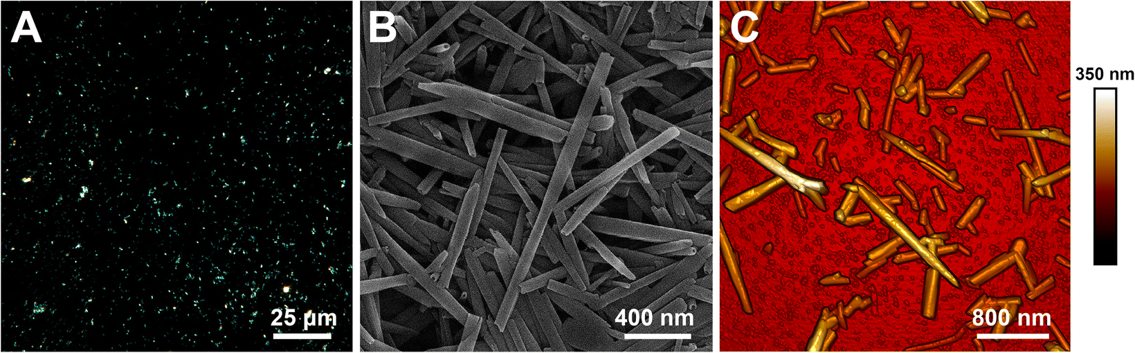
The research outline and a schematic diagram for fabrication of porous nanostructured scaffolds are illustrated in Fig. 1. Microscopy images of halloysite used as a dopant for the fabrication of nanomodified scaffold material and combined with MSCs for repair of the bone defect are presented in Fig. 2.
Sketch demonstrating the fabrication of the scaffold, differentiation of MSCs, and the implantation of scaffold with MSCs in the area of the bone defect

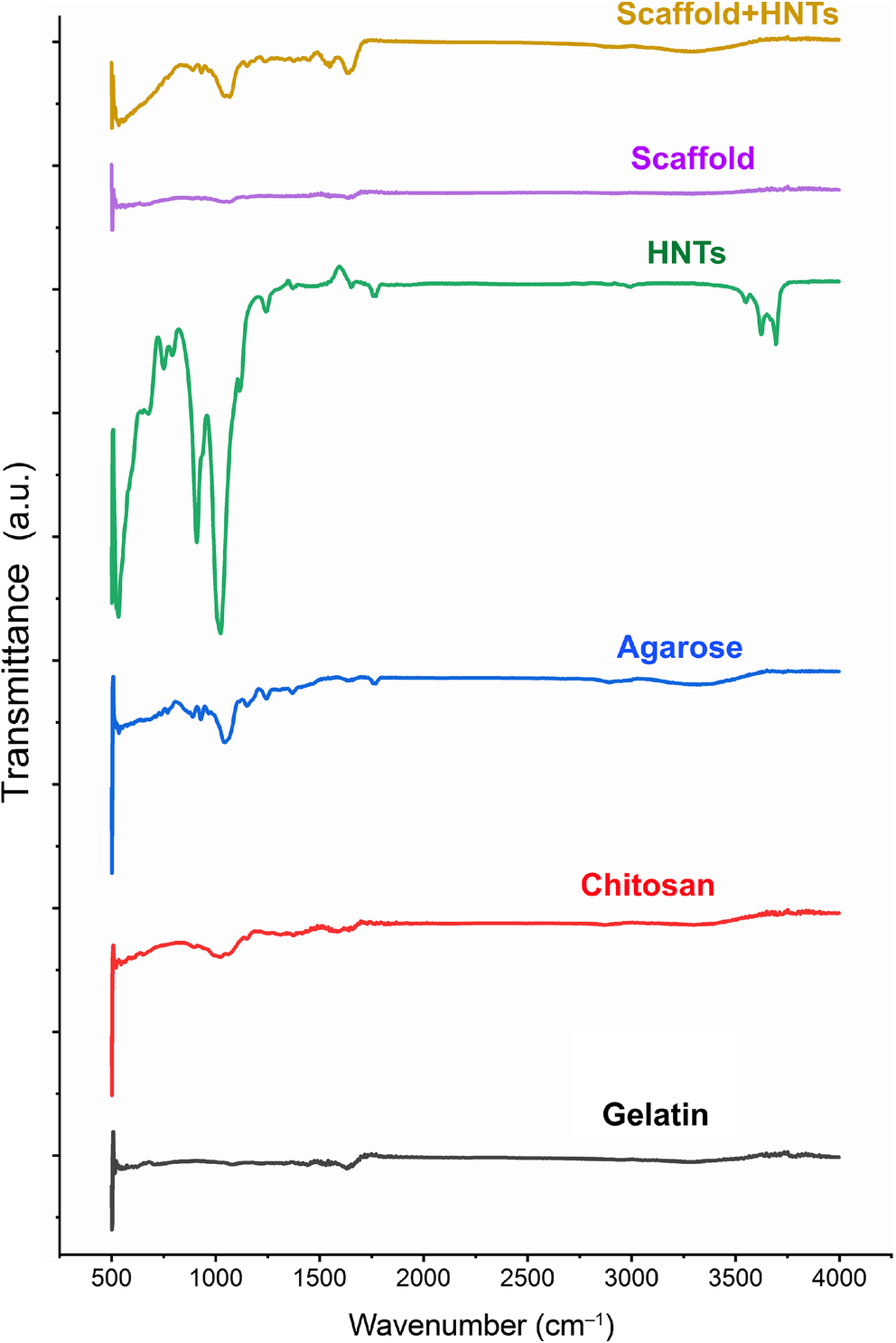
FTIR spectra of pure polymers, HNTs, and scaffolds

The main polymer of the fabricated scaffolds was agarose (2%) whereas the concentration of chitosan and gelatin was 1%. The FTIR spectrum of pure commercial agarose is presented in Fig. 3. The peak positions at 947 and 976 cm–1 are present in both the agarose and scaffold+HNTs samples. A subsequent characteristic decrease in the scaffold+HNTs curve along the y axis was also observed in the agarose sample. The peaks (C–O stretch) of scaffold+HNTs related to agarose are present in the range between 1100 and 1300 cm–1 (Singh et al., Reference Singh, Bhattacharya, Tomar, Singh and Singh2017), but with a shift in the peak position. At the same time the position of 1149 cm–1 in the scaffolds corresponded to the chitosan sample. The general profile of the FTIR spectra of the scaffold was most like that of agarose, related to its prevalence over other polymers. The observed shift in the agarose peaks in the scaffold appeared to be related to the presence of other biopolymers. The peaks related to the main material (agarose) were present in scaffold samples. Clearly, some shifting in the peak position in the biopolymer matrix indicate formation of the polyelectrolyte complex without addition of reinforcing substances as also described by Kulig et al. (Reference Kulig, Zimoch-Korzycka, Jarmoluk and Marycz2016) for chitosan and alginate. Generally, the wavenumber range between 87 cm–1 and 1300 cm–1 in scaffold samples can be considered as a fingerprint region for agarose.
Visualization of halloysite nanotubes using a dark field microscopy, b scanning electron microscopy, and c atomic force microscopy

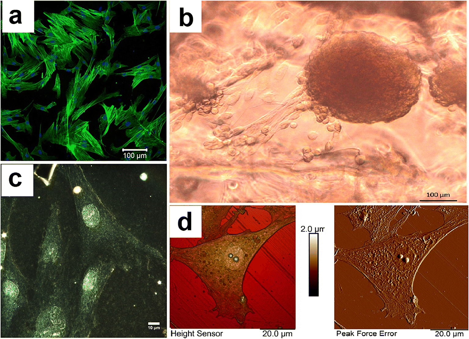
Cells obtained from the dog adipose tissue had the characteristic morphology of MSCs (Fig. 4a, c, d). During the cultivation time (72 h), MSCs on 3D porous matrix doped with HNTs formed spheroid-like structures spontaneously which were further attached to the surface of the matrix (Fig. 4b). This phenomenon demonstrated the ability of MSCs to form tissue-like constructs within the porous gelatine-chitosan-agarose scaffold with 6 wt.% of HNTs. Cells in spheroids are not uniform in their physiology, which corresponds more to the state of cells in the natural tissue (Lin & Chang, Reference Lin and Chang2008). Interaction of cells with their neighboring cells and extracellular matrix are also associated with modified diffusion speed (Achili et al., Reference Achili, Meyer and Morgan2012; Rozhina et al., Reference Rozhina, Batasheva, Gomzikova, Naumenko and Fakhrullin2019) All of these conditions promote the formation of tissue-mimicking structure from MSCs with high differentiation potential, which is manifested in the corresponding microenvironment in the presence of specific factors.
Morphology and cytoskeleton (F-actin) of allogenic MSCs (6th passage) obtained from a dog adipose tissue; b growth of dog MSCs on 3D porous biopolymer matrix doped with HNTs; note the spontaneous formation of 3D cell clusters – spheroids; cdark-field microscopy image of dog MSCs monolayer; and datomic-force images of single MSC

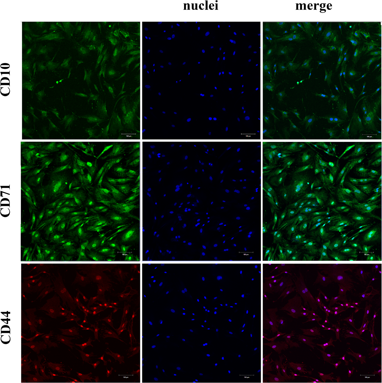
Using immunocytochemical staining and fluorescent microscopy analysis, cells isolated from adipose tissue of a dog have been shown to act as markers of MSCs (CD 44, CD 10, CD 71) (Fig. 5).
Positive staining of MSCs on stem-cells markers (CD10, CD71, CA44). Scale bar = 100 μm

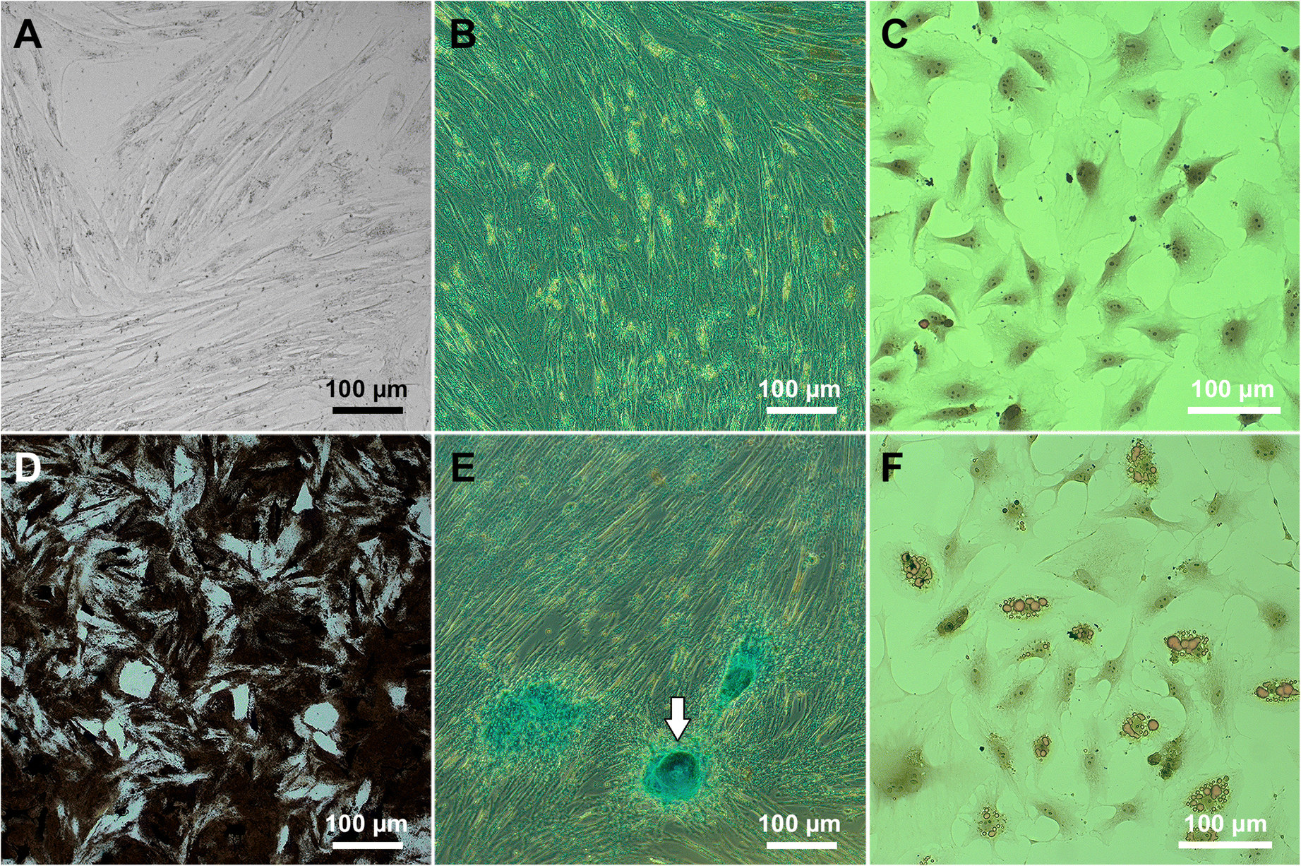
To determine the differentiation potential of the isolated cells, the specific media containing the growth factors which transform cells into specialized cells were used. Subsequent staining for specific products indicated the differentiation potential of MSCs in three directions (with relevant controls which had no specific growth factors): osteogenic (Fig. 6a, d), chondrogenic (Fig. 6b, e), and adipogenic (Fig. 6c, f).
Differentiation of MSCs: a, b, c control (cells grown without specific growth factors); d cells grown in osteogenic differentiation medium (von Kossa staining; black color indicates the mineralization zones); e cells grown in chondrogenic differentiation medium (Alcian blue staining; white arrow marks the cartilage formation zone with blue-colored acid mucopolysaccharides); and f cells, grown in adipogenic differentiation medium, Sudan 3 staining (note the oil drops inside the cells)

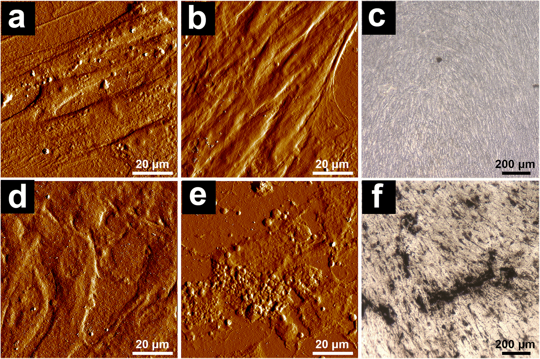
The opportunity for MSCs to differentiate in the osteogenic direction was examined on the surface of native polymeric matrices and nanostructured scaffolds with 6 wt.% of HNTs (Fig. 7). Osteogenic differentiation of MSCs was observed on the nanostructured matrix material surface (Fig. 7e, f) after 14 days. In the case of a native scaffold, it probably takes more time; after 14 days, only modest morphological changes in cells were noted. The addition of halloysite imparts osteoconductive properties to the matrix material, therefore.
Osteodifferentiation of MSCs on the biopolymer scaffolds. a control cells grown on α-MEM on the scaffold without HNTs; b control cells grown on α-MEM on the scaffold with 6 wt.% HNTs; c von Kossa staining of cells grown on α-MEM on the scaffold with 6 wt.% HNTs; d cells grown on osteodifferentiation cultural medium on the scaffold without HNTs; e cells grown on osteodifferentiation cultural medium on the scaffold with 6 wt.% HNTs; and f von Kossa staining of cells grown on osteodifferentiation cultural medium on the scaffold with 6 wt.% HNTs

Using AFM, Young’s modulus (DMT model) was shown to be greater in the case of osteodifferentiation on the scaffold with HNTs than in the same case but without nanomodification (Table 1). On the other hand, cells and extracellular space on the control scaffold without HNTs and a medium without osteogenic factors were stiffer than those grown on the unmodified matrices. This observation was related to the stage of differentiation process.
Mechanical characteristics of cells and extracellular space after osteogenic differentiation (14 days)

Porous biodegradable hydrogel scaffolding doped with nanoparticles can be used, therefore, not only as a passive carrier for stem cells for cell therapy, but also as a conductive material for the formation of tissue-like structures.
In one clinical case example, a 5 year-old Kurtshaar dog (male, body weight 30 kg) was operated on. This dog had a burdened anamnesis according to its owner: a year previously the dog had been injured (a closed comminuted diaphyseal fracture of the left humerus bone) in a traffic accident and was operated on as in the above-mentioned pathological process. A bone osteosynthesis with overlapping of the plate was performed. After 1 month, the rejection of the fixation construction began; the plate was removed and replaced with Kirshner wires. After another 30 days, the rejection of the Kirshner wires began, which were removed before the required time but the screw remained in the leg (Fig. 8a) and was probably the cause of the hypersensitivity and sensitization.
X-ray images of the damaged limb: a complete chronic diaphyseal fracture of the left humerus with an offset at an angle complicated by neoarthrosis and athrogenic elbow joint contracture; note a foreign body in the area of the proximal fragment (fixation screw). b the state of the limb 7 days after the operation, and c 30 days after the operation. Scale bar = 5 cm

Thus, the function of the left thoracic limb was impaired and this was manifested by intermittent, strong-degree lameness; a breach of skin integrity and pain during palpation were absent; mobility outside the joint, atrophy of extensor muscles and flexors of the elbow joint were noted. During the operation, the foreign body (screw) was removed, and the granulation tissue surrounding the fracture zone was excised. Complete repositioning of fragments was not achieved due to contracture of the elbow joint. A porous biopolymer matrix with allogenic dog MSCs was placed between the ends of the fragments. Fixation of the fragments was performed by retrograde fitting of two Kirschner wires with a diameter of 3 mm. Additional immobilization was provided by the application of the SoftCast bandage.
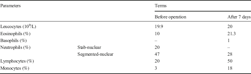
Clinical blood tests were within the normal range before and after the operation, but a slight degree of anaemia arose (Table 2).
Change of main hematological parameters in the early post-operative period

At the 7th day after surgery, a radiographic examination was performed (Fig. 8b). The fracture line on the roentgenogram was fuzzy, pronounced periosteal reaction along the bone length was observed, and the cortical layer was thinned and indistinct. Then, 30 days after the operative intervention, a periosteal corn bounded by a zone of fracture was formed, the fracture line was not clear, the lower fragment was osteoporotic, and an intermittent endosteal callus formed on the posterior surface of the bone (Fig. 8c).
In general, the course of the post-operative period was favorable, but after a month, the Kirshner wires had to be removed due to the development of oedema. Infusion with saline was performed. After the removal of the Kirshner wires, the general state of the dog improved and the oedema decreased. Blood analysis revealed an increase in the level of alkaline phosphatase and stab neutrophils, which is probably an indicator of an allergic reaction due to individual intolerance to metals (Tables 3, 4).
Changes in Leukogram in the early postoperative period

Changes in biochemical parameters of blood serum in the early post-operative period

Finally, the removal of the Kirshner wires led to the normalization of the limb state; the dog began to use the limb during walking. The use of porous biopolymer matrix together with stem cells contributed to a more active formation of bone callus and fracture fusion, which was not achieved during the previous operation using only metal-supporting constructions.
Discussion
In general, regeneration of bone is a much slower process than repair of bone defects. Several key factors have been identified for the successful repair of bone: the defect zone must be fully occupied by the implant material to inhibit dysfunctional fibrosis; MSCs must migrate into or be placed within the boundaries of the bone defect; these boundaries must be occupied by a cell-delivery vehicle that either is resorbed or becomes functionally integrated into the new structure. Several varieties of biomaterials have been studied and used clinically for bone repair and regeneration applications (Dimitriou et al., Reference Dimitriou, Jones, Mcgonagle and Giannoudis2011; Tan et al., Reference Tan, Yu, Wan and Yang2013; Ou et al., Reference Ou, Huang, Fu, Huang, Fang, Gu, Wu and Wang2020; Pereira et al., Reference Pereira, Cengiz, Silva, Reis and Oliveira2020).
A previous study demonstrated that stem-cell therapy accelerated the bone-defect regeneration that was effective for the treatment of osteonecrosis of the femoral head in the complex therapy (Wu et al., Reference Wu, Sun, Liu, Grottkau, He, Zou and Ye2020). A combination of allogeneic and autogenous MSCs with scaffolds on the base of fibrinogen and collagen demonstrated the efficacy of cell-based therapies for the treatment of bone defects by growth of newly formed cartilaginous tissue at the site of the original physeal defect (Planka et al., Reference Planka, Gal, Kecova, Klima, Hlucilova, Filova, Amler, Krupa, Kren, Srnec, Urbanova, Lorenzova and Necas2008). As demonstrated by Song et al. (Reference Song, Jia, Minami, Hill, Nakanishi, Shrestha and Ariga2020), highly aligned, fullerene nanowhisker, nanopatterned surfaces contribute to the preservation of multipotency and enhance regenerative capacity in MSCs. Soft scaffolds mimicking extracellular matrix on the base of fibronectin were fabricated by Jia et al. (Reference Jia, Minami, Uto, Chang, Hill, Nakanishi and Ariga2020). These scaffolds represent tunable adaptive materials, composed of a protein monolayer assembled at a liquid–liquid interface, which adapt dynamically to cell-traction forces and can influence the stem-cell differentiation and enable dynamic control of stem-cell behavior. Using Von Kossa staining methods and AFM in the present study, the ability of dog MSCs to differentiate in an osteogenic direction on the surface of hydrogels doped with HNTs under osteogenic conditions was noted. AFM was also used to determine alteration in the stiffness and elasticity of samples during osteodifferentiation. To find the Young's modulus of a surface, selecting a suitable model of the mechanics of contact between the AFM probe and the sample surface is necessary. The most commonly used models were developed by Hertz (Reference Hertz1881), Johnson-Kendall-Roberts (JKR) (Johnson et al., Reference Johnson, Kendall and Roberts1971), and Derjaguin-Mueller-Toporov (DMT) (Derjaguin et al., Reference Derjaguin, Muller and Toporov1975). The DMT model was chosen; this refines the simplest model (Hertz), taking into account adhesion outside the contact area, and is more applicable to small cantilevers (Bruker ScanAsyst-Air probes, Billerica, Massachusetts, Bruker, USA) (nominal length 115 μm, tip radius 2 nm, spring constant 0.4 Nm–1)) in contact with rigid specimens with low adhesion (Heinz & Hoh, Reference Heinz and Hoh1999).
In previous studies by the current authors, combinations of natural biopolymers, chitosan, agarose, gelatin, and hyaluronic acid were used to produce porous tissue engineering scaffolds. These biopolymer compositions were doped with natural nanofiller (halloysite nanotubes) to enhance both the biological compatibility and the mechanical properties of scaffolds (Suner et al., Reference Suner, Demirci, Yetiskin, Fakhrullin, Naumenko, Okay, Ayyala and Sahiner2019; Naumenko & Fakhrullin, Reference Naumenko and Fakhrullin2019). The surface irregularities of the scaffold pores due to the insoluble nanotubes promoted the best adhesion of the cells on scaffold materials. Moreover, they increased the strength of the composites. HNTs have been used successfully in several studies as nanomodifiers (Sun et al., Reference Sun, Zhang, Shen and Jia2010; Zheng & Wang, Reference Zheng and Wang2010; de Silva et al., Reference de Silva, Pasbakhsh, Goh, Chai and Ismail2013; Okamoto & John, Reference Okamoto and John2013; Liu et al., Reference Liu, Wu, Jiao, Xiong and Zhou2013) and showed no significant toxicity. Previous work (Naumenko et al., Reference Naumenko, Guryanov, Yendluri, Lvov and Fakhrullin2016) demonstrated that the addition of even small amounts of nanofiller (3–6 wt.%) led to significant improvement in mechanical stability and wettability of the biopolymer scaffolds and did not disturb the cell growth in vitro. In vivo study has demonstrated the biocompatibility of nanocomposite scaffolds studied in the organisms of rats, with a slight inflammatory effect but without rejection of implants. The approximate period of degradation was ~6 weeks, which was confirmed by histological examination. The blood supply of the implanted area was restored fully in six weeks. On the other hand, the addition of a significant amount of HNTs (up to 50 wt.%) to biopolymer matrix did not disturb the cell proliferation and hemocompatibility (Suner et al., Reference Suner, Demirci, Yetiskin, Fakhrullin, Naumenko, Okay, Ayyala and Sahiner2019). Data obtained in the present study as well as in previous experiments suggest that the HNTs-modified biopolymer composites show great potential in tissue engineering and can be used as stem-cell carriers for bone repair in vivo.
Conclusions
The use of biopolymer matrices in combination with stem cells promoted normal regeneration of bone tissue even in the case of complex fractures with incorrect primary fusion of bones. In addition, manifestations of rejection were observed. The bones of an animal prone to allergic reaction to metals were regenerated more efficiently after the removal of metal constructs; due to the lack of fixing time, however, the bone was displaced in the area of the callus formed. No inflammatory reaction was observed with respect to the nanocomposite biopolymer matrix and allogenic MSC. The combination of traditional surgery using metallic constructions with implantation of 3D-biopolymeric matrices can be effective in terms of bone regeneration.
ACKNOWLEDGMENTS
The present study was funded by the Russian Foundation for Basic Research (project number 20-015-00353) and partially supported by RFBR and the government of the Tatarstan Republic (grant number 18-415-160010).
Funding
Funding sources are as stated in the Acknowledgments.
Declarations
Conflict of Interest
The authors declare that they have no conflict of interest.