Folate (vitamin B9) and B12 (cobalamin) are essential B vitamins, which play critical roles as co-enzymes in the one-carbon metabolic pathway responsible for major cellular processes including DNA synthesis, DNA repair and amino acid metabolism(Reference Stover1–Reference Nazki, Sameer and Ganaie3). Folate is converted to tetrahydrofolate: (i) to produce thymidine and purines for DNA synthesis and (ii) for the methylation of homocysteine to methionine – a precursor to S-adenosylmethionine, which is involved in the methylation of DNA and proteins(Reference Stover1,Reference Nazki, Sameer and Ganaie3–Reference Crider, Yang and Berry5) . Folate deficiency affects all parts of the one-carbon metabolic pathway and can result in megaloblastic anaemia – large abnormal precursor erythrocytes – due to failure of DNA synthesis, cell division and erythropoiesis(Reference Stover1–Reference Nazki, Sameer and Ganaie3,Reference Bailey and Gregory6,Reference Koury and Ponka7) . B12 acts as a coenzyme in the conversion of homocysteine to methionine, which uses the methyl group donated by 5-methyl tetrahydrofolate to form tetrahydrofolate; therefore, B12 deficiency prevents the formation of tetrahydrofolate and causes homocysteine to increase and DNA synthesis to fail, resulting in megaloblastic anaemia and neurological disorders(Reference Stover1,Reference Lyon, Strippoli and Fang2,Reference Moll and Davis4,Reference Koury and Ponka7,Reference Sobczyńska-Malefora and Harrington8) .
The biological effects of folate and B12 deficiency could have implications for physically active individuals who need to maintain health and performance(Reference Woolf and Manore9,Reference Peeling, Sim and McKay10) . Basic military training is physically arduous and involves high volumes of endurance training(Reference O’Leary, Saunders and McGuire11,Reference O’Leary, Gifford and Knight12) . Folate and B12 deficiency could worsen endurance performance through impairing erythrocyte production and oxygen carrying capacity(Reference Woolf and Manore9,Reference Peeling, Sim and McKay10) . Basic military training can result in haematological changes in men and women, including a decrease in Hb(Reference O’Leary, Jackson and Izard13,Reference Moran, Heled and Arbel14) , although some studies report an increase in Hb in women(Reference McClung, Karl and Cable15,Reference McClung, Karl and Cable16) and a systematic review shows basic military training does not increase the prevalence of anaemia(Reference Carrasco, Navren and Larsson17). The haematological response to military training might be sex-specific with women in the British Army having a bigger decrease in Hb and a higher prevalence of macrocytosis (average erythrocyte size > 100 fL) than men at the end of training(Reference O’Leary, Jackson and Izard13). The decrease in Hb in women could potentially be due to poorer iron status(Reference O’Leary, Jackson and Izard13) and relatively higher training demands(Reference O’Leary, Saunders and McGuire11,Reference O’Leary, Saunders and McGuire18) of women; however, there is limited evidence for the effect of basic military training on serum folate and B12 status in women, which may contribute towards their reduced erythrocyte production and the macrocytosis. Epidemiological data show women have a three to seven-fold higher incidence of folate or B12 deficiency anaemia than men in the USA Army with B12 anaemia being the third most common nutritional deficiency(Reference Knapik, Farina and Fulgoni19). Data from the Israeli Defence Force show basic military training decreases serum folate and B12 after 4 months of training(Reference Moran, Heled and Arbel14). Women in British Army basic training have low folate intake(Reference Chapman, Roberts and Smith20) and commonly use hormonal contraceptives(Reference Myers, O’Leary and Knight21,Reference O’Leary, Evans and Close22) , which are associated with lower plasma and erythrocyte folate(Reference Shere, Bapat and Nickel23), increasing the risk of folate deficiency in training. Low serum folate and B12 may also result in lower bone mineral density (BMD) and higher fracture risk due to the negative effect of elevated circulating homocysteine on bone(Reference Clarke, Ward and Strain24). Women in basic military training are at higher risk of bone stress injuries than men(Reference Wentz, Liu and Haymes25,Reference O’Leary, Wardle and Rawcliffe26) but there are limited data exploring relationships between serum folate and B12 with BMD or bone metabolism outcomes in young women.
The primary aim of this study was to explore changes in serum folate and B12 status in women in basic military training. These data are secondary analyses from a large observational trial(Reference O’Leary, Jackson and Izard13,Reference O’Leary, Evans and Close22,Reference Carswell, Oliver and Wentz27–Reference Carswell, O’Leary and Swinton31) , but all hypotheses were made a priori. We hypothesised that serum folate and B12 would decrease with training and serum folate would be lower in women who took hormonal contraceptives. Secondary aims were to explore associations between serum folate and B12 and endurance performance, areal BMD and circulating markers of bone metabolism. We hypothesised that lower serum folate and B12 would be associated with lower areal BMD and higher circulating markers of bone turnover.
Methods
Participants
This study is secondary analyses from a larger observational trial exploring micronutrient deficiencies and health and performance outcomes in male and female British Army recruits(Reference O’Leary, Jackson and Izard13,Reference O’Leary, Evans and Close22,Reference Carswell, Oliver and Wentz27–Reference Carswell, O’Leary and Swinton31) . Serum folate and B12 were measured as a secondary outcome in women only, based on the observation of macrocytosis at the end of training in women but not men(Reference O’Leary, Jackson and Izard13). This study was advertised to female British Army trainees from two different training populations to provide a representative sample of women entering the British Army: soldier (non-infantry and non-officer) recruits at Army Training Centre, Pirbright, UK and officer cadets at Royal Military Academy, Sandhurst, UK. Participants were recruited during week 1 of their basic military training between April 2013 and July 2017. Prior to initiation of training, all participants passed an initial military medical assessment, which confirmed them to be injury-free, not pregnant and not possess any medical conditions that excluded them from military service. Each participant had the study procedures and risks fully explained verbally and in writing. Written informed consent was obtained from all participants. All procedures were approved by the Ministry of Defence Research Ethics Committee (ref: 165/Gen/10).
Experimental design
This study was an observational prospective cohort study. Venous blood samples were drawn at the start (week 1) and end (week 13) of basic military training for the analysis of serum folate and B12 and haematological outcomes; circulating markers of bone metabolism were analysed from the week 1 sample. Aerobic endurance performance was measured at week 1 and 13. Body mass, height and whole-body areal BMD were measured at the start of training. Self-reported alcohol intake, smoking status and hormonal contraceptive use (solider recruits at Army Training Centre, Pirbright only) were recorded in custom-designed questionnaires at the start and end of training; only data from the start of training were used in this study. Soldier recruits completed the 14-week British Army soldier basic military training course and officer cadets completed the first 14 weeks of their 44-week officer commissioning course, which is similar to soldier training. This initial block of training is designed to develop basic military skills alongside physical fitness, through aerobic endurance training, strength and conditioning, military-specific fitness training (obstacle course and circuit training), military drill, progressive loaded marching and basic military skills (field exercise and weapon handling). As trainees prepare for their basic component of training in week 14, there is a significant reduction in typical military activities; therefore, post-training measurements were recorded at week 13.
Questionnaires
Alcohol intake (units per month) was calculated from the following questions: ‘Do you drink alcohol? (Yes/No)’, ‘Generally, how many times per month do you drink alcohol? (Once/2 to 4/5 to 12/13 +)’ and ‘Generally, how many units do you drink on each occasion at home and when socialising (one pint of beer = 3 units/one glass of wine = 2 units/one shot of spirits = 1 unit/one bottle of alcopop = 1 unit)? (1 to 5/6 to 10/11 to 15/16 to 20/20 +)’. Smoking status (Yes/No/Previous) was determined from the question ‘Do you smoke tobacco (cigarettes, roll-ups, cigars, or pipe) (Yes/I occasionally smoke/I used to smoke regularly/I used to smoke occasionally/I have never smoked)? Participants were asked to select their hormonal contraceptive from non-user, combined oral contraceptive pill, progestin-only pill, implant and injection. Progestin-only pill, implant and injection users were pooled as progestin-only users. Supplement use was determined from the question ‘Do you currently take any of the following vitamins, mineral or medicinal supplements? (Multi-vitamin/Vitamin D and calcium combined/Calcium/Vitamin D/Iron/Vitamin B 12 /Cod liver oil/Anti-inflammatory/I do not take supplements/Other).
Blood collection and biochemical analyses
A venous blood sample was collected either in the morning (∼09.00 to 11.00 h) after breakfast (06.00−07·00 h) or early afternoon (∼13.00−15.00 h) after lunch (12.00−13.00 h). Follow-up (week 13) measurements were taken at the same time of day. Venous blood was withdrawn from a vein in the antecubital fossa and collected in serum separator and EDTA Vacutainer® tubes (Becton Dickinson). Serum samples were left to clot for 1 h at room temperature. Hb, erythrocyte count, mean corpuscular volume (MCV) and erythrocyte distribution width (RDW) were measured in EDTA whole blood within 30 min of collection using the COULTER Ac·T diff 2 Analyzer (Beckman Coulter, California, USA). Anaemia was defined as Hb < 12 g ∙ dl−1, and macrocytosis was defined as MCV > 100 fL(Reference O’Leary, Jackson and Izard13). Blood samples were centrifuged at 1500 g and 4°C for 10 min, and plasma and serum were aliquoted and stored in universal tubes at −80°C until analysis. No samples were exposed to prior freeze-thaw cycles before analyses. Serum folate and B12, and plasma procollagen type I N-terminal propeptide (PINP) and c-telopeptide cross-links of type I collagen (βCTX) were analysed by electro-chemiluminescence immunoassays on the COBAS c601 (Roche Diagnostics) platform. Folate and B12 status were classified using laboratory assay-specific reference ranges. Folate deficiency and insufficiency were taken as < 4·5 nmol·l−1 and 4·5−8·7 nmol·l−1. B12 deficiency and insufficiency were taken as < 73 pmol·l−1 and 73−144 pmol·l−1. PINP inter-assay CV was < 3 % between 20 and 600 µg·l−1 with a sensitivity of 8 µg·l−1. βCTX inter-assay CV was < 3 % between 200 and 150 µg·l−1 with a sensitivity of 10 ng·l−1. Serum total 25-hydroxyvitamin-D (25(OH)D) was measured by liquid chromatography tandem mass spectrometry(Reference Tang, Nicholls and Piec32). The 25(OH)D3 and 25(OH)D2 assays were calibrated using the National Institute of Science and Technology standard reference material SRM972a. Total 25(OH)D was calculated from the sum of 25(OH)D3 and 25(OH)D2. Total 25(OH)D inter-assay CV was < 8·5 % between 0·1 and 200·0 nmol·l−1. All biochemical analyses (excluding Hb, erythrocyte, RDW and MCV analyses) were undertaken by the Good Clinical Laboratory Practice and Vitamin D External Quality Assessment Scheme certified Bioanalytical Facility at the University of East Anglia, Norwich, UK.
Endurance performance
Endurance performance was assessed as the time to complete a maximal effort 2·4 km run on a standardised running course at each training site. Participants completed an 800 m warm-up, and completion time was recorded to the nearest second. The time to complete a 2·4 km run is indicative of maximal aerobic capacity and is a military field test assessed during selection, training and throughout a military career. All participants were accustomed to performing this test from selection, before commencing military training.
Whole-body areal bone mineral density
Whole-body areal BMD was assessed by dual energy X-ray absorptiometry (DXA) (Lunar iDXA, GE Healthcare) wearing light clothing. Officer cadets did not have a scan performed due to a lack of scanner availability at the Royal Military Academy, Sandhurst (total n for DXA = 99).
Statistical analyses
These data were secondary analyses(Reference O’Leary, Jackson and Izard13,Reference O’Leary, Evans and Close22,Reference Carswell, Oliver and Wentz27–Reference Carswell, O’Leary and Swinton31), and so no a priori sample size was calculated. All data were analysed using the R programming language (v.4.4.1). Based on a sample size of 137, the smallest effect size we could detect was a Cohen’s d of 0·24 with a power of 80 % and αof 0·05 (pwr package v.1·3–0). Descriptive statistics are presented as mean (sd) or n and percentage. The distribution of the data was checked using frequency distribution histograms. The week 1 and week 13 values for serum folate, serum B12, Hb, erythrocyte count, MCV, RDW and 2·4 km run time were determined using paired samples t tests or Wilcoxon signed rank tests for data non-normally distributed (serum folate and B12; long-tailed distributions). Multiple linear regression was used to test the association between alcohol intake and hormonal contraceptive use (non-users as the reference group) with serum folate and B12 at week 1 and week 13 (hormonal contraceptive use only) controlling for age, height and body mass. Multiple linear regression was used to test the association between serum folate, serum B12, Hb, erythrocyte count, and 2·4 km run time at week 1 controlling for age, height, body mass, smoking, alcohol intake and Army training course (standard entry or officer)(Reference Carswell, Jackson and Swinton30). Army training type was included to account for differences in endurance performance entry standards between sites. Multiple linear regression was used to test the association between serum folate and B12 with bone outcomes at week 1 (whole-body areal BMD, βCTX and PINP) controlling for height and body mass. Each predictor and outcome variable in the multiple linear regression models was centred and scaled (scale function) to standardise the coefficients. Variance and normality of the residuals for simple and multiple linear models were checked visually by plotting the residuals against the fitted values and from Q-Q plots. Partial regression plots were made showing the relationship between the scaled predictor and scaled response variables whilst holding the other covariates constant (avPlots function, car package (v.3.1–3)). Plots were drawn in the ggplot2 package (v.3.4.0). Significance was accepted as P ≤ 0·05.
Results
Participants
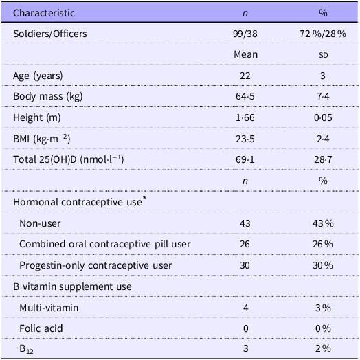
A total of 581 female British Army trainees volunteered to participate in the overarching study at week 1, with 137 women (soldiers, n 99; officers, n 38) completing both week 1 and week 13 testing and having blood test results available for serum folate and B12 (Table 1). Loss to follow-up at week 13 was due to failure to complete training (due to discharge from service or voluntary withdrawal), voluntary withdrawal from the study, or not selected for week 13 follow-up (not all training intakes were selected for follow-up testing). Non-availability of serum folate and B12 data was due to insufficient blood sample following testing of the main outcomes presented elsewhere. Officers did not have a DXA scan or did not complete the hormonal contraceptive questionnaire resulting in a total sample of 99 for DXA and hormonal contraceptive use. Run time data were missing for 37 participants resulting in a total sample of 100.
Participant characteristics (n 137). Data are mean (sd) or n (%)

Table 1 Long description
The table presents characteristics of 137 female British Army trainees who completed both week 1 and week 13 testing. It includes data on soldiers and officers, age, body mass, height, BMI, and total 25(OH)D levels. The table also details hormonal contraceptive use and B vitamin supplement use. The table has 12 rows and 4 columns. Column headers are Characteristic, n, Mean, and %. Row labels include Soldiers/Officers, Age (years), Body mass (kg), Height (m), BMI (kg/m^2), Total 25(OH)D (nmol/l), Hormonal contraceptive use, and B vitamin supplement use. Notable data points include the mean age of 22 years, mean body mass of 64.5 kg, mean height of 1.66 m, mean BMI of 23.5 kg/m^2, and mean total 25(OH)D of 69.1 nmol/l. Hormonal contraceptive use is categorized into non-users, combined oral contraceptive pill users, and progestin-only contraceptive users. B vitamin supplement use includes multi-vitamin, folic acid, and B12.
Missing data: age, body mass, height and BMI, n 2.
* Not recorded for officers at Royal Military Academy Sandhurst (n 38).
The effect of training on serum folate, serum B12 and haematological status
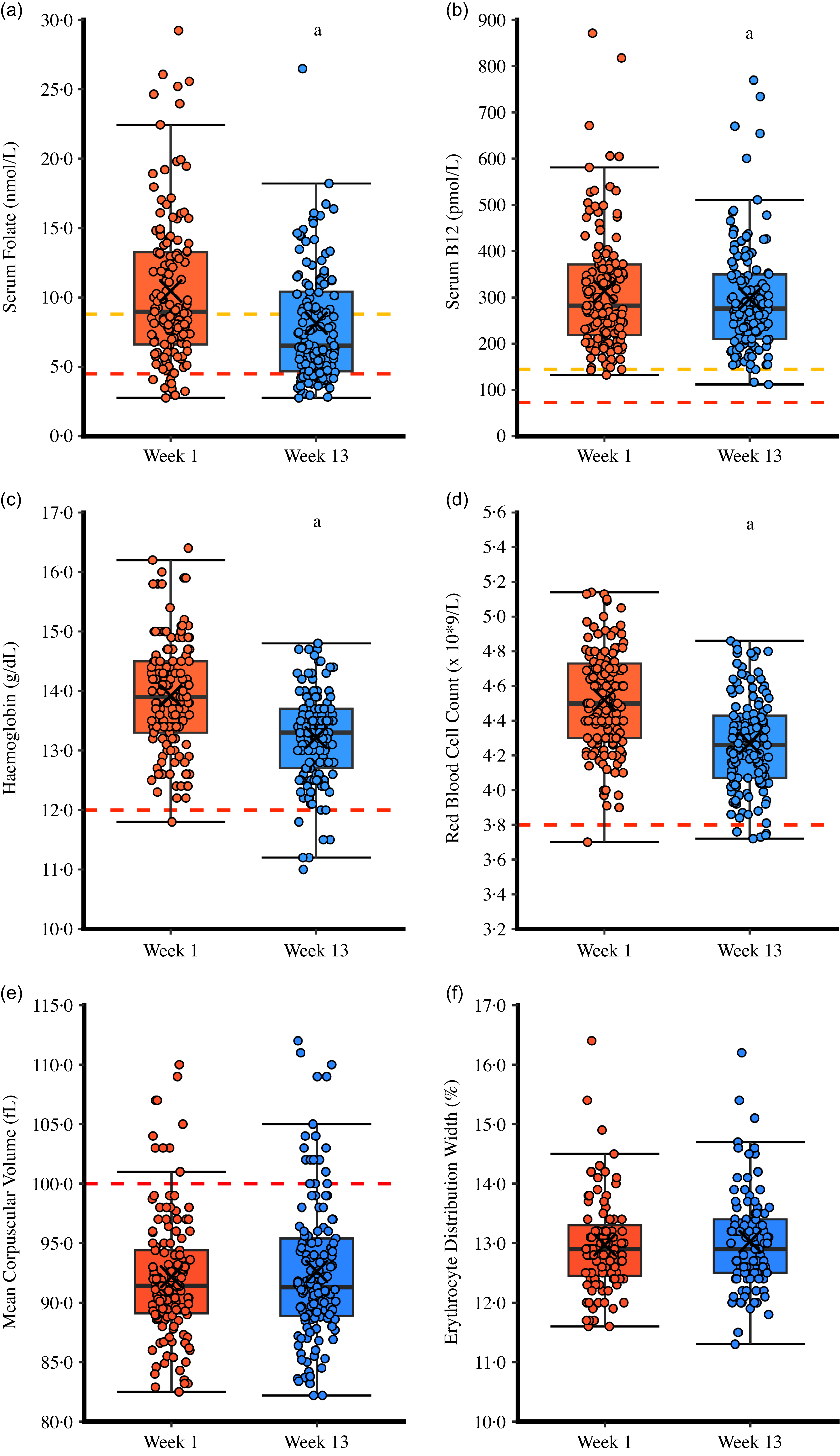
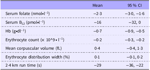
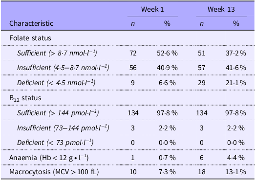
Serum folate, serum B12 and haematological status are presented in Figure 1, with mean absolute changes presented in Table 2. Training decreased serum folate (P < 0·001), B12 (P = 0·042), Hb (P < 0·001), and erythrocyte count (P < 0·001), but had no effect on MCV (P = 0·438) or RDW (P = 0·088). Training decreased 2·4 km run time (week 1 = 682 (sd 56); week 13 = 652 (sd 51); P < 0·001). The prevalence of serum folate and B12 deficiency, anaemia and macrocytosis is presented in Table 3. Of the fifty-one women who were sufficient in serum folate at week 13, forty-six were sufficient and seven were insufficient at week 1. Of the fifty-seven women who were insufficient in serum folate at week 13, a total of twenty-five, twenty-seven, and five were sufficient, insufficient and deficient in serum folate at week 1. Of the twenty-nine women who were deficient in serum folate at week 13, a total of three, twenty-two, and four were deficient at week 1. Of the 3 women who were insufficient in serum B12 at week 13, two were sufficient and one was insufficient at week 1. All anaemic women had normal serum B12, but three had insufficient serum folate and one had deficient serum folate. None of the women who had macrocytosis at week 1 had insufficient or deficient serum folate or B12 or anaemia. Two women with macrocytosis at week 13 had insufficient serum folate and two women had anaemia, but none were deficient in serum folate, and all had normal serum B12. Alcohol intake and hormonal contraceptive use were not associated with serum folate or B12 after controlling for age, height and body mass at week 1 (P ≥ 0·100). Hormonal contraceptive use was associated with serum B12 at week 13, with progestin-only users having lower serum B12 than non-users (β (95 % CI) = −63 (−126, −1), P = 0·047); there was no association between hormonal contraceptive use and serum folate (P ≥ 0·818).
The effect of military training on serum folate, serum B12 and haematological status in women. Individual data are represented by circles. Box plots represent median, interquartile range and range. Crosses represent mean. Amber dotted horizontal lines represent insufficient serum folate and B12. Red dotted horizontal lines represent deficient or lower bound of normal range (except mean corpuscular volume where the line represents upper bound of normal range). a P < 0·05 v. week 1.

Mean absolute change (95 % CI) in serum folate, serum B12, markers of haematological status and 2·4 km run time from week 1 to week 13 of military training in women

Table 2 Long description
The table presents the mean absolute change and 95 percent confidence interval for various health markers and a 24 km run time over a 13-week military training period in women. The table has 8 rows and 3 columns. The columns are labeled Mean and 95 percent CI. The rows are labeled with different health markers and the 24 km run time. Row 1: Serum folate (nmol per L), Mean: -2 to -3, 95 percent CI: -3.0, -1.6. Row 2: Serum B12 (pmol per L), Mean: -16, 95 percent CI: -32, 0. Row 3: Hemoglobin (g per dL), Mean: -0.7, 95 percent CI: -0.9, -0.5. Row 4: Erythrocyte count (per 10 to the power of 9 per L), Mean: -0.2, 95 percent CI: -0.3, -0.2. Row 5: Mean corpuscular volume (fL), Mean: 0.4, 95 percent CI: -0.4, 1.3. Row 6: Erythrocyte distribution width (percent), Mean: 0.1, 95 percent CI: -0.1, 0.2. Row 7: 24 km run time (s), Mean: -29, 95 percent CI: -36, -22.
Prevalence of serum folate and B12 deficiencies, anaemia and macrocytosis. Data are n (%)

Table 3 Long description
The table presents the prevalence of serum folate and B12 deficiencies, anaemia, and macrocytosis over two weeks. It has 4 rows and 6 columns. The columns are labeled as Characteristic, Week 1 n, Week 1 %, Week 13 n, and Week 13 %. The rows are labeled as Folate status, B12 status, Anaemia, and Macrocytosis. The folate status row is further divided into Sufficient, Insufficient, and Deficient categories. The B12 status row is also divided into Sufficient, Insufficient, and Deficient categories. The table shows the number and percentage of individuals in each category for both Week 1 and Week 13. Notable trends include a decrease in the number of individuals with sufficient folate status and an increase in those with insufficient and deficient folate status from Week 1 to Week 13. The B12 status remains relatively stable, with most individuals maintaining sufficient levels. There is a slight increase in the prevalence of anaemia and macrocytosis from Week 1 to Week 13.
MCV, mean corpuscular volume.
Associations between serum folate, serum B12, Hb and erythrocyte count with 2·4 km run time
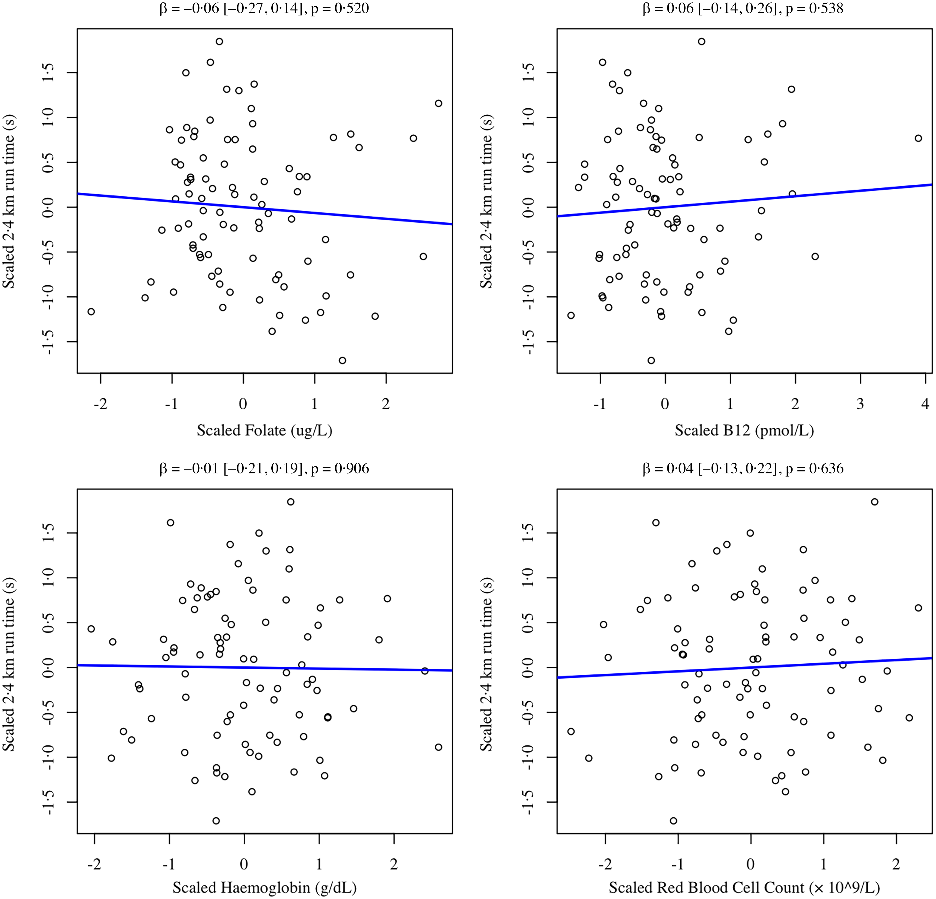
Associations between serum folate, serum B12 and Hb with 2·4 km run time (seconds) are presented in Figure 2. There was no association between serum folate, serum B12, Hb or erythrocyte count with 2·4 km run time (seconds) when controlling for age, height, body mass, smoking, alcohol intake and Army training type (P ≥ 0·520).
Partial regressions between serum folate, serum B12, Hb and erythrocyte count with 2·4 km run time (s) at week 1 controlling for age, BMI, smoking status, alcohol intake and Army training course. Data are mean-centred and scaled. β, scaled coefficients with 95 % CI from partial effects from multiple linear regression.

Figure 2 Long description
Panel A: A scatter plot showing the relationship between scaled folate (ug/L) on the horizontal axis and scaled 24 km run time (s) on the vertical axis. The blue line represents the regression line with a beta value of -0.06, a 95 percent confidence interval of [-0.27, 0.14], and a p-value of 0.520. Panel B: A scatter plot showing the relationship between scaled B12 (pmol/L) on the horizontal axis and scaled 24 km run time (s) on the vertical axis. The blue line represents the regression line with a beta value of 0.06, a 95 percent confidence interval of [-0.14, 0.26], and a p-value of 0.538. Panel C: A scatter plot showing the relationship between scaled haemoglobin (g/dL) on the horizontal axis and scaled 24 km run time (s) on the vertical axis. The blue line represents the regression line with a beta value of -0.01, a 95 percent confidence interval of [-0.21, 0.19], and a p-value of 0.906. Panel D: A scatter plot showing the relationship between scaled red blood cell count (x 10^9/L) on the horizontal axis and scaled 24 km run time (s) on the vertical axis. The blue line represents the regression line with a beta value of 0.04, a 95 percent confidence interval of [-0.13, 0.22], and a p-value of 0.636.
Associations between serum folate and B12 with bone mineral density and circulating markers of bone metabolism
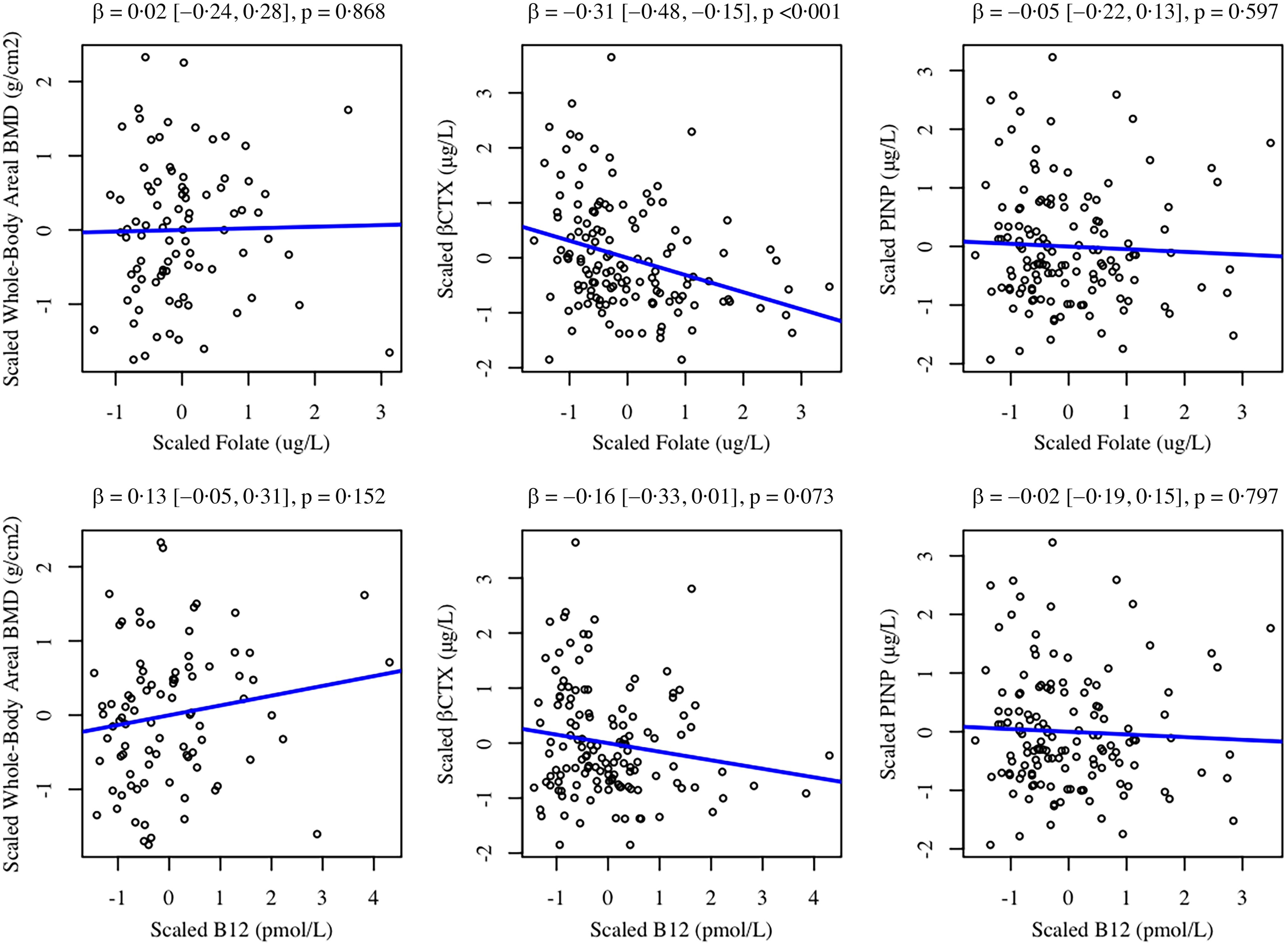
Associations between serum folate and B12 with whole-body areal BMD and circulating markers of bone metabolism controlling for height and body mass are presented in Figure 3. There was no association between serum B12 and whole-body areal BMD or circulating markers of bone metabolism at week 1 (P ≥ 0·073). Higher serum folate was associated with lower βCTX (P < 0·001) but was not associated with whole-body areal BMD or PINP (P ≥ 0·597). A one sd increase in serum folate was associated with a 0·31 sd decrease in βCTX.
Partial regressions between serum folate and B12 with whole-body areal bone mineral density and circulating markers of bone metabolism at week 1 controlling for height and body mass. Data are mean-centred and scaled. β, scaled coefficients with 95 % CI from partial effects from multiple linear regression; βCTX, β C-telopeptide cross-links of type 1 collagen; BMD, bone mineral density; PINP, procollagen I N-terminal propeptide.

Discussion
Serum folate and B12 decreased from before to after basic military training resulting in a high prevalence of folate deficiency at the end of training in women. Hb and erythrocyte count were also lower after basic military training. Lower serum folate was associated with higher circulating concentrations of a marker of bone resorption, but serum folate and B12 were not associated with endurance performance or whole-body areal BMD. We have previously shown military trainees are at risk of vitamin D(Reference Carswell, Oliver and Wentz27,Reference Carswell, Jackson and Swinton30,Reference Carswell, O’Leary and Swinton31) and iron(Reference O’Leary, Jackson and Izard13) deficiency. Compared with men in military training, women have lower aerobic fitness(Reference O’Leary, Jackson and Izard13) and are at increased risk of bone stress injuries(Reference Wentz, Liu and Haymes25,Reference O’Leary, Wardle and Rawcliffe26) ; therefore, these data have important implications for informing nutritional strategies to support the health and performance of women in the British Army as well as other physically active young populations. These data also provide novel insight into folate and B12 status, and their association with skeletal outcomes, in young active women.
The effect of training on serum folate, serum B12 and haematological status
Serum folate and B12 decreased from pre- to post-training. A decrease in serum folate with basic military training is consistent with previous findings in a study in men in the Israeli Defence Force(Reference Moran, Heled and Arbel14) but this study provides the first evidence from women in the military, who are at greater risk of folate and B12 deficiency than men(Reference Knapik, Farina and Fulgoni19). The decrease in serum folate was larger (−17·0 %) than the decrease in serum B12 (−1·2 %), consistent with the known metabolism of these B vitamins; serum folate reflects recent folate intake(Reference Snow33,Reference Devalia, Hamilton and Molloy34) and correlates with erythrocyte folate and circulating homocysteine(Reference Sobczyńska-Malefora and Harrington8), whereas body B12 stores can last for several years in the presence of reduced intake due to liver stores and efficient recycling(Reference Socha, DeSouza and Flagg35,Reference Andres36) . Decreased folate intake is the most common cause of folate deficiency, but decreased absorption and increased folate requirements are other potential causes(Reference Sobczyńska-Malefora and Harrington8,Reference Socha, DeSouza and Flagg35) . Serum folate can decline within a few days of low folate intake(Reference Snow33,Reference Eichner and Hillman37) – despite tissue folate levels being normal(Reference Snow33) – and estimated folate intake in basic military training correlates with post-training serum folate(Reference Lutz, Nakayama and Karl38). Folate intake has been shown to be below recommended daily intakes in active women(Reference Woolf and Manore9), women in Israeli Defence Force basic training(Reference Israeli, Merkel and Constantini39) and women in British Army basic training (where B12 intake is also inadequate)(Reference Chapman, Roberts and Smith20). Therefore, based on these previous studies, the decrease in serum folate in this study was potentially due to low folate intake in military training, which can occur because of reduced consumption of folate-rich food sources or destruction of folate during cooking.
The prevalence of folate deficiency (< 4·5 nmol·l−1) was 6·6 % and 21·1 % at week 1 and week 13 of training and the prevalence of folate deficiency or insufficiency (< 8·7 nmol·l−1) was 47·5 % and 63·5 % at week 1 and week 13 of training. We found no evidence of B12 deficiencies (serum B12 deficiency < 73 pmol·l−1; serum B12 insufficiency = 73–144 pmol·l−1). The prevalence of folate deficiency and sufficiency at the start of training was similar to children and adults in the UK population (approximately 50 %)(Reference Jones, Collins and Meadows40), but by the end of training was higher than the general UK population; comparisons between studies must be made with caution due to differences in serum folate assays and thresholds for deficiency. Alcohol intake was not associated with serum folate or B12. High intake of alcohol is a risk factor for folate deficiencies due to decreased folate intestinal absorption, reduced folate uptake into the liver and impaired folate metabolism(Reference Moll and Davis4,Reference Shulpekova, Nechaev and Kardasheva41,Reference Halsted, Villanueva and Devlin42) , but alcohol intake assessed by questionnaires was not estimated to be excessive in this population. Hormonal contraceptive use is also associated with lower plasma and erythrocyte folate(Reference Shere, Bapat and Nickel23) – potentially due to reduced absorption and increased metabolism and excretion(Reference Basciani and Porcaro43) – and lower serum B12, potentially due to a decrease in binding capacity or a redistribution of B12 (Reference Berenson and Rahman44,Reference Wilson, Bivins and Russell45) . Progestin-only contraceptive users had a 63 pmol·l−1 lower B12 than non-users at week 13, after controlling for age, height and body mass, but there was no difference between combined oral contraceptive pill users and non-users; it is possible we only observed an association at week 13 and not week 1 because many women (31 %) chose to change hormonal contraceptive for the start of basic training(Reference Chapman, Roberts and Smith20). These data are in contrast to observational data showing a greater decline in circulating B12 after starting the combined oral contraceptive pill compared with progestin-only methods (injection)(Reference Berenson and Rahman44). Progestin-only contraceptives do not provide a form of exogenous oestradiol compared with combined oral contraceptives, but the reason for the lower serum B12 in progestin-only contraceptive users only is not clear. Nevertheless, the effect of progestin-only contraceptives was small and not clinically significant.
There was no change in MCV or RDW from pre- to post-training, but the prevalence of macrocytosis (MCV > 100 fL) was 7 % and 13 % at week 1 and 13 of training (as reported previously in these women(Reference O’Leary, Jackson and Izard13)). All the woman with macrocytosis at week 1 had normal serum folate and B12 and Hb, and two of the 18 women with macrocytosis at week 13 had insufficient serum folate and two had anaemia (Hb <12 g ∙ l−1). Macrocytosis is a key indicator of megaloblastic anaemia(Reference Socha, DeSouza and Flagg35), but these data suggest that the folate deficiency did not contribute to the macrocytosis and the macrocytosis did not usually present with anaemia. However, MCV can increase before Hb decreases, MCV can often remain in the normal range with folate or B12 deficiency – particularly with iron deficiency – and MCV has poor sensitivity for detecting folate and B12 deficiency(Reference Snow33). We have previously shown a high proportion of these women have low iron stores (ferritin) in training and training reduces ferritin(Reference O’Leary, Jackson and Izard13), which may have reduced cell size and resulted in normal MCV in those with folate deficiency. Macrocytosis can also be caused by high levels of alcohol intake and hypothyroidism(Reference Snow33,Reference Nagao and Hirokawa46) – due to decreased direct effects of thyroid hormone on erythropoiesis(Reference Nagao and Hirokawa46) – and there is evidence that British Army basic training decreases thyroxine and increases thyroid-stimulating hormone in women(Reference O’Leary, Gifford and Knight12). Hb (as reported previously(Reference O’Leary, Jackson and Izard13)) and erythrocyte count decreased from pre- to post-training, which could be caused by plasma volume expansion with endurance training; however, we have previously shown a decrease in ferritin in the women in this study, which may have contributed to the decrease in Hb(Reference O’Leary, Jackson and Izard13). British Army basic military training can worsen iron status due to high physical activity levels, high daily distances travelled on foot and prolonged periods of load carriage(Reference O’Leary, Saunders and McGuire11,Reference O’Leary, Gifford and Knight12) , which may result in inflammation, gastrointestinal bleeding, haematuria and haemolysis from ground impact forces and eccentric muscle contraction(Reference O’Leary, Jackson and Izard13,Reference McClung, Karl and Cable15,Reference McClung, Karl and Cable16,Reference Lippi and Sanchis-Gomar47) . Haemolysis may increase folate requirements(Reference Snow33,Reference Socha, DeSouza and Flagg35), and it is not clear whether the arduous and prolonged exercise in this study increased folate requirements and contributed to increased risk of deficiency. However, there is no strong evidence of an association between the amount or type of exercise and folate requirements; circulating folate and B12 were not different between highly active and sedentary women (although highly active women ate more folate and B12)(Reference Woolf, Hahn and Christensen48) or between athletes who compete in short distance v. long-distance events(Reference Habte, Adish and Zerfu49) and were not associated with level of physical activity(Reference Baart, Balvers and De Vries50).
Associations between serum folate, serum B12, Hb and erythrocyte count with endurance performance
There was no evidence of associations between serum folate, serum B12, Hb or erythrocyte count and 2·4 km run time. The 2·4 km run is used to test aerobic fitness both upon entry and in service and is related to maximal oxygen uptake(Reference Burger, Bertram and Stewart51). Hb and erythrocyte are important contributors to oxygen carrying capacity and maximal oxygen uptake. The lack of association between serum folate or B12 and performance was likely because the deficiencies were not associated with megaloblastic anaemia and the decrease in serum folate with training had likely not yet manifested in any decrease in tissue or cell folate(Reference Lukaski52). In agreement with this finding, supplementation studies show that folate supplementation and acute declines in serum folate have no impact on endurance exercise performance in female runners(Reference Matter, Stittfall and Graves53).
Associations between serum folate and B12 with bone mineral density and markers of bone metabolism
There was no evidence of serum B12 being associated with whole-body areal BMD or markers of bone metabolism. Lower serum folate was associated with higher βCTX – a marker of type I collagen breakdown and bone resorption – but was not associated with whole-body areal BMD or PINP – a marker of type I collagen formation. Our βCTX data should be interpreted with caution because our blood samples were not fasted first morning samples, and βCTX is affected by feeding and renal clearance and displays a circadian rhythm. Low serum folate increases circulating homocysteine(Reference Nazki, Sameer and Ganaie3), which can increase osteoclast activity(Reference Herrmann, Widmann and Colaianni54,Reference Herrmann, Schmidt and Umanskaya55) through increasing reactive oxygen species(Reference Koh, Lee and Kim56). Low serum folate has been associated with higher serum homocysteine, and higher serum homocysteine has been associated with higher βCTX in older women(Reference Gerdhem, Ivaska and Isaksson57). Lower circulating folate has also been associated with increased osteoporotic fracture risk(Reference McLean, Jacques and Selhub58,Reference Van Meurs, Dhonukshe-Rutten and Pluijm59) and lower whole-body areal BMD in older women(Reference Kalimeri, Leek and Wang60,Reference Bailey, Looker and Lu61) , but evidence for a relationship between folate status and BMD is mixed(Reference Van Wijngaarden, Doets and Szczecińska62,Reference Bailey and Van Wijngaarden63) . However, most of these data are on older women and the relevance to young women is not clear. Homocysteine may also interfere with collagen cross-bridging affecting the structure and strength of bone that is not detected by DXA(Reference Clarke, Ward and Strain24,Reference Swart, Van Schoor and Lips64) . We only had DXA data available from a subset of participants, which means our whole-body areal BMD analyses had lower power than our bone metabolism marker analyses. Data from men in the Israeli Defence Force show serum folate or serum folate changes with basic military training were not different between those who developed a stress fracture compared with those who did not(Reference Moran, Heled and Arbel14), and so the clinical implications of folate deficiency for military training are not clear. To our knowledge, there are limited data on serum folate, serum B12 and bone in young active women, who are a population at risk of bone stress injuries in military training; further research is required to better understand the implication of low serum folate and high βCTX on bone health outcomes in this population.
Limitations
We did not measure circulating homocysteine or erythrocyte folate, which could help provide further mechanistic and clinical insight into the effect of lower serum folate; low serum folate does not necessarily equate to high homocysteine and requires erythrocyte folate deficiency. Our blood samples were not fasted, and some of our circulating markers follow a circadian pattern, but we standardised the time post-meal, and follow-up blood samples were matched for time of day and time post-meal, and so our interpretation of changes with training was minimally impacted. We did not ask participants to track menstrual bleeding during the study, which can affect markers of iron status and anaemia. We did not measure folate or B12 intake during training, which should be assessed in future studies to help identify the cause of changes in circulating concentrations. We did not correct for plasma volume changes with training in our analysis of Hb or erythrocyte count and cannot confirm there were changes in total Hb, but we used clinically valid assessments of anaemia. The study monitored changes over 12 weeks, which is unlikely to be sufficient time to detect changes in serum B12 or for some of the clinical effects of folate and B12 deficiency to manifest, and longer duration studies are warranted to investigate the effect of arduous military training on these outcomes. Due to logistical challenges, we did not measure lumbar spine or hip areal BMD, which are more clinically relevant sites for detecting low bone mass; future studies should aim to include these outcomes to more fully understand the effect of folate and B12 deficiencies on bone. Our study was underpowered for detecting associations with stress fractures, and so we are unable to determine the clinical risk of low serum folate.
Conclusion
Serum folate decreased from pre- to post-basic military training in women, possibly due to low folate intake, resulting in a high prevalence of folate deficiency at the end of training. This acute decline in serum folate was not associated with anaemia or impairments in endurance performance, but low serum folate was associated with higher bone resorption, the implications of which are unclear.
Acknowledgements
The authors thank Dr Daniel Kashi, Xin Hui Aw Yong, Mark Ward, Claire Potter and Dr Laurel Wentz for their assistance with data collection.
This study was funded by the UK Ministry of Defence (Army).
S. J., R. M. I., N. P. W. and J. P. G. designed the study. T. J. O., S. J., N. P. W., A. T. C. and S. J. O. collected the data. J. C. Y. T. and W. D. F. analysed the biochemical samples. T. J. O., H. L. W. and C. V. C. produced the manuscript and performed the data analysis. All authors edited the manuscript and approved the final version.
The authors have no competing interests to declare.
All data are available from the corresponding author pending approval of public release from UK Ministry of Defence.