Introduction
Echinoparyphium recurvatum is a 45-collar-spined echinostome digenean parasite of a wide range of wild aquatic birds, those recorded in Britain including tufted duck (Aythya fuligula), mallard (Anas platyrhynchos), moorhen (Gallinula chloropus) and mute swan (Cygnus olor) (Beverley-Burton, Reference Beverley-Burton1972). The adult fluke was first described from the gut of scaup, Anas marila ( = Aythya marila), in Germany (Von Linstow, Reference von Linstow1873; Lühe, Reference Lühe1909), and the species has since been reported in the literature from a variety of bird and mammal hosts from a wide range of geographical locations worldwide. However, it has long been suspected that the apparently cosmopolitan E. recurvatum may, in reality, be composed of several morphologically very similar, yet biologically quite distinct, species. Odening (Reference Odening1964) suggested that E. recurvatum cercariae occur as two distinct types (‘lymnaeidarum’ and ‘planorbidarum’) in Germany. In Britain, McCarthy (Reference McCarthy1990) provided experimental life cycle evidence that E. recurvatum may represent a species complex of morphologically similar yet biologically discrete species. Kiselienė & Grabda-Kazubska (Reference Kiselienė and Grabda-Kazubska1990) and Grabda-Kazubska & Kiselienė (Reference Grabda-Kazubska and Kiselienė1991), working with material from Poland and Lithuania, differentiated two of these species: Echinoparyphium pseudorecurvatum and Echinoparyphium mordwilkoi, respectively. Grabda-Kazubska & Kiselienė (Reference Grabda-Kazubska and Kiselienė1989) described the life cycle of E. recurvatum from material utilizing the lymnaeid snails Radix ( = Lymnaea) pereger and Radix auricularia in Poland and Lithuania. They regarded this form developing in lymnaeid snails to be E. recurvatum sensu stricto and the present author is in agreement. More recently, Sohn (Reference Sohn1998) described the life cycle of E. recurvatum from Korea, where the parasite was found to be utilizing lymnaeid snails (Radix auricularia coreana) as first intermediate host.
To date only one published study, that of Sohn et al. (Reference Sohn, Woo and Hong2002), has attempted to describe the surface topography of the adult parasite using scanning electron microscopy. These authors described E. recurvatum from chicks experimentally infected with metacercarial cysts obtained from naturally infected lymnaeid snails, Radix auricularia coreana, from Sunamchon, Korea, Eastern Asia, a location obviously remote from the type locality of E. recurvatum (Von Linstow, Reference von Linstow1873) sensu stricto in Germany, Europe. This present study set out to investigate, using scanning electron microscopy, the surface topography of adult E. recurvatum sensu stricto obtained experimentally in mallard ducks (Anas platyrhynchos) from a population of the parasite utilizing the lymnaeid snail Lymnaea peregra ( = Radix pereger) as first intermediate host in West Sussex, England, a site much closer to the type locality of the species.
The study presented here has three objectives. First, it sets out to describe in detail the surface structure of adult E. recurvatum originating from Britain, a location in the same bio-geographic region as the European type locality for this species, the Western Palaearctic. Second, it aims to compare the surface morphology with that of adult worms described by Sohn et al. (Reference Sohn, Woo and Hong2002) from Korea, East Asia (Eastern Palaearctic). Third, it aims to provide information that may be used to differentiate E. recurvatum from other 45-collar-spined species in the genus Echinoparyphium, such as E. pseudorecurvatum and E. mordwilkoi from Europe and Echinoparyphium flexum from North America.
Materials and methods
Adult E. recurvatum (15 days old) were obtained as previously described by McCarthy (Reference McCarthy1989), via infection-free laboratory-bred hosts, beginning with 45-collar-spined cercariae emitted from naturally infected L. peregra snails collected from Harting Pond, West Sussex, England. A total of 18 adult worms obtained from the intestine of experimentally infected mallard (A. platyrhynchos) were rinsed briefly in avian saline (0.75% NaCl) at 40°C, and then in a single rapid wash of phosphate buffer at pH 7.4. They were then fixed in cold (20°C) 2.5% glutaraldehyde solution buffered to pH 7.4 for 2 h and were then post-fixed for a further 2 h in 1% osmium tetroxide buffered to pH 7.4 with sodium cacodylate. Worms were then dehydrated through a graded ethanol series to acetone and critical point dried using carbon dioxide in a Samdri-780 critical point drier. The specimens were coated with platinum in the presence of argon using an ion-sputtering apparatus and were then examined using a Hitachi S-510 scanning electron microscope, and a JEOL 100-CX transmission electron microscope with AS14D scanning facility, at an accelerating voltage of 25 kV.
Results
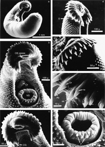
The adult E. recurvatum (15 days old) is shown in fig. 1a, lying in the classic curved position that this echinostome assumes on fixation. The anterior third of the parasite is shown in fig. 1b from the ventral aspect. The most conspicuous feature of the parasite is the anterior spine-bearing collar, at the centre of which is located the simple sub-terminal oral sucker. The collar, shown en face in fig. 1c and in lateral aspect in fig. 1d, is incomplete. A distinct ventral gap is present just below the oral sucker. The collar spines of E. recurvatum are slightly curved, pointed and are arranged around the collar according to the formula 4 − 4 − 3 − 3 − 31 = 45. The dorsal spines of the oral tier are slightly smaller than those of the aboral tier and this observation is supported by light microscope studies in which both tiers of spines have been viewed in the same plane (see McCarthy, Reference McCarthy1990, Fig. 3B). A distinct corner group of four angle spines, slightly larger and more robust in form than those of the rest of the collar, is present at the edge of the collar on each side of the ventral gap (fig. 1c). This observation verifies that of Von Linstow (Reference von Linstow1873) in the original description of this echinostome. A line of three lateral spines is present on each side of the collar. These appear to be smaller in size than the angle spines, but larger than the dorsal spines. Each collar spine is individually accommodated in a sheath of collar tegument, which shows fine longitudinal striations (fig. 1f). The collar spines appear to be retractile, a feature of adult echinostomatids recently commented upon by Fried et al. (Reference Fried, Kanev and Reddy2009).
Adult Echinoparyphium recurvatum, 15 days old: (a) entire worm; (b) anterior third of adult showing oral sucker (OS), ventral sucker (VS) and cirrus (Cr); (c) en face view of collar showing corner group of four spines (CG); (d) lateral view of spined collar, note the area of smooth tegument immediately posterior to the collar (S); (e) dorsal view of collar showing oral (Or) and aboral (Ab) tiers of spines; (f) retractile collar spines showing smaller oral (Or) and larger aboral (Ab) spines accommodated in sheaths of striated collar tegument, partially retracted spine (re) and extended spine (ex); (g) ventral sucker with indented rim and aciliate papillae (p).

The ventral sucker of the mature adult lies in the anterior third of the body (fig. 1a and b), and is shown in detail in fig. 1g. The general tegument of the sucker on both its inner and outer surfaces is aspinose and smooth. The rim of the sucker appears to be regularly indented around its circumference. Large knob-like aciliate, presumed sensory, papillae are present on the outer lip of the sucker in the specimen shown in fig. 1g, but these structures have been observed at other locations around the sucker rim on other specimens examined.
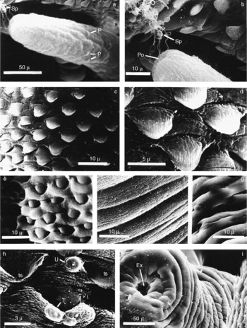
The highly protrusible cirrus of E. recurvatum is located just anterior to the ventral sucker (fig. 1b). It is cylindrical in general form and possesses a single tip, at the end of which opens a simple terminal pore (fig. 2a). The filamentous spermatozoa of E. recurvatum may be seen in fig. 2b exiting the cirrus tip. The cirrus tegument is aspinose and is covered with longitudinal ridges with irregularly indented margins. Simple aciliate sensory papillae are present on the middle and distal regions of the cirrus, but are absent from the extreme tip (fig. 2a).
Adult Echinoparyphium recurvatum, 15 days old: (a) distal region of protrusible cirrus with ridged tegument and sessile aciliate sensory papillae (P), filamentous sperm (Sp) are discharged from the tip; (b) cirrus tip in detail showing the terminal pore (po) and filamentous sperm (Sp); (c) and (d) dorsal body spines; (e) ventral body spines; (f) and (g) ventral body tegument; (h) single (U) and group (T) of uniciliate sensory papillae from anterior ventral tegument among body spines (ts); (i) posterior tip of adult worm showing terminal excretory pore (Ex).

Rows of rounded, scale-like spines (see fig. 2c, d and e) cover both the dorsal and ventral body tegument of E. recurvatum from a point just posterior to the anterior collar to a region just posterior to the ventral sucker (see fig. 1b). Here the spinose tegument gives way to the smooth, folded, aspinose tegument of the posterior two-thirds of the body (see fig. 2f and g). The spines of the ventral anterior tegument are similar in shape to those of the dorsal surface, but the most anterior rows appear to be partly embedded in the folded tegument. The most common sensory structures found on the general body surface of the parasite are the uniciliate dome papillae shown in fig. 2h. These papillae, which occur either singly or in groups of two or three, are found most commonly over the collar and ventral tegument of the anterior regions. They are not found on the smooth tegument of the posterior part of the body. The extreme posterior of E. recurvatum comes to a bluntly rounded point, at the end of which is located a simple terminal excretory pore, (fig. 2i).
Discussion
The tegumental features of adult E. recurvatum of British origin revealed in the present study appear to be very similar to those described for E. recurvatum adults from Korea by Sohn et al. (Reference Sohn, Woo and Hong2002). In particular, collar spine morphology and distribution seem to correspond closely, and the shape, size and distribution of body spines of the adults show no significant differences when comparing adults of equivalent age (15 days post-infection). Likewise, the form of the oral and ventral suckers of the worms from the two different geographical origins are also very much alike in terms of general form and the type and distribution of sensory papillae. It is therefore interesting to note the morphological stability of E. recurvatum across such a wide geographic span from Western to Eastern Palaearctic. However, it would be interesting to compare such biological characteristics as temperature-related transmission dynamics of free-swimming larval stages which might be expected to differ significantly between E. recurvatum of British/European and Korean origins. Information on temperature-related cercarial transmission dynamics for E. recurvatum of British origin is already available (McCarthy, Reference McCarthy1999). It is also possible that there may be a range of other biological characteristics that could differ between these forms.
Collar and body spination are important features in echinostome identification and systematics. The collar spines of E. recurvatum of British and Korean origin are very similar in form and accord well with the light microscope observations for E. recurvatum contained in Grabda-Kazubska & Kiselienė (Reference Grabda-Kazubska and Kiselienė1989, Reference Grabda-Kazubska and Kiselienė1991) and Kiselienė & Grabda-Kazubska (Reference Kiselienė and Grabda-Kazubska1990). However, the collar spines of E. recurvatum appear to be significantly shorter and more pointed than those of E. mordwilkoi (Grabda-Kazubska & Kiselienė, Reference Grabda-Kazubska and Kiselienė1991). The present study and that of Sohn et al. (Reference Sohn, Woo and Hong2002) have found that for E. recurvatum the body spines are generally round-ended in form, described as rounded and scale-like in the present paper and spade- or tongue-shaped by Sohn et al. (Reference Sohn, Woo and Hong2002). In both studies the body spines were found to extend just beyond the posterior border of the ventral sucker. This accords well with Kiselienė & Grabda-Kazubska (Reference Kiselienė and Grabda-Kazubska1990) who state that E. recurvatum, and also E. pseudorecurvatum, have scale-like spines only slightly overpassing the ventral sucker. Interestingly, however, the closely related E. mordwilkoi differs by having thorn-shaped tegumental spines extending posteriorly to the level of the second testis (Kiselienė & Grabda-Kazubska, Reference Kiselienė and Grabda-Kazubska1990; Grabda-Kazubska & Kiselienė, Reference Grabda-Kazubska and Kiselienė1991).
To date, E. recurvatum is the only species of the genus Echinoparyphium to have been examined using scanning electron microscopy. Further studies on other members of this taxonomically intriguing genus, such as E. mordwilkoi and E. pseudorecurvatum from Europe, E. dunni (see Lie & Umathevy, Reference Lie and Umathevy1965) from South-East Asia, and Echinoparyphium elegans (see Mouahid & Moné, Reference Mouahid and Moné1988) from Sardinia and North Africa, could be expected to yield valuable morphological information. In particular, a scanning electron microscope study of the adult of the North American species Echinoparyphium flexum (Linton 1892), for which the life cycle was described by Najarian (Reference Najarian1954), could be expected to help clarify its taxonomic position in relation to other members of the genus, especially with regard to E. recurvatum. Recently, Kanev et al. (Reference Kanev, Fried and Radev2008) have produced an interesting discussion highlighting identification problems with species in the E. recurvatum complex from physid snails in the USA, of which E. flexum is one. It is hoped that the study on E. recurvatum of British origin presented here may form a basis for a comparison with the tegumental topography of the North American species E. flexum.