Introduction
Increasingly, patients who have undergone organ transplantation, or those with diabetes or acquired immunodeficiency syndrome, are treated with the extensive use of anti-tumour drugs, broad-spectrum antibiotics and immunosuppressants; among these patients, fungal infectious diseases are common.Reference Granville, Chirala, Cernoch, Ostrowski and Truong 1 Fungal rhinosinusitis, the most common head and neck fungal infectious disease, is classified into two types: invasive and non-invasive. Invasive fungal rhinosinusitis includes acute invasive, chronic invasive and invasive granuloma types. The invasion of fungi into the peripheral mucous membrane, blood vessels, nerves and sclerotin can lead to the destruction of peripheral tissues and sclerotin, similar to the clinical manifestations and imaging characteristics of malignant tumours. Fungal ball and allergic fungal rhinosinusitis are included in the non-invasive fungal rhinosinusitis types.Reference Hora 2 , Reference deShazo, O'Brien, Chapin, Soto-Aguilar, Gardner and Swain 3 Patients with acute invasive fungal rhinosinusitis who are not treated in time have a short life expectancy.Reference Ribes, Vanover-Sams and Baker 4 The protracted course of the disease and expanded lesion area can result in chronic invasive and granuloma-type fungal rhinosinusitis in the absence of timely treatment. Thus, elucidating the mechanism of invasion in invasive fungal rhinosinusitis and developing a means for rapid diagnosis is essential.
Serine proteinase is a pathogenic factor of fungi, and plays an important role during the invasion of fungi into the deeper layers of host tissue. Serine proteinases destroy the elastin, collagen protein and fibronectin of the host, induce inflammatory chemokine expression, and affect intercellular adhesion and communication between the cytoskeleton and cells,Reference Knutsen, Hutchinson, Albers, Consolino, Smick and Kurup 5 , Reference Ayala, Vasquez, Wetzler and So 6 which leads to the destruction of local sclerotin and peripheral tissues.
The mammary serine protease inhibitor maspin exerts a significant anti-tumour effect by obstructing the infiltration and metastasis of breast tumour cells.Reference Sheng, Carey, Seftor, Dias, Hendrix and Sager 7 Studies related to bladder cancer and breast cancer have shown that maspin expression gradually decreases as the tumour progresses through clinical stages.Reference Umekita, Ohi, Sagara and Yoshida 8 , Reference Beecken, Engl, Engels, Blumenberg, Oppermann and Camphausen 9 However, most recent studies on maspin have focused on tumour-related diseases; few have evaluated the expression of maspin in the nasal membrane of patients with invasive fungal rhinosinusitis.
Studying the expression of maspin in such patients can help us to explore its contribution to the development and progression of invasive fungal rhinosinusitis. Additionally, it has potential application in immunohistochemical staining for the detection of invasive fungal rhinosinusitis.
Materials and methods
Specimens
Specimens of the mucous membranes of 169 cases with fungal rhinosinusitis were collected, which were preserved in the Pathology Department of PLA 118 Hospital. Only 42 cases with complete clinical data were selected for further analysis. The experimental group comprised pathological specimens of the affected mucous membranes of patients’ nasal sinuses. Fungal rhinosinusitis was verified by immunohistochemical staining with haematoxylin and eosin, methenamine silver, and a polyclonal antibody against aspergillus. Of the selected cases, 12 were invasive cases and 30 were non-invasive cases. For the control group, 30 mucous membrane specimens were collected from chronic rhinosinusitis patients treated by functional nasal endoscopic surgery from January 2013 to December 2014.
Of the 42 cases in the fungal rhinosinusitis group, 18 were men and 24 were women. Their ages ranged from 22 to 70 years, and the average age was 42.6 years. Of the 30 control individuals, 16 were men and 14 were women. Their ages ranged from 24 to 66 years, and the average age was 44.2 years.
This study was carried out in accordance with the declaration of Helsinki, and was conducted with approval from the Ethics Committee of PLA 118 Hospital. Written informed consent was obtained from all participants.
Immunohistochemical staining
Paraffin-embedded tissue blocks of the specimens were used to produce serial sections with a thickness of 2 µm. The sections were baked in a drying oven at 80 °C for 1.5 hours, conventionally dewaxed for hydration, and soaked in 3 per cent hydrogen peroxide for 10 minutes to eliminate the activity of endogenous peroxidases. The sections were washed and cooled for 15 minutes with running water after thermal repair under high pressure in the presence of ethylenediaminetetraacetic acid (pH 8.0).
A primary antibody (rabbit anti-maspin polyclonal antibody, purchased from Santa Cruz Biotechnology, Dallas, Texas, USA) was added and diluted 1:100 according to the results of a pre-experiment. Sections were incubated overnight with the antibody at 4 °C. The secondary antibody solution (enzyme-labelled mouse and rabbit universal secondary antibody, purchased from Beijing Zhongshan Goldbridge Biotechnology, Beijing, China) was added drop-wise to the sections.
These sections were then incubated at 37 °C for 15 minutes, developed with 3, 3′-diaminobenzidine (DAB) (condensed DAB kit ZLI-9019, purchased from Beijing Zhongshan Goldbridge Biotechnology), counterstained with haematoxylin for 6 minutes, and conventionally dehydrated using a gradient series of alcohol. The slices were sealed after they became transparent. Phosphate-buffered solution was used to replace the rabbit anti-maspin polyclonal antibody in the negative control, whereas a known positive section was used as the positive control.
Determination of results
The results of immunohistochemical staining for anti-maspin polyclonal antibody were observed under a microscope. Maspin protein was expressed on the mucous epithelia of nasal sinuses and glandular epithelial cells, and positive staining was primarily located in the cytoplasm and/or nucleus. For the percentage of positive cells, 0–5 per cent, 6–25 per cent, 26–50 per cent, 51–75 per cent and over 75 per cent were identified as grades 0, 1, 2, 3 and 4, respectively. In terms of the intensity of staining, no staining, pale yellow, pale brown and brown were counted as grades 0, 1, 2 and 3, respectively.
The results of immunohistochemical staining were scored based on the percentage of positive cells and the intensity of staining. Five 400× high-power fields were randomly selected in each slice and graded for the percentage of positive cells. The intensity of staining was also determined in each visual field. The two grading scores were multiplied, and the average value for the five high-power fields was considered as the final grade of the slice.
Statistical analysis
SPSS® version 17.0 software was used for statistical analysis. One-way analysis of variance tests, t-tests and Bonferroni tests were used for the comparison of mean values. A p-value of less than 0.05 indicated that differences were statistically significant.
Results
Immunohistochemical staining results
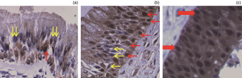
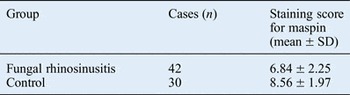
We investigated immunohistochemical staining for anti-maspin polyclonal antibody. The differences in maspin expression in the various groups are shown in Figure 1, and in Tables I and II. Maspin expression was significantly down-regulated in the fungal rhinosinusitis group as compared to the chronic rhinosinusitis group (t = −3.367, p < 0.05). Maspin expression among the invasive fungal rhinosinusitis, non-invasive fungal rhinosinusitis and control groups was also compared, and the inter-group difference was statistically significant (F = 23.377, p < 0.05). The overall score was lowest for the invasive fungal rhinosinusitis group compared with the other two groups, and the differences were statistically significant (t = −3.390, p < 0.05; t = −4.143, p < 0.05, respectively). The difference between the non-invasive fungal rhinosinusitis group and the control group was not statistically significant (t = 0.753, p > 0.05).
Immunohistochemical staining for maspin protein (×100), in mucous epithelia of: (a) an invasive fungal rhinosinusitis case, (b) a non-invasive fungal rhinosinusitis case and (c) nasal sinuses in the control group. Red arrowheads indicate positive expression and yellow arrowheads indicate negative expression.

Invasive fungal rhinosinusitis diagnosis
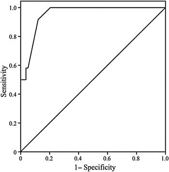
We also investigated the diagnosis of invasive fungal rhinosinusitis according to maspin immunohistochemical staining grade. Based on the receiver operating characteristic curve (Figure 2), the critical immunohistochemical staining grade for maspin was 5.70. The sensitivity of this value for invasive fungal rhinosinusitis was 91.7 per cent, the specificity was 88.3 per cent and the Youden index was 0.80.
Receiver operating characteristic curve for maspin diagnosis of invasive fungal rhinosinusitis.

Discussion
Fungi are generally present throughout the environment, and are found even in the nasal cavities of patients.Reference Kim, Sohn, Kim, Choi, Byun and Lee 10 Nevertheless, under normal conditions, fungal diseases can be avoided because of the protection provided by the local physical barrier of the mucous epithelia of the nasal cavity and nasal sinuses, and by the immune system of the host. The important anatomical factors for the development of fungal rhinosinusitis include a moist internal environment in the nasal sinuses and the obstruction of the ostiomeatal complex. Moreover, the status of the host's immune system and the corresponding interactions of micro-organisms in the host play an important role in the development and progression of fungal rhinosinusitis.Reference Soler and Schlosser 11 Multiple extracellular enzymes and toxins, which are regarded as the pathogenic agents in fungal rhinosinusitis that induce an immune response in the host, can be produced in the human body after being infected by fungi.
Serine proteinases are one of many types of proteinases secreted by fungi, which can lead to the destruction of host tissues. A new anti-oncogene (i.e. the maspin gene) has been isolated in normal mammary epithelial tissues and cells, by means of subtractive hybridisation technology, in a comparative study between normal mammary tissue and mammary cancer tissue conducted in 1994.Reference Zou, Anisowicz, Hendrix, Thor, Neveu and Sheng 12 That study reported that maspin served as an inhibitor of serine proteinase. Since then, numerous studies have investigated the expression of maspin in tumour-related diseases. Maspin has been found to be highly expressed in normal mammary tissues and in fibrous cystic epithelium, although the expression is significantly decreased in breast ductal carcinoma in situ, invasive breast cancer and the different stages of the disease accompanying lymphatic metastasis.Reference Maass, Teffner, Rösel, Pawaresch, Jonat and Nagasaki 13 Moreover, the expression of maspin is gradually decreased with the progression of the clinical breast cancer stage.Reference Umekita, Ohi, Sagara and Yoshida 8
Furthermore, studies of maspin expression in bladder cancer cells have found that the expression varies with the degree of bladder cancer differentiation. Non-expression and low expression of maspin have been observed in cell lines with relatively higher malignancy, whereas high maspin expression has been detected in cell lines with relatively lower malignancy levels and in atypical hyperplastic tissue. Moreover, the proliferation rate of bladder cancer cells in mice has been negatively correlated with maspin expression.Reference Beecken, Engl, Engels, Blumenberg, Oppermann and Camphausen 9
The extracellular histological barrier of the human body is composed of a basal membrane and extracellular matrix. The nasal membrane is a structure that is jointly composed of type IV collagen, laminin, contactin and fibronectin. A study has demonstrated that breast cancer cell lines expressing maspin can pass through type IV collagen; additionally, laminin was reduced by 96 per cent.Reference Odero-Marah, Khalkhali-Ellis, Chunthapong, Amir, Seftor and Seftor 14 The study further showed that this effect could be blocked by an antibody against maspin. Intercellular adhesive action can be maintained by adhesion molecules, which are primarily composed of cadherin, immunoglobulin, integrin and selectin, whereas the expression of E-cadherin can be affected by maspin. Thus, maspin plays an important role in preventing cells from penetrating the basal membrane and maintaining intercellular adhesiveness. Factors that may be involved in the process of fungal invasion include those involved in the destructive effects of the serine proteinase secreted by fungi. These factors include facilitation of the penetration through the basal part of the nasal sinus mucous membrane and the abnormal intercellular adhesiveness resulting from the decrease in maspin expression.
In fungal and invasive rhinosinusitis, fungi can spread towards deep tissues, similar to malignant tumours, whereas the extent of the lesion in non-invasive disease is relatively limited. Hence, examining maspin expression is extremely important when exploring the mechanisms underlying the development of invasive fungal rhinosinusitis and fungal invasion in general.
In this study, the expression of maspin in invasive fungal rhinosinusitis was explored in a preliminary manner on a clinical basis. Maspin expression was down-regulated in the fungal rhinosinusitis group; this was most significant in the invasive fungal rhinosinusitis group. However, the specific mechanism by which maspin expression is down-regulated remains unclear. The toll-like receptors belong to the family of transmembrane signal transduction receptors that connect human non-specific immunity and specific immunity.Reference Lin, Verma and Hodgkinson 15 Furthermore, toll-like receptors 2, 4 and 9 have been reported to be capable of binding to fungal components.Reference Wuthrich, Deepe and Klein 16 , Reference Newton and Dixit 17 Once the human body is infected by fungi, surface components of the fungal thalli bind to toll-like receptors to activate the signalling protein nuclear factor kappa B, which results in the release of multiple inflammatory cytokines, leading to complex biological reactions.Reference Kim, Choi, Jeon, Cha, Hwang and Chung 18 The down-regulation of maspin expression in the invasive fungal rhinosinusitis group was closely related to this process, which needs further study. Other studies have indicated that intracellular Iκα kinase IKKα in the respiratory epithelial cells of mice could be activated upon induction of the mycelia of Aspergillus fumigatus, and that down-regulation of maspin expression can also be induced.Reference Zhang, An and Zhao 19
-
• Maspin expression was down-regulated in the fungal rhinosinusitis group as compared to the chronic rhinosinusitis group
-
• This down-regulation is important in: invasive fungal rhinosinusitis development, mucous membrane fungal invasion and bone destruction
-
• A maspin staining score of 5.70 was the critical value for invasive fungal rhinosinusitis diagnosis, with sensitivity and specificity of 91.7 and 88.3 per cent, respectively
-
• Maspin may be an important biomarker for rapid diagnosis of invasive fungal rhinosinusitis
The expression of maspin was significantly down-regulated in the invasive fungal rhinosinusitis group in this study. This indicates that the down-regulation of maspin expression plays an important role in: the development of invasive fungal rhinosinusitis, the invasion of fungi into the mucous membrane and the induction of bone destruction. Relatively high sensitivity and specificity for the diagnosis of invasive fungal rhinosinusitis can be achieved using the maspin immunohistochemical staining grade. Hence, maspin may be an important biomarker for the rapid diagnosis of invasive fungal rhinosinusitis.
Maspin expression in fungal rhinosinusitis and control groups

SD = standard deviation
Maspin expression in invasive and non-invasive fungal rhinosinusitis and control groups

SD = standard deviation