Introduction
Black spot disease in fish is caused by metacercariae of digenetic trematodes from at least seven families (Kurochkin and Biserova Reference Kurochkin and Biserova1996). Studies on the dynamics and distribution of this disease have become increasingly relevant in recent years. Results have shown that the disease is widely distributed among freshwater and marine fishes, many of which are commercially valuable (Duflot et al. Reference Duflot, Cresson, Julien, Chartier, Bourgau, Palomba, Mattiucci, Midelet and Gay2023; Marković et al. Reference Marković, Ćirković, Aleksić, Milošević, Olivera, Dragana, Aksentijević and Radojičić2012). As emphasised, many of the causative agents of this disease are potentially zoonotic; some have even been found in humans (Chai and Jung Reference Chai and Jung2017; Chai and Jung Reference Chai, Jung, Toledo and Fried2024).
Infection of the host fish occurs as follows: trematode cercariae penetrate the fish’s skin, become localised in the skin and subcutaneous tissue, and develop into encysted metacercariae (Duflot et al. Reference Duflot, Midelet, Bourgau, Buchmann and Gay2021; Flores-Lopes Reference Flores-Lopes2014; Iqbal et al. Reference Iqbal, Shukerova and Minhas2014; Schaaf et al. Reference Schaaf, Kelson, Nusslé and Carlson2017). A capsule consisting of several layers of host cells is then formed around the parasites’ cyst. Encapsulated metacercariae stimulate migration of pigment cells (melanocytes) to the sites of their localisation, which results in characteristic, well-discernible pigment spots on the fishʼs skin surface, facilitating the ease of detection and diagnosis of this disease (Duflot et al. Reference Duflot, Cresson, Julien, Chartier, Bourgau, Palomba, Mattiucci, Midelet and Gay2023; Flores-Lopes and Thomaz Reference Flores-Lopes and Thomaz2011; Ondračková et al. Reference Ondračková, Šimková, Gelnar and Jurajda2004; Teixeira-de Mello and Eguren Reference Teixeira-de Mello and Eguren2008; Tobler and Schlupp Reference Tobler and Schlupp2008).
Several studies have examined the pathogenic effects of various trematode species responsible for black spot disease in fish. The pathology is manifested as loss in body weight, growth retardation, superficial skin and muscle lesions, mass mortality of juveniles, among other effects (Charo-Karisa et al. Reference Charo-Karisa, Ali, Marijani, Ibrahim, Trinh, Chadag and Benzie2021; Lemly and Esch Reference Lemly and Esch1984; Markovic and Krsmanovic Reference Markovic and Krsmanovic2008). The morphology of capsules forming around the encysted metacercariae was also examined in multiple studies, which also included descriptions of the capsule formation process in some trematode species (Berezantsev and Dobrovolsky Reference Berezantsev and Dobrovolsky1968; El-Mayas and Kearn Reference El-Mayas and Kearn1995; Pulsford Reference Pulsford1980). Despite extensive data on black spot disease pathogenesis, further studies are needed to detail the specific impacts of these trematodes on their fish hosts.
Taui Bay in the northern Sea of Okhotsk is characterised by a rich fauna of marine and freshwater fish species. Many are known to have pigmented spots on their body surface, yet the causative agents of these spots remain unknown in most cases. Only two species have been studied in this regard: the stone cockscomb Alectrias alectrolophus (Pallas, 1814) and the Middendorff’s eelpout Hadropareia middendorffii Schmidt, 1904. These studies have shown that the black spots visible on these fishesʼ skin surface are a result of parasitisation by trematode metacercariae (Atrashkevich et al. Reference Atrashkevich, Orlovskaya, Regel, Mikhailova and Pospekhov2005; Skorobrekhova Reference Skorobrekhova2009). However, no other studies of this fish disease have been conducted in the region to date.
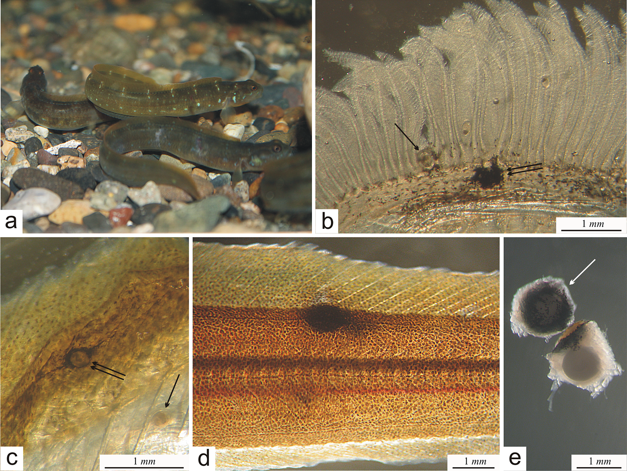
Middendorff’s eelpout is a small (maximum length of 25 cm) endemic fish inhabiting the intertidal zone at depths of 0–30 m (Figure 1a) (Chereshnev et al. Reference Chereshnev, Volobuev, Khovanskii and Shestakov2001). Two species of trematodes that can stimulate host pigment production have been isolated from tissues of this fish and identified as Cryptocotyle lingua (Heterophyidae) and Liliatrema skrjabini (Liliatrematidae) (Atrashkevich et al. Reference Atrashkevich, Orlovskaya, Regel, Mikhailova and Pospekhov2005). Both parasite species have a trixenous life cycle. Adult trematodes parasitise the intestines of fish-eating birds. On the coast of Taui Bay, C. lingua have been found in the slaty-backed gull Larus schistisagus Stejneger, 1884, black-throated diver Gavia arctica (Linnaeus, 1758), greater scaup Aythya marila (Linnaeus, 1761), and white-winged scoter Melanitta deglandi (Bonaparte, 1850). Trematodes of the L. skrjabini species have also been found in the slaty-backed gull, as well as the pelagic cormorant Phalacrocorax pelagicus Pallas, 1811 (Atrashkevich et al. Reference Atrashkevich, Orlovskaya, Regel, Mikhailova and Pospekhov2005). While the first intermediate host for C. lingua in this region has been identified to be the winkle Littorina sitkana, the first intermediate mollusc species host of L. skrjabini remains unknown. The second intermediate host of both trematode species are fish, in which metacercariae are localised in the oral cavity, under the skin, and in other tissues (Skorobrekhova Reference Skorobrekhova2009; Wakabayashi Reference Wakabayashi1997).
Middendorff’s eelpout, H. middendorffii, infected by metacercariae of C. lingua and L. skrjabini. (a) General view of Middendorff’s eelpout. (b) Encapsulated metacercariae of C. lingua partially (indicated by arrow) and completely (double arrow) pigmented on the fish’s gills. (c) An encapsulated metacercaria of C. lingua partially pigmented under the fishʼs skin (double arrow), and an unpigmented metacercaria capsule in the fishʼs muscles (arrow). (d) Aggregation of pigment around a L. skrjabini metacercaria located under the fish’s skin. (e) Encapsulated metacercariae of L. skrjabini extracted from the fishʼs muscles. One of the trematodeʼs capsules (arrow) is completely covered with pigment deposits.

In this study, we aimed to estimate the prevalence and intensity of infection in Middendorffʼs eelpout by metacercariae of the trematodes Cryptocotyle lingua and Liliatrema skrjabini. We also aimed to quantify infection in different parts of the fishʼs body and to examine the thin and ultrathin structure of the capsules surrounding these metacercariae.
Materials and methods
The trematode species under investigation were identified by O.M. Orlovskaya, a researcher from the Laboratory of Helminth Ecology, Institute of Biological Problems of the North (IBPN), Far Eastern Branch, Russian Academy of Sciences (FEB RAS), based on morphological characters (Atrashkevich et al. Reference Atrashkevich, Orlovskaya, Regel, Mikhailova and Pospekhov2005). Under her supervision, trematode capsules were first sorted by species and then counted. Next, specimens were selected for analysis of capsule morphology. It was found that capsules containing encysted metacercariae of C. lingua differed significantly from those containing L. skrjabini in terms of size, natural colour, and morphological features of the parasites (e.g., the oral sucker structure).
In June and July 2024, we examined 47 specimens of fish caught at low tide in the coastal waters of Nagaev Bay, Sea of Okhotsk. The proximity to the scientific institution, situated on the Sea of Okhotsk coast, allowed us to carry out dissection and examination of fresh fish immediately on the sampling day. The fish were caught in small batches of two to three specimens each. On shore, the fish were euthanised by cutting the spinal cord immediately behind the head with a sharp knife, in accordance with the approval of the Bioethics Commission, IBPN FEB RAS (Protocol No. 002/025). The fish were then transported to the laboratory on ice. The total body length from the snout tip to the caudal fin tip (TL) was measured, and the sex was determined for each specimen.
To assess the distribution of parasites, each fish’s body was divided into three parts: the head, the trunk, and the fins. Using a scalpel, we made transverse incisions on the body, cutting down to the spinal column, and examined each dissected piece of muscle tissue together with the skin under an MBS-10 microscope using the compression technique. After examining the musculature, we viewed the exposed spinal column on both sides. We examined the fish’s fins both with the naked eye and under a microscope. We crushed the fish’s head with scissors and then examined small portions of tissues from the obtained mass. When dissecting each fish, we counted the number of trematode capsules of both species in all three body regions.
When analysing the material, we used two parameters of fish infection by trematode metacercariae: prevalence of infection (the number of host individuals infected by a certain parasite species divided by the total number of hosts examined), and intensity of infection (the number of individuals of a certain parasite species in each infected host) (Pronin et al. Reference Pronin, Zhaltsanova, Pronina, Nekrasov, Rinchino, Rusinek, Sanzhieva, Dugarov, Belyakova and Kudryashev1991). The correlation between the number of parasites and the host’s parameters (total length and sex), if present, was estimated using MS Excel 2007.
To study the fine morphology of the parasites, we dissected the encapsulated metacercariae from the fish’s skin and muscles and fixed them in a 2% glutaraldehyde solution in 0.1 M phosphate buffer. After fixation, the material was post-fixed in an osmium tetroxide (OsO4) solution in 0.2 M phosphate buffer for 12 h, dehydrated, and embedded in an Epon-Araldite (Fluka, Switzerland) mixture. During dehydration, the specimens were stained with a saturated uranyl acetate solution in 70% ethanol overnight. Semithin (1 μm) sections, cut on an LKB microtome (LKB Bromma, Stockholm, Sweden), were stained with methylene blue according to Morgenstern (Reference Morgenstern1969), and viewed under an Olympus CX41 microscope (Olympus Corporation, Tokyo, Japan). Ultrathin (70 nm) sections, cut on a PT-PC ultramicrotome (Boeckeler Instruments, Tucson, Arizona, USA), were viewed under a JEM-1400Plus transmission electron microscope (JEOL, Tokyo, Japan).
Results
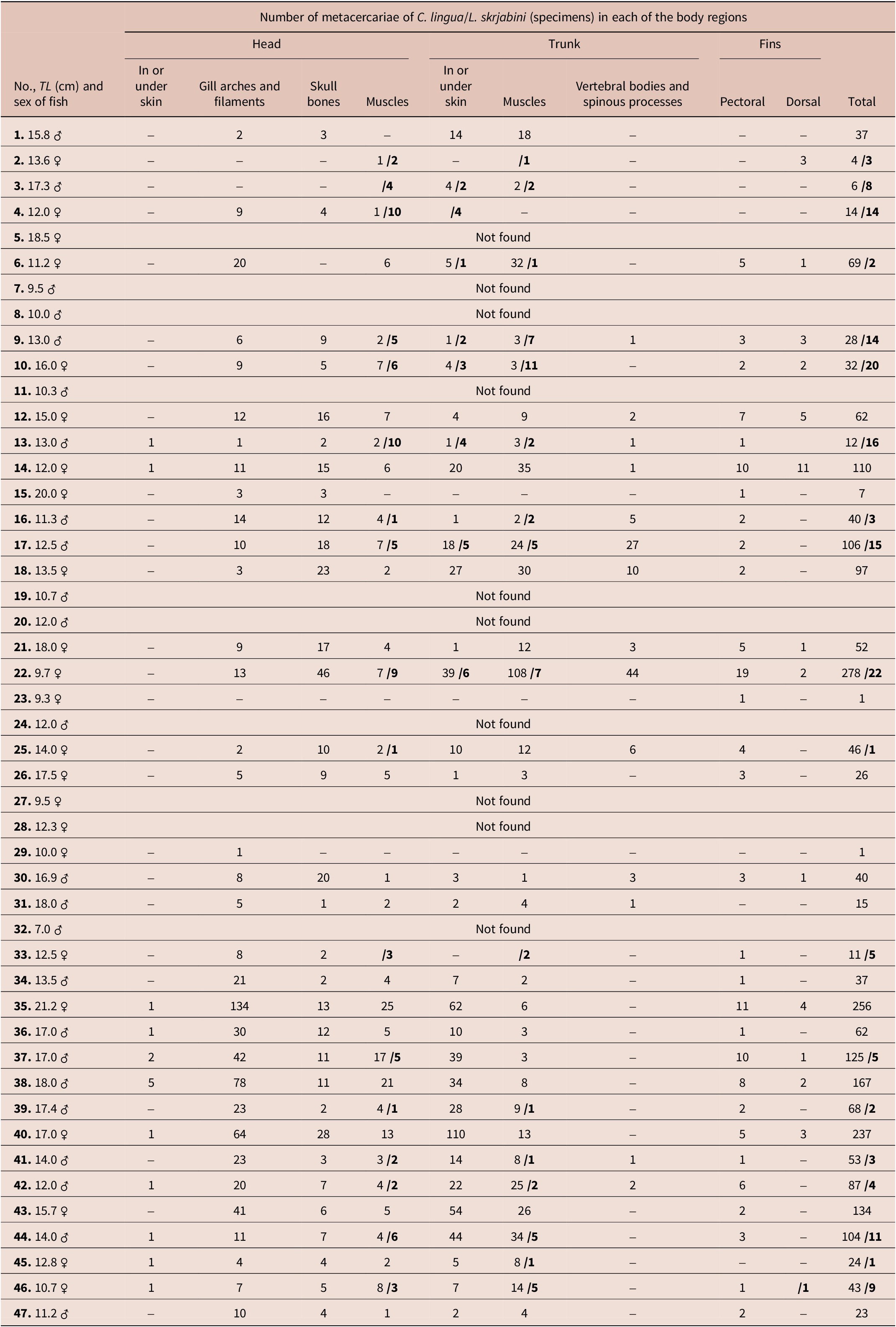
The examined Middendorff’s eelpout individuals were infected either by metacercariae of both trematode species or only C. lingua. There were no cases in which fish were parasitised by L. skrjabini larvae only. The results of the fish dissection and the distribution of metacercariae in different parts of the body are presented in Table 1. The prevalence of infection by C. lingua was high (as much as 79%), with an intensity of infection ranging from one to 278 larvae per infected fish (average = 53). Of the 47 fish examined, 19 (40%) were infected by L. skrjabini larvae. The intensity of infection ranged from one to 22 larvae per infected fish (average = eight).
Intensity of infection of Middendorff’s eelpout by metacercariae of Cryptocotyle lingua and Liliatrema skrjabini and their distribution on the fish’s body

Note: Value before slash (in normal font) is number of C. lingua metacercariae; value after slash (in bold font) is number of L. skrjabini metacercariae.
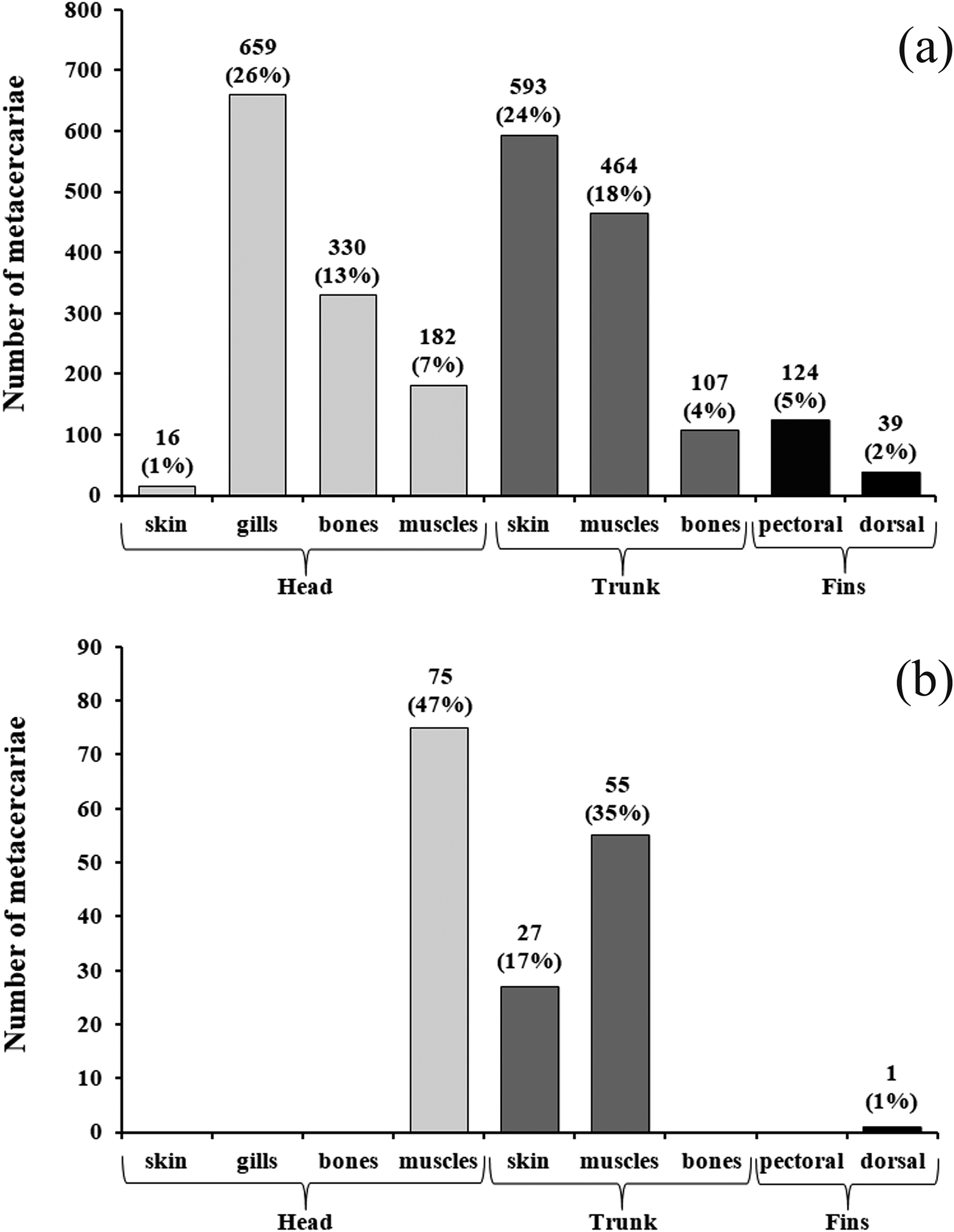
Of the total number of specimens (2,514) of C. lingua larvae extracted from Middendorff’s eelpout, a substantial proportion (93%) was found in the head (47%) and trunk (46%) regions of the examined fish (Figure 2a). Of the larvae found in the head, the highest amount were located on the surfaces of the gill arches and filaments (26%). Approximately half as many (13%) were infested in bones of the cranium, jaw, and opercula. Encapsulated metacercariae were located both on the surfaces of bones and in their cavities. Of the total number of larvae found in the fish’s trunk region, the highest proportion were located in the dermal layer of the skin or under the skin (24%) (Figure 2a). The muscle tissue of the fish was similarly affected (18%), with trematode larvae distributed fairly evenly in both the upper and deeper layers of the muscles. A small number of metacercariae were attached to the surfaces of spinous processes and vertebral bodies (4%). The pectoral and dorsal fins were infected to a much lesser extent (7%) than the head and trunk, with trematode larvae located on the fin rays. No trematode larvae were found in the other fins. Of the total number of L. skrjabini metacercariae (158 specimens), 52% were detected in the trunk section and 47% in the head section; only one specimen was found in the dorsal fin (Figure 2b). Our correlation analysis did not show any significant relationship between the intensity of infection of the host and its body length or sex.
Number of C. lingua (a) and L. skrjabini (b) metacercariae in different regions of a fish’s body.

Metacercariae of both species were surrounded by two easily separable envelopes: an inner transparent cyst, and an outer relatively dense capsule. Encapsulated metacercariae of C. lingua were characterised by a slightly elongated shape with a size of ca. 0.2 × 0.3 mm. Capsules of L. skrjabini were globe-shaped and reached a diameter of 0.7–1.3 mm. Signs of pigmentation were found on the surfaces of the trematode capsules, but the intensity of pigmentation varied markedly from one specimen to another, which might be related to different durations of parasites’ residency in the fishes’ tissues. In some cases, distinct clusters of pigment cells were observed on the capsule surface, with the encysted larva visible between them (Figures 1b, c). In other cases, metacercariae were covered by a continuous, dark layer of pigment cells, through which encysted larva were not visible (Figures 1b, d, e). Some of metacercariae had no signs of melanisation (Figure 1c).
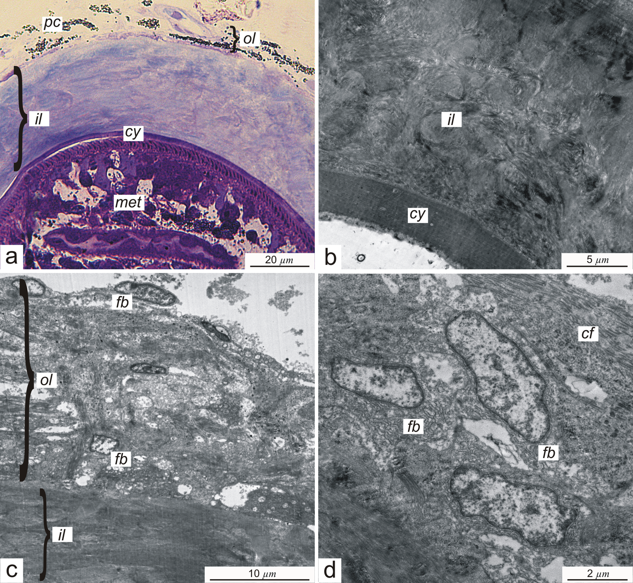
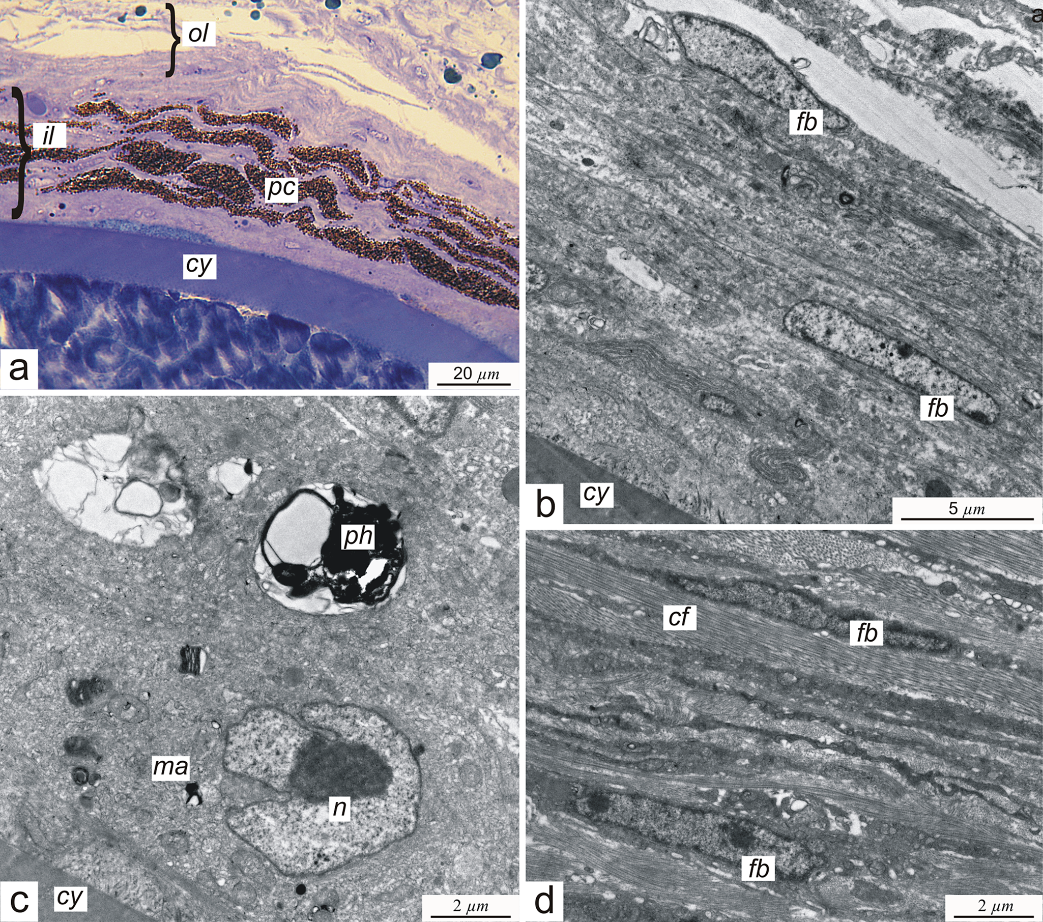
In semithin sections, the metacercarial cyst of both species appeared as a thin, dark, homogeneous envelope, tightly adjacent to the parasite’s surface. The capsules consisting of host cells closely surrounded the encysted larvae. Capsules of C. lingua, regardless of their localisation, had a similar organisation. There were two distinct layers in the capsule structure: the inner fibrous, light-coloured layer and the outer cellular layer (Figure 3a). The inner layer was thicker (30–40 μm) and consisted of randomly oriented, tightly arranged collagen fibres (Figures 3b, c); their characteristic transverse striation became apparent in the ultrathin sections. The superficial outer layer of the capsule (maximum thickness of 20 μm) consisted of several rows of branch-shaped fibroblasts and a high amount of collagen fibres (Figures 3c, d). Aggregations of pigment cells were localised on the periphery of the capsule and extended between adjacent myofibres (Figure 3a). The capsule of L. skrjabini metacercaria was 60–70 μm thick. In its structure, two layers were identified; they were approximately equal in thickness, with an indistinct boundary between them (Figure 4a). The inner layer of the capsule was formed by densely organised cells and aggregations of collagen fibres in intercellular spaces (Figures 4b, c). In some cases, the cells were too densely arranged to allow their boundaries to be discerned. Fibroblasts and macrophages were identified among the cells of this layer. The more abundant fibroblasts were characterised by diverse shapes, ranging from elongated to rounded (Figure 4b), and long processes. Their nuclei were characterised by sparse karyoplasm and a small nucleolus. The synthesising apparatus in the form of numerous cisterns of the rough endoplasmic reticulum was developed in the cytoplasm. Macrophages (Figure 4c) were larger, with a rounded nucleus and a large amount of cytoplasm that included numerous lucent vacuoles and putative phagolysosomes. The capsule’s outer layer, unlike the inner layer, was composed only of loosely organised fibroblasts and collagen fibres (Figure 4d), as well as pigment cells. In some metacercariae, pigment cells were located not only in the capsule’s outer layer, but also deeper, almost reaching the cyst’s surface (Figure 4a). All examined capsules were permeated by blood vessels.
Structure of a capsule around metacercariae of C. lingua in tissues of Middendorff’s eelpout, H. middendorffii. (a), Semithin section through a fragment of the parasiteʼs tegument, the cyst, and the capsule surrounding it; (b), Ultrathin section through the inner capsule layer; (c), Ultrathin section through the inner and outer capsule layers; (d), Ultrathin section of fibroblasts in the outer capsule layer. Letter designations: cy – cyst, cf – collagen fibres, fb – fibroblasts, il – inner capsule layer, met – metacercaria, ol – outer capsule layer, pc – pigment cells.

Structure of a capsule around metacercariae of L. skrjabini in tissues of Middendorff’s eelpout, H. middendorffii. (a), Semithin section through the cyst and the capsule surrounding the parasite; (b), Ultrathin section through the inner capsule layer; (c), Ultrathin section of a macrophage in the outer capsule layer; (d), Ultrathin section through the outer capsule layer. Letter designations: cy – cyst, cf – collagen fibres, fb – fibroblasts, il – inner capsule layer, ma – nucleus of the macrophage, ol – outer capsule layer, pc – pigment cells, ph – phagolysosome.

Discussion
This is the first study to investigate the infection parameters and features of localisation of metacercariae causing black spot disease in fish from the Sea of Okhotsk. The results show that the trematode C. lingua is the main causative agent of this disease in Middendorff’s eelpout. In particular, larvae of this trematode were found to be relatively evenly distributed in the fishesʼ muscles. This does not align with the data presented in earlier literature, which suggests that the causative agents of black spot disease are concentrated mainly in the skin and subcutaneous layer of the musculature (Borges et al. Reference Borges, Skov, Bahlool, Møller, Kania, Santos and Buchmann2015; Duflot et al. Reference Duflot, Midelet, Bourgau, Buchmann and Gay2021).
Most trematode species that cause black spot disease are from the families Diplostomidae and Heterophyidae (Kurochkin and Biserova Reference Kurochkin and Biserova1996). The trematode C. lingua that was studied here is one of the most widely distributed and best-studied species of the family Heterophyidae and is a typical causative agent of this disease (Duflot et al. Reference Duflot, Midelet, Bourgau, Buchmann and Gay2021). The other trematode species, L. skrjabini, belongs to Liliatrematidae, a less widespread and low-abundance family (Frolov Reference Frolov2010; Gubanov Reference Gubanov and Petrov1953). Previously, metacercariae of this species were found in shorthorn sculpin Myoxocephalus scorpius near the Kuril Islands (Gubanov Reference Gubanov and Petrov1953), in Schlegelʼs rockfish (Sebastes schlegelii) near Hokkaido Island (Ohbayashi and Konno Reference Ohbayashi and Konno1966; Wakabayashi Reference Wakabayashi1997), in stone cockscomb (Alectrias alectrolophus) from coastal waters of the Sea of Okhotsk (Skorobrekhova Reference Skorobrekhova2009), and in Dybowskiʼs blenny (Pholidapus dybowskii) in the northwestern Pacific Ocean near Sakhalin Island (Sokolov et al. Reference Sokolov, Frolov, Novokreshchennykh and Atopkin2021). Researchers reported that larvae of L. skrjabini were localised in fishʼs muscles and were covered with pigment deposits. Thus, the Middendorffʼs eelpout extends the list of known second intermediate hosts for L. skrjabini whose metacercariae cause black spot disease in this fish.
Previous studies have shown that trematode metacercariae parasitise different parts of fishesʼ bodies unevenly (Duflot et al. Reference Duflot, Cresson, Julien, Chartier, Bourgau, Palomba, Mattiucci, Midelet and Gay2023; Paradis and Chapleau Reference Paradis and Chapleau1994; Teixeira-de-Mello and Eguren 2008). The distribution of metacercariae in a fishʼs body may depend on environmental conditions, and on the behavioural characteristics of the parasite and the host (Ondračková et al. Reference Ondračková, Jurajda and Gelnar2002). Flores-Lopes (Reference Flores-Lopes2014) reported that in banded astyanax (Psalidodon fasciatus), the maximum numbers of trematode metacercariae from the family Diplostomidae were found on the ventral and dorsal surfaces of the body. As the author assumes, the finding of many diplostomid trematodes on the ventral surface of the fishes’ bodies may be explained by the initial development of cercariae in a benthic mollusc, with their subsequent migration to the ventral side of the fish. Moreover, the presence of pigmented spots on the dorsal body surface, caused by parasitic metacercariae, makes it easier for the definitive host (fish-eating bird) to find infected fish. On the other hand, fins are preferred sites of invasion for other trematode species (Golovin et al. Reference Golovin, Ivanov, Ivanova, Rybkina and Lajus2021; Korniychuk and Martinenko Reference Korniychuk and Martinenko2009; Paradis and Chapleau Reference Paradis and Chapleau1994). In particular, Golovin et al. (Reference Golovin, Ivanov, Ivanova, Rybkina and Lajus2021) estimated the number of Cryptocotyle spp. metacercariae by the number of black spots on the body surface in three-stickleback (Gasterosteus aculeatus). The authors concluded that the number of larvae in the caudal fin was approximately three-fold higher than the average for the body. They explained the high degree of infection of the caudal fin by its fast movements and delicate structure. They further suggested that this structure, along with the locomotor features of this fin, are favourable for the successful attachment of cercariae on the fishʼs skin and their further invasion into the body. On the contrary, infection on the fins of Atlantic cod (Gadus morhua) is insignificant; Lysne et al. (Reference Lysne, Hemmingsen and Skorping1994) suggested that frequent fin movement may be one explanation for this finding. In the Middendorff’s eelpout examined here, we found C. lingua and L. skrjabini metacercariae mainly in the head and trunk regions of the body. It is noteworthy that despite the significantly smaller area of the head compared to the body, the numbers of metacercariae in these regions were approximately equal, and this finding applied to both trematode species. Metacercariae of both species were also found in the fins, however with a minimal intensity of fin infection compared to other regions of the body. We assume that the insignificant degree of infection of the fins, especially the dorsal one, is due to their anatomical features. As in many zoarcids, the dorsal fin in Middendorff’s eelpout merges with the caudal and anal fins, forming a single, dense fin margin (Poezzhalova-Chegodaeva Reference Poezzhalova-Chegodaeva2020). Such a structure provides fish with high swimming performance near the bottom where manoeuvrability and stability under currents are particularly important. It is likely that the rigid structure of the fins makes it difficult for trematode cercariae to invade them.
Fish of many species can be infected by trematodes that cause black spot disease (De Wit and Johnson Reference De Wit and Johnson2024; Duflot et al. Reference Duflot, Cresson, Julien, Chartier, Bourgau, Palomba, Mattiucci, Midelet and Gay2023; Johnson et al. Reference Johnson, Malawauw, Piaskowy, Calhoun, Kohl, Horst and Zelmer2024; Sándor et al. Reference Sándor, Molnár, Gibson, Székely, Majoros and Cech2017). However, large body size of fish and the high degree of infection often make it difficult to reliably quantify their infection parameters. Researchers have applied various methods to identify and count metacercariae over the fishʼs body. A common approach is to count them by the number of black spots formed around encysted trematode larvae on the fishʼs body surface (Korniychuk and Martinenko Reference Korniychuk and Martinenko2009; Ondračková et al. Reference Ondračková, Jurajda and Gelnar2002). For example, metacercariae of Cryptocotyle spp. on the body surface and the caudal fin of three-spined stickleback were counted by analysing a digital image of the fishʼs left flank (Golovin et al. Reference Golovin, Ivanov, Ivanova, Rybkina and Lajus2021). However, that study might have missed trematodes that had recently migrated through the skin and did not have enough time to deposit pigment in the surrounding capsules (McQueen et al. Reference McQueen, Mackenzie, Roberts and Young1973). Another approach to estimating the number of parasites is by digesting pieces of fishʼs muscle in a pepsin/HCl/saline solution. Duflot et al. (Reference Duflot, Midelet, Bourgau, Buchmann and Gay2021) digested both small skin fragments and whole fillet of whiting (Merlangius merlangus) to isolate metacercariae of the genus Cryptocotyle from the fishesʼ tissues. However, the tissue crushing and pepsin digestion could also damage part of the metacercariae, thereby underestimating their actual number (Duflot et al. Reference Duflot, Midelet, Bourgau, Buchmann and Gay2021). In our study, the infection of Middendorffʼs eelpout by trematode metacercariae was quantified in freshly caught fish whose tissues were examined using the compression method. This method allowed us to count all unpigmented metacercariae, as well as those located deep in the muscle tissue and on the surface of bones.
As was previously mentioned, trematodes that are causative agents of black spot disease in fish first appear in the form of cysts that eventually become surrounded by a capsule of a hostʼs immune cells, covered by several layers of pigment cells (Flores-Lopes Reference Flores-Lopes2014; Quist et al. Reference Quist, Bower and Hubert2007; Wittrock et al. Reference Wittrock, Bruсe and Johnson1991). Our observation showed that encysted metacercariae of C. lingua and L. skrjabini in the Middendorffʼs eelpout were localised inside fibrous capsules either completely or partially covered with pigment deposits. A distinguishing feature of the metacercariae capsule in C. lingua was the thick inner fibrous layer consisting of densely arranged collagen fibres, which was the main constituent. The same layer was found in the metacercariae capsule of this species which naturally infects European plaice (Pleuronectes platessa) and stone cockscomb (Alectrias alectrolophus) (Pulsford Reference Pulsford1980; Skorobrekhova Reference Skorobrekhova2009). As a result of experimental infection of thicklip grey mullet (Chelon labrosus) by C. lingua, a similar layer was synthesised by capsule fibroblasts on day 55 (Pulsford Reference Pulsford1980). The capsules of the L. skrjabini metacercariae that we examined lacked the layer formed exclusively by collagen fibres, but their composition was dominated by fibroblasts, and collagen fibres were only present in small quantities.
As previous data has shown, the encapsulated forms of some trematode species can cause severe tissue damage in fish (Berezantsev and Dobrovolsky Reference Berezantsev and Dobrovolsky1968). For example, metacercariae of Uvulifer sp. form depressions on the operculum surface in Catostomidae fish species and can even perforate it (Quist et al. Reference Quist, Bower and Hubert2007). Dennis et al. (Reference Dennis, Izquierdo, Conan, Johnson, Giardi, Frye and Freeman2019) showed that metacercariae of Scaphanocephalus located in the skin of fishes of the genus Acanthurus cause dermatitis due to rubbing of the body against the substrate. The pathological process caused by the presence of metacercariae in fishesʼ tissues is often accompanied by leukocyte infiltration of the capsule wall surrounding them and the hostʼs muscle or connective tissue in the immediate vicinity of the parasites (Dennis et al. Reference Dennis, Izquierdo, Conan, Johnson, Giardi, Frye and Freeman2019; Flores-Lopes Reference Flores-Lopes and Thomaz2011). In eastern mosquitofish (Gambusia holbrooki), several layers of epithelioid cells form the inner layer of the capsule adjacent to the cyst surface of Neascus metacercariae (Horth et al. Reference Horth, Gauthier and Vogelbein2013). In our study, the presence of a few leukocytes (macrophages) in capsules was characteristic only of L. skrjabini, and the localisation of these cells tended to be in the inner capsule layer, similar to trematodes of the genus Neascus.
Overall, the causative agents of black spot disease in the Middendorff’s eelpout examined in this study were determined to be two trematode species that infect fish to a high (in the case of C. lingua) or moderate (in the case of L. skrjabini) degree. Further study of the distribution of black spot disease and the effects of the causative agents on fish is important for understanding their patterns in relation to the dynamics of fish populations and may also have implications for the development of effective anthelmintic treatments at aquaculture farms.
Acknowledgements
The author expresses deep gratitude to O.M. Orlovskaya (Institute of Biological Problems of the North FEB RAS) for her valuable help in the species identification of metacercariae.
Financial support
Not applicable.
Competing interests
The author declares no competing interests.
Ethical standard
All applicable institutional, national, and international guidelines for the care and use of animals were followed.