1. Introduction
Reptilian eggs have a rigid mineralized external calcareous layer which is homogeneous as in Gekkonidae, has needle-like aragonite crystallites radiating from the inner core as in turtles, or consists of clearly defined cylindrical shell units arising from the outer surface of the shell membrane as in crocodiles and dinosaurs (Mikhailov, Reference Mikhailov1991). This assortment of ultrastructural units of eggshell is underlain by an organic shell membrane which consists of proteinaceous fibres and is known as Membrana Testacea (MT) (Grellet-Tinner, Reference Grellet-Tinner2005; Schweitzer, Reference Schweitzer2011; Yang et al. Reference Yang, Chen, Wiemann, Spiering and Sander2018). In living reptiles, the MT is divided into two layers: the inner thin layer comprising organic fibres disposed in a sub-parallel fashion and the outer thick layer with interlacing fibres (Packard & DeMarco, Reference Packard, DeMarco, Deeming and Ferguson1995). The two layers of the MT are difficult to identify in fossil eggshells. The MT exists as a plane where zones of mineralized mammillae consisting of loosely assembled crystals are present in the centre (Cuif et al. Reference Cuif, Dauphin and Sorauf2010). These zones become nucleation points for the formation of eggshell units (Grellet-Tinner, Reference Grellet-Tinner2005). The biological function of MT was considered as a filter for microorganisms (Krampitz & Graser, Reference Krampitz and Graser1988). It has been observed that the MT does not bind Ca ions, hence it does not play any role in the initial phase of calcification of eggshells and its mesh-like structure supports calcium carbonate crystals (Krampitz & Graser, Reference Krampitz and Graser1988).
While the outer rigid mineralized eggshell layer is generally found in fossils, the MT because of its soft anatomy is rarely preserved in fossils, specifically when exposed to oxidizing environments. However, there is evidence for the preservation of MT in the geological record. Some of the previous reports on preservation of MT in fossil state, such as the Pliocene turtle eggshells from the Canary Islands (Rothe & Klemmer, Reference Rothe and Klemmer1991) and the Upper Cretaceous dinosaur eggshells from Canada (Tazaki et al. Reference Tazaki, Aratani, Noda, Currie and Fyfe1994), were later rejected (Kohring, Reference Kohring1999). There are some undisputed records of fossilized MT. These include the Early Jurassic records in Massospondylus eggshells of Rooidraai locality in South Africa (Stein et al. Reference Stein, Prondvai, Huang, Baele, Sander and Reisz2019), Lufengosaurus eggs of China (Reisz et al. Reference Reisz, Huang, Roberts, Peng, Sullivan, Stein, LeBlanc, Shieh, Chang, Chiang and Yang2013) and Mussasaurus eggshells of Argentina (Reisz et al. Reference Reisz, Huang, Roberts, Peng, Sullivan, Stein, LeBlanc, Shieh, Chang, Chiang and Yang2013; Stein et al. Reference Stein, Prondvai, Huang, Baele, Sander and Reisz2019); the Late Cretaceous Megaloolithus eggshells from the Tremp Basin, Spain (Kohring, Reference Kohring1999; Sellés et al. Reference Sellés, Bravo, Delclòs, Colombo, Martí, Ortega-Blanco, Parellada and Galobart2013; Bravo & Gaete, Reference Bravo and Gaete2015); the Late Cretaceous dinosaur eggshells of the Gobi desert (Sochava, Reference Sochava1969; Kolesnikov & Sochava, Reference Kolesnikov and Sochava1972); the Late Cretaceous Triprismatoolithus (probable alvarezsaurid) eggshells from Montana, USA (Yang et al. Reference Yang, Chen, Wiemann, Spiering and Sander2018); the eggshells of titanosaurid clutches with embryonic skeletons from the Late Cretaceous Auca Mahuevo site in Patagonia, Argentina (Grellet-Tinner et al. Reference Grellet-Tinner, Chiappe and Coria2004; Grellet-Tinner, Reference Grellet-Tinner2005); the Late Cretaceous Megaloolithus siruguei (Megaloolithidae) eggshells from Haţeg Basin, Romania (Grigorescu & Oana-Claudia, Reference Grigorescu and Oana-Claudia2017–18); crocodilian eggshells from the Eocene (Lutetian) of Geiseltal, Germany (Krokolithes helleri Kohring & Hirsch, Reference Kohring and Hirsch1996); Early Miocene avian eggshells from Dolnice, Czech Republic (Kohring, Reference Kohring1998); and avian eggshells from the Late Miocene of Nördlinger Ries, Germany (Kohring & Sachs, Reference Kohring and Sachs1997).
The Late Cretaceous dinosaur fauna has been extensively documented from the Upper Cretaceous Lameta Formation or infratrappean sedimentary beds that stratigraphically underlie the lava flows of the Deccan Traps in western and central India. The fauna comprises at least six species of abelisaurid theropod dinosaurs and about three taxa of titanosaurid sauropods (see Prasad, Reference Prasad2012; Prasad & Sahni, Reference Prasad and Sahni2014; Khosla & Bajpai, Reference Khosla and Bajpai2021 for references). In addition to body fossils, widespread occurrence of clutches, eggs and eggshells of sauropod dinosaurs has also been reported from the Lameta outcrops of Madhya Pradesh, Gujarat and Maharashtra (Srivastava et al. Reference Srivastava, Mohabey, Sahni and Pant1986; Sahni et al. Reference Sahni, Tandon, Jolly, Bajpai, Sood, Srinivasan, Carpenter, Hirsch and Horner1994; Khosla & Sahni, Reference Khosla and Sahni1995; Mohabey, Reference Mohabey1998; Srivastava & Mankar, Reference Srivastava and Mankar2015; H. Dhiman, unpub. PhD thesis, Delhi Univ., 2021; Dhiman et al. Reference Dhiman, Dutta, Kumar, Verma and Prasad2021). Additionally, some fragmentary dinosaur bones are also known from the Upper Cretaceous Kallamedu Formation (see Prasad et al. Reference Prasad, Verma, Flynn and Goswami2013) and the Upper Cretaceous Mahadek Formation of Meghalaya (Mishra & Sen, Reference Mishra and Sen2001). However, for the latest Cretaceous, dinosaur fauna from India is scarcely known and is represented by a few isolated occurrences of body fossils and fragmentary and isolated eggshells from the Upper Cretaceous (Late Maastrichtian) intertrappean beds that occur intercalated with the Deccan volcanic flows (see Prasad, Reference Prasad2012; Prasad & Sahni, Reference Prasad and Sahni2014). No clutches or eggs have been reported so far from the Deccan intertrappean beds, and possibly the dinosaurs of India entered the waning phase by the time of their preservation in the intertrappean beds (Prasad & Sahni, Reference Prasad and Sahni2014). Dinosaur eggshells were previously reported from the intertrappean beds of Asifabad, Nagpur (Sahni et al. Reference Sahni, Rana and Prasad1984; Vianey-Liaud et al. Reference Vianey-Liaud, Jain and Sahni1987), Mohgaonkalan (Srinivasan, Reference Srinivasan1996), Kisalpuri (Khosla et al. Reference Khosla, Prasad, Verma, Jain and Sahni2004) and Gujri (Kshetrimayum et al. Reference Kshetrimayum, Parmar, Lourembam and Prasad2021). Of these sites, Gujri and Kisalpuri intertrappean beds yield both sauropod and ornithoid eggshells.
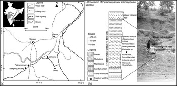
Here we report the exceptional preservation of a fossilized MT layer in titanosaurid dinosaur eggshells from the intertrappean beds of Piplanarayanwar in Chhindwara District, Madhya Pradesh (Fig. 1). The documentation of a MT layer from titanosaur eggshell adds to the few previously known reports and this is the first report of MT from the Mesozoic deposits of India. The discovery also helps us to understand possible environmental conditions that facilitated the preservation of the soft organic layer.
Location map of the Deccan intertrappean section of Piplanarayanwar, Chhindwara District, Madhya Pradesh, India.

2. Geological context
The Deccan Traps, representing one of the largest continental flood basalts of the Earth, cover an area of c. 500 000 km2 in central, western and south-central India. The Deccan lava flows in general occur unconformably over the Precambrian basement rocks or Gondwana sediments or at many places over the Upper Cretaceous sediments formally known as the Lameta Formation or informally as the infratrappean beds. The Lameta Formation in the Lameta Ghat type section near Jabalpur attains a maximum thickness of 70 m and is composed of basal Green Sandstone, Lower Limestone, Mottled Nodular Bed and Upper Calcareous Sandstone (Tandon et al. Reference Tandon, Sood, Andrews and Dennis1995) in this order of superposition. Sedimentary beds are present not only below the Deccan lava flows, but also occur intercalated with the volcanic flows. Such sedimentary beds are relatively thin and are generally known as intertrappean beds, referring to their position sandwiched between the volcanic flows. Both the Lameta Formation and the intertrappean beds have been a great source of several vertebrate groups (see Khajuria et al. Reference Khajuria, Prasad and Manhas1994; Prasad & Khajuria, Reference Prasad and Khajuria1995; Khosla & Sahni, Reference Khosla and Sahni2003; Prasad, Reference Prasad2012; Prasad & Sahni, Reference Prasad and Sahni2014). In the past, palaeontological evidence has been used to indicate that the Lameta Formation is Turonian in age (Sahni, Reference Sahni1972) and the intertrappean beds range in age from Upper Cretaceous to Early Eocene (Sahni, Reference Sahni1934; Sastry & Lahiri, Reference Sastry and Lahiri1981; Bande et al. Reference Bande, Chandra, Venkatachala, Mehrotra and Venkatachala1986) or even Oligocene (Mathur, Reference Mathur1968; Shivarudrappa, Reference Shivarudrappa1976–7). However, later studies have shown that the Lameta Formation is Maastrichtian in age based on fish remains, palynofossils and on comparison of dinosaur fauna with that of Madagascar (Courtillot et al. Reference Courtillot, Besse, Vandamme, Montigny, Jaeger and Cappetta1986; Buffetaut, Reference Buffetaut1987; Dogra et al. Reference Dogra, Singh and Kulshrestha1994; Khajuria et al. Reference Khajuria, Prasad and Manhas1994). The intertrappean beds are now regarded as Latest Cretaceous to Early Palaeocene in age on the basis of vertebrate fossils, microfossils and palynofossils (Prasad, Reference Prasad1989; Raju et al. Reference Raju, Ravichandran, Dave, Jaiprakash and Singh1991; Jaiprakash et al. Reference Jaiprakash, Singh and Raju1993; Khajuria et al. Reference Khajuria, Prasad and Manhas1994; Prasad & Khajuria, Reference Prasad and Khajuria1995; Sahni et al. Reference Sahni, Venkatachala, Kar, Rajnikanth, Prakash, Prasad and Singh1996; Singh et al. Reference Singh, Kar and Prasad2006; Keller et al. Reference Keller, Adatte, Bhowmick, Upadhyay, Dave, Reddy and Jaiprakash2012; Samant et al. Reference Samant, Mohabey, Srivastava and Thakre2014).
The Deccan volcanic province of India is divided into four sub-provinces: Main Deccan Province, Malwa Plateau, Mandla Lobe and Saurashtra. The intertrappean sedimentary section under reference falling within the Mandla Lobe is exposed along a road 1.20 km SW of Piplanarayanwar village (21° 34′ 45.50″ N, 78° 42′ 56.26″ E) in Chhindwara District, Madhya Pradesh (Fig. 1a). The sedimentary sequence of this section consists of sandy marlstone, marlstone and mudstone (Fig. 1b) and measures 1.6 m in thickness. This intertrappean section preserves only the upper basaltic flow. However, the lower basaltic flow can be observed at the lower slopes of the hill along the road where the intertrappean beds are exposed. A total of 500 kg of bulk samples were collected from different sedimentary units of this intertrappean section and all the samples were screen-washed using microvertebrate fossil recovery techniques to recover vertebrate microfossils. A rich assemblage of vertebrate microfossils represented by Igdabatis indicus Prasad & Cappetta, Reference Prasad and Cappetta1993, ?Rhombodus sp., cf. Lepisosteus indicus, Pycnodontidae gen. et sp. indet., Osteoglossidae gen. et sp. indet., Enchodus sp., Anura indet., Crocodilia indet., Indophis sahnii Rage and Prasad (Reference Rage and Prasad1992) (a nigerophiid snake), and dinosaur eggshell fragments was recovered from this intertrappean section. Eggshell fragments referred in this work were recovered from the marlstone beds. These beds also yielded ostracod fauna represented by Paracypretta jonesi (Bhatia & Rana, Reference Bhatia and Rana1984), Cyclocypris amphibolos (Whatley et al. Reference Whatley, Bajpai and Srinivasan2002) and Mongolianella cylindrica (Sowerby, Reference Sowerby1840) and a charophyte flora represented by Chara sp., Platychara raoi (Bhatia & Mannikeri, Reference Bhatia and Mannikeri1976) and Dughiella obtusa (Feist-Castel, Reference Feist-Castel1975). The genus Igdabatis (Igdabatis sigmodon) was first reported from the Maastrichtian deposits of Niger (Cappetta, Reference Cappetta1972). The second documentation of Igdabatis outside India was from the Campanian–Maastrichtian deposits of Spain (Soler-Gijón & López-Martínez, Reference Soler-Gijón and López-Martínez1998; Kriwet et al. Reference Kriwet, Soler-Gijón and López-Martínez2007). In India, until now, this genus has been described from the Upper Cretaceous (Maastrichtian) Lameta Formation of Pisdura in Maharashtra (Jain & Sahni, Reference Jain, Sahni and Maheshwari1983), Jabalpur in Madhya Pradesh (Courtillot et al. Reference Courtillot, Besse, Vandamme, Montigny, Jaeger and Cappetta1986), Marepalli in Telengana (Prasad & Cappetta, Reference Prasad and Cappetta1993), and the Upper Cretaceous (Maastrichtian) intertrappean beds of Asifabad (Prasad & Sahni, Reference Prasad and Sahni1987; Prasad & Cappetta, Reference Prasad and Cappetta1993) and Naskal in Telengana (Prasad & Khajuria, Reference Prasad and Khajuria1990), Nagpur in Maharashtra (Gayet et al. Reference Gayet, Rage and Rana1984), Lotkheri (Kapur et al. Reference Kapur, Bajpai, Saravanan and Das2006) and Kisalpuri in Madhya Pradesh (Khosla et al. Reference Khosla, Prasad, Verma, Jain and Sahni2004; Verma et al. Reference Verma, Khosla, Kaur and Prashanth2016). So far, Igdabatis has not been reported from the Palaeocene strata of India. Though its alleged presence in the Upper Cretaceous – Palaeocene Fatehgarh Formation in Barmer Basin, Rajasthan, western India, was reported (Mathur et al. Reference Mathur, Mathur and Loyal2006), the batoid fish fauna of the Fatehgarh Formation is quite distinct from that of Deccan intertrappean beds and no form related to Igdabatis is present in the vertebrate fauna of this formation (GVRP pers. observ.). It can also be argued that the Igdabatis-yielding intertrappean section of Piplanarayanwar is Campanian in age as this genus occurs in Campanian–Maastrichtian deposits of Spain (Soler-Gijón & López-Martínez, Reference Soler-Gijón and López-Martínez1998). We do not subscribe to this argument as the oldest Deccan lava flows were dated at 67.4 ± 0.3 Ma (see Kale et al. (Reference Kale, Dole, Shandilya and Pande2019) for a synthesis of geochronology of the Deccan Traps) and the intertrappean sedimentary beds occur intercalated with the volcanic flows at younger stratigraphic levels than the oldest volcanic flows. Therefore, based on the presence of age-diagnostic myliobatid fish Igdabatis indicus (Prasad & Cappetta, Reference Prasad and Cappetta1993) and associated Maastrichtian ostracod fauna, the intertrappean beds of Piplanarayanwar are considered as Late Cretaceous (Maastrichtian) in age (Lourembam et al. Reference Lourembam, Prasad and Grover2017).
3. Materials and methods
Bulk screen-washing of 500 kg samples using the combined kerosene–water immersion technique led to the recovery of vertebrate microfossils and associated microfossils consisting of ostracods and charopohytes from the Deccan intertrappean sediments of Piplanarayanwar. Following the sorting of screen-washed residue under a stereoscopic binocular microscope, only five eggshell fragments described here were recovered along with various other vertebrate microfossils. The specimens were cleaned with an ultrasonic vibrator before Scanning Electron Microscope (SEM) imaging was done. The chemical composition of different layers of the eggshells was measured using the Energy-Dispersive X-Ray (EDS) attachment to the SEM model Zeiss EVO MA10 at the Department of Entomology, Indian Agricultural Research Institute, New Delhi.
Institutional Abbreviation: DUGF stands for the fossil catalogue numbers of the Department of Geology, University of Delhi, Delhi.
4. Systematic palaeontology
Fusioolithidae Fernández and Khosla (Reference Fernández and Khosla2015)
Fusioolithus baghensis (Khosla & Sahni Reference Khosla and Sahni1995) Fernández and Khosla (Reference Fernández and Khosla2015)
Referred material. DUGF/162–166
Stratigraphic horizon and locality. Intertrappean beds exposed 1.20 km NW of Piplanarayanwar village, Chhindwara District, Madhya Pradesh, India.
Age. Maastrichtian (Late Cretaceous).
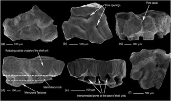
Description. The external surface morphology of the eggshells is undulating, with moderately developed, coarse circular nodes (Fig. 2a, b, f). These nodes have a diameter ranging between 145 and 500 µm and coalesce to form linear, coarse ridges. The surface morphology is rough, and depressed areas are present between the ridges. Tubular pores with circular openings extending to the internal surface and measuring from 0.13 mm to 0.20 mm in diameter are observed on the external surface of the eggshell at the base of coarse and raised nodes or ridges (Fig. 2a, b, f). Elongated, cone-shaped pore canals are also observed in the radial section (Figs 2c–e, 3e).
SEM photomicrographs of the eggshell specimens of Fusioolithus baghensis (Khosla & Sahni, Reference Khosla and Sahni1995; Fernández & Khosla, Reference Fernández and Khosla2015) from the intertrappean beds of Piplanarayanwar. (a) External surface of DUGF/164 showing ridged and tuberculated morphology. (b) External surface of DUGF/163 showing undulating tubercles separated by depressed regions in between them and also exhibiting pore canal openings. (c) Radial section of DUGF/163 with broad shell units, pore canals and basal mammillary knobs. (d, e) Radial sections of DUGF/165 exhibiting the inner organic layer (Membrana Testacea), overlying mammillary knobs with interconnected pore canals, and outer shell units. (f) Tuberculated and undulating external surface of DUGF/162 with a pore opening.

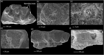
SEM photomicrographs of the eggshell specimens of Fusioolithus baghensis (Khosla & Sahni, Reference Khosla and Sahni1995; Fernández & Khosla, Reference Fernández and Khosla2015) from the intertrappean beds of Piplanarayanwar. (a) Internal surface of DUGF/166 consisting of circular knobs that are connected to each other at some places and are composed of criss-crossing calcite strands. (b, c) Enlarged views of a resorption crater (squared area in (a)) showing calcite crystals radiating from the centre. (d) Enlarged view of one of the basal knobs displaying calcite strands oriented perpendicular to the long axis of the shell units. (e) Radial section of DUGF/165. The shell units are wider than tall at places separated by cone-shaped pore canals and show acicular lines radiating from an organic core and well-developed basal knobs underlain by a layer of Membrana Testacea. (f) Enlarged view of Membrana Tesatcea (DUGF/165).

The height and width of individual shell units are c. 0.53 and 0.33 mm, respectively, with a height–width ratio of 1.60:1. The shell units are short, broad and fan-shaped, having flared out from both sides of the vertical axis of the nucleation centre. From one-third of their height, the lateral margins of shell units still flare out but to a lesser extent. This makes them appear parallel at this level. The concentric growth lines are not observed in SEM photographs. Numerous radial lines representing acicular calcite crystallites commence from the calcium reserve body at the base of the shell units. A horizontal network of pore canals is seen at the base of shell units (Figs 2d, e, 3e). They seem to have formed at the intersection of mammillary knobs at the base of shell units.
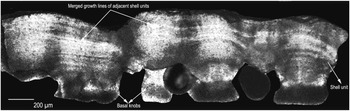
The radial thin-section of the eggshells clearly shows shell units flaring upwards and fused with the adjacent shell units, resulting in arched and continuous growth lines (Fig. 4). The growth lines show concavity between adjacent shell units. The basal end knobs are also characteristically present (Fig. 4).
Thin-section of Fusioolithus baghensis eggshell (DUGF/162) showing broad, short and partially fused shell units with well-developed basal knobs. The growth lines of adjacent shell units merge with each other, forming concave surfaces where they merge with their counterparts on the adjacent shell unit.

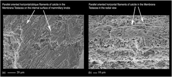
In the radial section of the eggshells, a thin (c. 245 µm) fibrous layer of compact calcite is present on the inner surface of the eggshell units (Figs 2d, e, 3e, f). SEM study of this part in DUGF/165 demonstrated the presence of disorganized, filament-like structures that measure 240–280 µm in length (Fig. 5a, b). We interpret this layer as the mineralized Membrana Testacea (MT). The MT proximal to the shell units is more compact than the one distal to them or occurring on the inner surface of the mammillary knobs. The change of MT’s texture from more compact to somewhat aerated form has also been observed in the eggshells from egg-level 3 of Auca Mahuevo (Grellet-Tinner et al. Reference Grellet-Tinner, Chiappe and Coria2004) which recalls the condition observed in modern non-avian reptiles and birds (Packard & DeMarco, Reference Packard, DeMarco, Deeming and Ferguson1995). Horizontally disposed parallel to oblique filamentous structures of calcite with specks of amorphous calcium carbonate on the surface (discernible at higher magnification) (Fig. 5b) and oriented perpendicular to the long axis of the shell units are present throughout the thickness of the MT layer (Figs. 3d–f, 5a, b). The individual strands are 4 to 5 µm in diameter.
(a) Enlarged view of basal knobs showing parallel or obliquely oriented calcite filaments on the internal surface (DUGF/166). (b) Enlarged view of parallel calcite filaments in the MT layer oriented perpendicular to the long axis of the shell units (DUGF/165). Specks of amorphous calcium carbonate can be seen on the surface of these filaments at higher magnification.

The MT is succeeded externally by widely spaced cylindrical knobs representing the mammillary or basal knobs. The mammillary knobs, spherical or ridged in cross-section, have a diameter ranging between 180 and 370 µm and a height of c. 120 µm (Fig. 3a). The inner surface of the basal knobs is covered by long and criss-crossing or braided strands of calcite (Fig. 3d). Some of these knobs merge with each other (Fig. 3a). A few of them also exhibit central pits with radiating calcite crystals which may possibly represent the resorption craters (Fig. 3a–c). The distance between these knobs is variable. Above the basal knobs lie the individual fan-shaped, broad and short shell units which are set apart from each other by interstitial space or partially fused (Figs 2c–e, 3e).
Remarks: Fernández and Khosla (Reference Fernández and Khosla2015) erected a new oofamily Fusioolithidae in which the eggshells are characterized by dinosauroid–spherulitic basic type, tubospherulitic morphotype, tubocanaliculate pore system, compactituberculate ornamentation, growth lines crossing the boundary between shell units starting at the inner one-third of the eggshell thickness and sometimes continuing to the external surface, and fan-shaped eggshell units partially fused without clearly demarcated boundary lines. Many of the oospecies earlier described under the genus Megaloolithus were transferred to the new genus Fusioolithus (Fernández and Khosla, Reference Fernández and Khosla2015). These authors synonymized Megaloolithus baghensis (Khosla & Sahni, Reference Khosla and Sahni1995) and Megaloolithus balasinorensis (Mohabey, Reference Mohabey1998) from India, Megaloolithus pseudomamillare from France (Vianey-Liaud et al. Reference Vianey-Liaud, Hirsch, Sahni and Sigé1997), Patagoolithus salitralensis from Argentina (Simón, Reference Simón2006) and Megaloolithus cf. baghensis (Sellés et al. Reference Sellés, Bravo, Delclòs, Colombo, Martí, Ortega-Blanco, Parellada and Galobart2013) from Spain with Fusioolithus baghensis (Khosla & Sahni, Reference Khosla and Sahni1995), the type species for the genus Fusioolithus.
Eggshells from egg-level 3 of the Upper Cretaceous Auca Mahuevo site, Argentina, were initially referred to the oofamily Megaloolithidae (Chiappe et al. Reference Chiappe, Coria, Dingus, Jackson, Chinsamy and Fox1998). Grellet-Tinner et al. (Reference Grellet-Tinner, Chiappe and Coria2004) referred them to Megaloolithus patagonicus, an oospecies previously described from the Upper Cretaceous rocks of Nequén Basin, Argentina, by Calvo et al. (Reference Calvo, Engelland, Heredia and Salgado1997). However, Fernández et al. (Reference Fernández, Vila, Moreno-Azanza, Otero, Carballido and Diego2022) observed that the Auca Mahuevo eggshells are indistinguishable from those of Patagoolithus salitralensis (Simón, Reference Simón2006) known from the Salitral Moreno area in Argentina. The latter is quite distinct from M. patagonicus in eggshell microstructure and ultrastructure. Fernández et al. (Reference Fernández, Vila, Moreno-Azanza, Otero, Carballido and Diego2022) assigned Auca Mahuevo eggshells from egg-level 3 along with P. salitralensis to F. baghensis.
Fusioolithus includes two oospecies F. baghensis and F. berthei (Khosla & Sahni, Reference Khosla and Sahni1995). The latter differs from the former in having thicker eggshells (2.4–2.9 mm) and greater height–width ratio (3.27:1). The microstructural and ultrastructural features of Piplanarayanwar eggshells clearly indicate that these eggshell fragments belong to the dinosauroid–spherulithic type with tubocanaliculate pore system. The radial thin-section of the eggshells shows fan-shaped shell units flaring upwards and partially fused with the adjacent shell units, resulting in arched and continuous growth lines (Fig. 4). The growth lines showing concavity in between two shell units and basal end knobs are also characteristically present. Such fused shell units, merged growth lines and swollen basal-end caps strongly support the assignment of Piplanarayanwar eggshells to the oospecies Fusioolithus baghensis.
In general morphology of having fibrous bundles perpendicular to the long axis of the shell units and on comparison with the earlier reports of MT in dinosaur eggshells (Kohring, Reference Kohring1999; Grellet-Tinner et al. Reference Grellet-Tinner, Chiappe and Coria2004; Sellés et al. Reference Sellés, Bravo, Delclòs, Colombo, Martí, Ortega-Blanco, Parellada and Galobart2013; Bravo & Gaete, Reference Bravo and Gaete2015), the calcitic layer at the base of shell units is identified as an MT layer. However, these eggshell fragments are clearly different from other Megaloolithus and Fusioolithus oospecies of Khosla and Sahni (Reference Khosla and Sahni1995), Mohabey (Reference Mohabey1998) and Fernández and Khosla (Reference Fernández and Khosla2015) in being thin and lacking more pronounced cylindrical shell units and highly nodose external surface ornamentation. Though the wide shell units resemble those of the eggshell of turtles, the presence of basal knobs and the horizontal network of pore canals identify them as sauropod eggshells (Grellet-Tinner et al. Reference Grellet-Tinner, Chiappe and Coria2004; Dhiman et al. Reference Dhiman, Prasad and Goswami2019).
5. Elemental composition of the eggshell layers using Energy-Dispersive X-Ray Spectroscopy
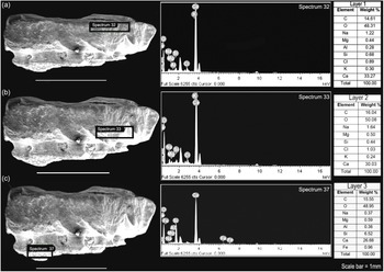
Elemental analysis by Energy-Dispersive Spectroscopy (EDS) was performed at the top and base of the eggshell units and below mammillary knobs where the Membrana Testacea is preserved. The EDS analysis showed that the mineralogical composition of the top and bottom levels of the eggshell units are essentially the same, consisting of carbon (C), calcium (Ca), oxygen (O) and silicon (Si) as the main components, in addition to small quantities of sodium (Na), magnesium (Mg), potassium (K) and chlorine (Cl) (Fig. 6a, b) which are characteristic of fossil amniote eggshells. EDS elemental analysis of the MT showed closely similar elemental values except for some minor variations (Fig. 6c). Although the more common elements such as Ca, O and C occur in more or less similar proportions throughout the preserved eggshell, as compared to the eggshell proper the MT layer has slightly reduced amounts of Ca and Na and slightly increased levels of Si. Additionally, it is observed that aluminium (Al) and iron (Fe) are also present, but Cl and K are absent in this layer. The slightly increased levels of Si and Al might have been introduced by clay minerals from the siliciclastic rocks in which the eggs were buried. Decreased Ca content, increased Si content and the introduction of Fe indicated that limited diagenetic replacement of calcium carbonate has taken place in the MT layer during the process of fossilization. Increased Si values in the MT layer may also point to the fact that organic matter may have formed a template for early silicification (Folk & Lynch, Reference Folk and Lynch2001) as was observed in the MT layer of Auca Mahuevo dinosaur eggs from Argentina (Grellet-Tinner, Reference Grellet-Tinner2005).
Elemental profiles of Fusioolithus baghensis eggshell (DUGF/165) from the intertrappean beds of Piplanarayanwar in the Energy Dispersive X-ray Spectroscopy (EDS) spectra. EDS spectra from the top (a) and base (b) of the eggshell units and the Membrana Testacea (c).

6. Discussion
6.a. Membrana Testacea
Though Rothe and Klemmer (Reference Rothe and Klemmer1991) reported the presence of MT in diagenetically altered chelonian eggshells from the Pliocene of the Canary Islands, based on the occurrence of the same material in the pore canals, on the edge of the shell fragments and in the supposed MT, Kohring (Reference Kohring1999) interpreted the porous structure below the shell units as diagenetically altered sediment attached to the shell. Similarly, Kohring (Reference Kohring1999) suggested that the putative MT identified on the inner surface of dinosaur eggshell from the Upper Cretaceous of Canada by Tazaki et al. (Reference Tazaki, Aratani, Noda, Currie and Fyfe1994) might be hyphae of fungi and, hence, a modern contamination. Later, Grellet-Tinner (Reference Grellet-Tinner2005) documented the presence of MT on the inner surface of the calcareous eggshells of megaloolithid dinosaurs recovered from the Campanian Anacleto Formation, Auca Mahuevo, Patagonia, Argentina. The eggshells described here are isolated in occurrence and no clutches were found. Hence their identification is essentially based on comparison to other known and undisputed titanosaurid eggs having embryonic bones such as Auca Mahuevo, Argentina (Grellet-Tinner et al. Reference Grellet-Tinner, Chiappe and Coria2004). Gross morphological features and comparison with known MT layers in previously reported dinosaur eggshells (Kohring, Reference Kohring1999; Grellet-Tinner, Reference Grellet-Tinner2005) favour the identification of the calcareous layer on the inner surface of dinosaur eggshells from the intertrappean beds of Piplanarayanwar, Chhindwara District, Madhya Pradesh, as the MT. The MT layer of Piplanarayanwar eggshells exhibits a mesh-like texture, and the ultrastructure exhibits fine tendrils and fibres of calcitic material that replicate the soft membrane, similar to those of previous reports (Kohring, Reference Kohring1999; Grellet-Tinner, Reference Grellet-Tinner2005). In the presence of external nodes and nodes coalescing into ridges, a similar pore diameter and horizontal pore canal network at the base of inner knobs of shell units, partially fused eggshell units, and the merging of growth lines of adjacent shell units starting from the inner one-third of the shell thickness, the Piplanarayanwar eggshells are somewhat comparable to the eggshells of the Auca Mahuevo site (Grellet-Tinner et al. Reference Grellet-Tinner, Chiappe and Coria2004; Grellet-Tinner, Reference Grellet-Tinner2005). However, the eggshell dimensions of Piplanarayanwar and Auca Mahuevo do not match, as the eggshells of the Indian site have imperfectly preserved outer shell surface. The thickness of the MT layer is c. 245 µm in the Indian eggshells, which is intermediate between the values reported for the MT layer of the dinosaur eggshells from egg-level 3 of Auca Mahuevo, Argentina (281 µm; Grellet-Tinner et al. Reference Grellet-Tinner, Chiappe and Coria2004), and Tremp Basin, Spain (300 µm; Sellés et al. Reference Sellés, Bravo, Delclòs, Colombo, Martí, Ortega-Blanco, Parellada and Galobart2013). In height–width ratio, the Piplanarayanwar eggshells (1.60:1) are closer to those of Tremp Basin (1.88:1) than to those of Auca Mahuevo (>2.0:1). On the other hand, the diameters of pore openings of Piplanarayanwar eggshells (0.13 to 0.20 mm) are within the range of Auca Mahuevo eggshell pore openings (0.15 to 0.29 mm).
6.b. Conditions of preservation
Preservation of micron-sized fibres of MT is not common in the fossil record and such rare preservations are possibly the result of some favourable conditions for preservation such as anoxic depositional environments and the metabolic activities of associated microbes (Bottjer et al. Reference Bottjer, Etter, Hagadorn, Tang, Bottjer, Etter, Hagadorn and Tang2002). Based on the study of Cretaceous sauropod dinosaur eggshells from Spain, Eocene crocodilian eggshells from Germany, Miocene avian eggshells from the Czech Republic and Germany, and modern bird eggshells, Kohring (Reference Kohring1999) concluded that the preservation of MT in these eggshells was due to the aerobic decay of the internal tissue prior to burial that allowed the formation of calcium-rich gel within the MT. An analysis of modern avian eggs showed resistance of MT to early stage of diagenesis and formation of gel composed of calcium and sulphur in between the organic fibrils of the MT (Kohring, Reference Kohring1999). However, later studies have shown that aerobic decay prior to burial is not necessary for bacterial mediation of the calcium carbonate precipitation onto the MT strands (Reid et al. Reference Reid, Visscher, Decho, Stolz, Bebout, Dupraz, Macyntyre, Paerl, Pinckney, Prufert-Bebout, Steppe and Des Marais2000; Visscher et al. Reference Visscher, Reid and Bebout2000). Subaerial exposure would result in the desiccation and removal of the MT from the eggshell (Hayward et al. Reference Hayward, Zelenitsky, Smith, Zaft and Clayburn2000). Soft tissue like the MT can be preserved as fossil only when it undergoes rapid replication by mineral precipitation, such as calcium carbonate (Allison, Reference Allison1988). As soft tissues get destroyed by their own enzymes such as those released by lysosomes under aerobic conditions, their preservation necessitates an anaerobic environment in which the bacteria replicate the original soft tissue structure in exquisite cellular detail before its destruction and then stimulate biomineralization of the replica (Raff et al. Reference Raff, Schollaerta, Nelsona, Donoghue, Ceri-Wyn, Turnera, Stein, Dong, Bengtsone, Huldtgren, Stampanoni, Chongyui and Raff2008). Grellet-Tinner (Reference Grellet-Tinner2005) suggested that lack of energy and no exposure to subaerial conditions are more important for the preservation of MT. As observed in the lithification of microbial mats such as stromatolites (Reid et al. Reference Reid, Visscher, Decho, Stolz, Bebout, Dupraz, Macyntyre, Paerl, Pinckney, Prufert-Bebout, Steppe and Des Marais2000; Visscher et al., Reference Visscher, Reid and Bebout2000), in an anaerobic depositional environment, sulphate- or sulphur-reducing bacteria would transform the inorganic and organic compounds of the eggshells into an energy source that leads to alkaline conditions congenial for the precipitation of calcium carbonate (Grellet-Tinner, Reference Grellet-Tinner2005).
The MT-bearing eggshells of the present study have been retrieved from the marlstone facies of the intertrappean unit that was deposited in a coastal-plain palaeoenvironment (Lourembam et al. Reference Lourembam, Prasad and Grover2017). The Piplanarayanwar intertrappean biota is represented predominantly by freshwater and terrestrial elements such as Lepisosteus, Osteoglossidae gen. et sp. indet., Siluriformes indet., indeterminate anurans, aquatic snake Indophis sahnii, crocodiles (Crocodilia indet.), sauropod dinosaur eggshells, coprolites, freshwater ostracods and charophytes, as compared to limited marine/brackish water elements like Igdabatis indicus, ?Rhombodus sp., Pycnodontidae gen. et sp. indet. and Enchodus sp. Composition-wise, the biota of these intertrappean beds constitute 40 % actinopterygians, 8 % chondrichthyans, 4 % amphibians, 16 % reptiles, 12 % ostracods and 20 % charophytes. The predominance of freshwater elements over brackish-water/marine forms suggests that the freshwater forms occupied a permanent body such as a lake or pond close to the sea coast intermittently connected with a river system and the marine/brackish water taxa were introduced into the lake when they moved upstream of the river (Sahni, Reference Sahni and Maheshwari1983; Díez-Canseco et al. Reference Díez-Canseco, Arz, Benito, Díaz-Molina and Arenillas2014; Verma et al. Reference Verma, Khosla, Kaur and Prashanth2016; Lourembam et al. Reference Lourembam, Prasad and Grover2017; Prasad et al. Reference Prasad, Farooqui, Murthy, Sarate and Bajpai2018). Fossils from these intertrappean beds are very well preserved, with intact tooth enamel, and do not show any evidence of long-distance transport with respect to other intertrappean beds of the Deccan Volcanic Province such as those of Naskal and Asifabad in south-central India, which were either subjected to surficial etching or long-distance transport. In the absence of well-preserved clutches in the studied intertrappean beds we can only speculate on the possible preservation medium that facilitated permineralization of the MT layer. Preservation of the MT in a permineralized condition at the base of the mammillary knobs of the dinosaur eggshells suggests that the palaeoenvironment was conducive for the replacement of the organic layer with calcium carbonate before its decomposition. Stagnant brackish water body might have inhibited the decomposition or large-scale diagenetic recrystallization. These eggshells might have been derived from eggs deposited close to the banks of a lacustrine system or transported into the water body from a nearby terrestrial clutch site soon after hatching and before the decomposition of organic material. The presence of well-preserved pore canals without sediment or mineral infilling, as evident from the circular pore openings at the base of external nodes of the shell (Fig. 2b, f) and from radial sections (Fig. 2c–e), point to the sufficiently porous nature of the eggshell that permitted the exchange of gases and vapour with the external environment. In the case of Piplanarayanwar eggshells, shielding of eggshells from the aerobic conditions through rapid burial by fine sediments possibly occurred soon after hatching of the eggs. As in the case of the titanosaurid eggs of Auca Mahuevo, Argentina (Grellet-Tinner, Reference Grellet-Tinner2005), and Haţeg Basin, Romania (Grigorescu & Oana-Claudia, Reference Grigorescu and Oana-Claudia2017–18), we believe that the preservation of the MT layer in the dinosaur eggshells of the Piplanarayanwar intertrappean beds was facilitated by bacterially mediated precipitation of calcium carbonate. Whether the permineralization or replacement of the MT by calcium carbonate took place before the segregation of eggshells from the clutch or after their burial by fine sediment is difficult to interpret in our current state of knowledge. However, neither the eggshells nor the associated fossils provide any evidence for long-distance transport to the depositional milieu.
7. Summary
Here we document for the first time the preservation of fossil Membrana Testacea, the organic layer that underlies the mammillary knobs of eggshells, from the Mesozoic rocks of India. The eggshells with preserved MT come from a marlstone facies of the Upper Cretaceous intertrappean beds of Piplanarayanwar in central India, and their microstructure places them within the titanosaurid oospecies Fusioolithus baghensis. The MT layer occurring on the internal surface of the eggshells is characterized by parallel strands of calcium carbonate oriented perpendicular to the long axis of the eggshell units. The preservation of soft organic layer such as MT in fossil state is rarely known and mostly restricted to fossil sites of exceptional preservation. Circumstantial evidence from associated fossils, the fine marly sediments in which the eggshells were preserved, and the state of preservation of pore canals and the MT layer all point to rapid burial in a humid environment under anaerobic conditions and calcium carbonate mineralization of the MT layer by bacterial mediation as was interpreted for eggshells of the Auca Mahuevo (Argentina) (Grellet-Tinner, Reference Grellet-Tinner2005) and Haţeg Basin (Romania) (Grigorescu & Oana-Claudia, Reference Grigorescu and Oana-Claudia2017–18) sites. The fossils of the Piplanarayanwar intertrappean beds represented by a higher number of freshwater than brackish/marine taxa point to the presence of lacustrine bodies in a coastal setting with intermittent connections to the sea, the banks of which were the possible clutch sites of dinosaurs. The stagnant brackish water bodies where the eggshells were deposited could have prevented early decomposition of the soft MT.
Acknowledgements
The authors are grateful to anonymous reviewers for their insightful comments and suggestions on the original manuscript which led to significant improvement in the presentation of data. LRS acknowledges the University of Delhi for University Fellowship. Field and laboratory components of this work were supported by funds from the research grant of the JC Bose National Fellowship to GVRP.
Conflict of interest
None.