Introduction
Bulimia nervosa (BN) is characterized in part by recurrent binge eating and compensatory behaviors (e.g. self-induced vomiting, fasting) intended to prevent weight gain (American Psychiatric Association, 2013). Binge eating is a complex behavior, but the subjective sense of ‘loss of control’ (LOC), or a ‘feeling that one cannot stop eating or control what or how much one is eating’ (American Psychiatric Association, 2013) has been shown repeatedly to be its most salient aspect (e.g. Mond, Latner, Hay, Owen, & Rodgers, Reference Mond, Latner, Hay, Owen and Rodgers2010). Independent of the amount consumed in eating episodes, LOC is associated with distress and psychosocial impairment across eating disorder diagnoses (Goldschmidt, Reference Goldschmidt2017). The eleventh edition of the International Classification of Diseases for Mortality and Morbidity Statistics (ICD) even defines binge eating in BN by LOC, not by episode size (World Health Organization, 2018), but very little is known about the pathophysiology of this experience. Although objectively large binge-eating episodes often decrease with treatment, smaller LOC eating episodes tend to persist (Hildebrandt & Latner, Reference Hildebrandt and Latner2006; Niego, Pratt, & Agras, Reference Niego, Pratt and Agras1998). A significant impediment to meeting the need for more effective interventions for BN (Linardon & Wade, Reference Linardon and Wade2018) is limited understanding of the neurocognitive processes that may contribute to and maintain key symptoms like LOC eating.
Some neuroimaging findings suggest that altered functioning of the cortico-striato-thalamo-cortical circuit involved in self-regulatory control may contribute to LOC eating. In healthy individuals, the lateral and medial prefrontal cortices, lateral orbitofrontal cortex, and anterior cingulate cortex are involved in the effortful control of behavior, thoughts, emotions, and cravings (Hampshire, Chamberlain, Monti, Duncan, & Owen, Reference Hampshire, Chamberlain, Monti, Duncan and Owen2010; Kober et al., Reference Kober, Mende-Siedlecki, Kross, Weber, Mischel, Hart and Ochsner2010; Meyer & Bucci, Reference Meyer and Bucci2016). In teens and adults with BN, youth at risk for BN, and adults with binge-eating disorder (BED), functional magnetic resonance imaging (fMRI) data indicate hypoactivation of these regions, particularly medial and lateral prefrontal cortices, during response inhibition on the Simon Spatial Incompatibility Task, Stroop Task, and Stop Signal Task (Balodis et al., Reference Balodis, Molina, Kober, Worhunsky, White, Sinha and Potenza2013; Bartholdy et al., Reference Bartholdy, O'Daly, Campbell, Banaschewski, Barker, Bokde and Stedman2019; Cyr, Yang, Horga, & Marsh, Reference Cyr, Yang, Horga and Marsh2018; Marsh et al., Reference Marsh, Steinglass, Gerber, O'Leary, Walsh and Peterson2009, Reference Marsh, Horga, Wang, Wang, Klahr, Berner and Peterson2011). However, deficient prefrontal activation during attempts to inhibit button pressing is also well-documented in other disorders of behavioral dysregulation (e.g. substance use disorders and attention-deficit hyperactivity disorder (ADHD); Morein-Zamir et al., Reference Morein-Zamir, Dodds, van Hartevelt, Schwarzkopf, Sahakian, Muller and Robbins2014; Morein-Zamir & Robbins, Reference Morein-Zamir and Robbins2015). Pinpointing the neurocognitive alterations that occur specifically during attempts to control eating could ultimately improve our understanding and treatment of LOC and eating-disorder-specific symptoms.
To date, this understanding has been constrained by limitations in tasks and technology. FMRI studies have examined brain response during the delivery of palatable solutions to participants' mouths (Bohon & Stice, Reference Bohon and Stice2011; Frank, Reynolds, Shott, & O'Reilly, Reference Frank, Reynolds, Shott and O'Reilly2011; Frank et al., Reference Frank, Wagner, Achenbach, McConaha, Skovira, Aizenstein and Kaye2006; Van den Eynde & Treasure, Reference Van den Eynde and Treasure2009), or during free v. restricted access to such solutions (Goldschmidt et al., Reference Goldschmidt, Dickstein, MacNamara, Phan, O'Brien, Le Grange and Keedy2018), but these taste tasks have not yet explicitly required participants to attempt to engage control. Classic inhibitory control paradigms have been adapted to include food picture stimuli (for review, see Berner, Winter, Matheson, Benson, & Lowe, Reference Berner, Winter, Matheson, Benson and Lowe2017); however, because these paradigms measure control over button-pressing in response to food images, the neural processes that may contribute to a lack of control over eating behavior in BN remain unidentified. In addition, because fMRI requires a supine position and is very sensitive to motion, fMRI tasks cannot require participants to repeatedly and rapidly initiate and stop naturalistic eating responses.
To begin to address these gaps in the literature, we combined a portable imaging technology with a new go/no-go task designed to assess the ability to control eating behavior. On no-go trials of this task, in lieu of inhibiting button pressing, participants are instructed to inhibit a pre-potent tendency to sip and swallow a palatable yogurt shake. While women with BN and group-matched healthy controls (HC) completed this novel task and a standard go/no-go task, functional near-infrared spectroscopy (fNIRS) measured prefrontal hemodynamic responses analogous to the blood oxygen level-dependent signal measured in fMRI (Moriguchi et al., Reference Moriguchi, Noda, Nakayashiki, Takata, Setoyama, Kawasaki and Hanakawa2017). FNIRS is an optical imaging technology with poorer spatial resolution than fMRI, and its penetration depth is limited to the cortex. However, fNIRS has several advantages: it is non-invasive, silent, low-cost, and it provides superior temporal resolution than fMRI and superior spatial resolution than electroencephalograpy. Since fNIRS is wearable, participants can sit upright and naturally during imaging, and motion from behavior like eating does not create problematic artifacts (Pinti et al., Reference Pinti, Tachtsidis, Hamilton, Hirsch, Aichelburg, Gilbert and Burgess2020). Further, fNIRS has been used across a wide range of psychiatric populations to document altered neurocognitive task-related activation (Ehlis, Schneider, Dresler, & Fallgatter, Reference Ehlis, Schneider, Dresler and Fallgatter2014), and cortical activation detected by fNIRS has been cross-validated with fMRI, specifically during go/no-go tasks (Cui, Bray, Bryant, Glover, & Reiss, Reference Cui, Bray, Bryant, Glover and Reiss2011).
We used these novel methods to test the hypotheses that relative to HC, women with BN would show eating-specific inhibitory control deficits, as indexed by commission errors, and prefrontal hypoactivation, particularly in the lateral prefrontal cortex (PFC). In addition, we predicted that hypoactivation would be most pronounced in women with severe symptoms. We conducted parallel analyses using a standard, button-pressing go/no-go task for comparison.
Materials and methods
Participants
Participants were right-handed females, aged 18–45, between 85% and 120% of ideal body weight (Metropolitan Life Insurance Company, 1959). The BN group included women who met Diagnostic and Statistical Manual of Mental Disorders, Fifth Edition (DSM-5) criteria for BN (American Psychiatric Association, 2013) for at least 6 months. To reduce variance in our BN sample, all participants with BN endorsed self-induced vomiting as a compensatory behavior (although others could also be endorsed). Women with lifetime ADHD and any current DSM-IV-TR Axis I disorder [apart from BN, major depressive disorder (MDD), or generalized anxiety disorder (GAD) in the BN group] were excluded. HC with any current or past eating disorder symptoms or taking any psychoactive medications, and women with BN regularly taking any psychoactive medications other than selective serotonin reuptake inhibitors (SSRIs) were excluded. Participants who rated liking of the shake < 6 on a 9-point Likert-type scale were excluded to ensure that all participants perceived the shake as palatable (see online Supplementary material for full list of exclusion criteria).
Procedure
Participation included phone screening and one in-person visit. The Drexel University Institutional Review Board approved the protocol. All participants provided written informed consent.
Participants were instructed to consume a standardized 300-kcal breakfast (1 English muffin, 1½ pats of butter, 250 g apple juice; Broft et al., Reference Broft, Shingleton, Kaufman, Liu, Kumar, Slifstein and Walsh2012; Zimmerli, Devlin, Kissileff, & Walsh, Reference Zimmerli, Devlin, Kissileff and Walsh2010) 4 h before the scheduled start of the eating task and to refrain from eating or drinking (except water) in this interim. Compliance with these instructions was verbally confirmed for all participants, height and weight were measured, and a taste test of the eating-task shake was completed. Participants next completed self-report measures and semi-structured interviews administered by bachelor's-level or higher study staff. Diagnostic items of the Eating Disorder Examination (EDE; Fairburn, Cooper, & O'Connor, Reference Fairburn, Cooper, O'Connor and Fairburn2008) established BN diagnosis. The Mini-International Neuropsychiatric Interview (M.I.N.I.; Sheehan et al., Reference Sheehan, Lecrubier, Sheehan, Amorim, Janavs, Weiller and Dunbar1998), was administered to assess DSM-IV-TR Axis I diagnoses. The two-subtest Wechsler Abbreviated Scale of Intelligence (WASI-II; Wechsler, Reference Wechsler2011) was used to estimate general intellectual functioning (FSIQ).
Finally, participants completed both go/no-go tasks with concurrent fNIRS. To avoid the potentially confounding influences of fullness, satiety, or arousal from food-stimulus exposure during the eating task on standard-task performance or activation, all participants completed the standard go/no-go task first.
Neuroimaging tasks
Go/no-go tasks
General inhibitory control was measured using a block-design go/no-go task adapted from Rodrigo et al. (Reference Rodrigo, Di Domenico, Ayaz, Gulrajani, Lam and Ruocco2014). Participants were instructed to press a button with their right index finger in response to go stimuli (non-X letters) but withhold this response to no-go stimuli (X; 14.3% of trials across the entire task). Stimuli were presented in a pseudorandom order (jittered interstimulus interval (ISI): 2–4 s). Four blocks included only go stimuli (‘GO’ blocks) and four blocks included intermixed go and no-go stimuli (‘NO-GO’ blocks; per block: 28 trials, 8 no-gos; Fig. 1a).
Schematic representation of block design and within-block trial sequences on the (a) standard go/no-go task and (b) eating go/no-go task. After every two blocks, a crosshair centered on a black screen was displayed for 15 s to allow the hemodynamic response to return to baseline.

Inhibitory control over eating responses was measured using a go/no-go task with properties identical to the standard task except for the orders of blocks, stimuli, and ISIs, to control for potential practice effects (Fig. 1b). Strawberry yogurt shakeFootnote †Footnote 1 (1500 g) in a clear container with an opaque lid and a vertically fixed straw was placed on a table in front of participants. The shake has been used in several prior laboratory studies of binge eating (0.7 kcal/g; Kissileff, Walsh, Kral, & Cassidy, Reference Kissileff, Walsh, Kral and Cassidy1986; Kissileff, Zimmerli, Torres, Devlin, & Walsh, Reference Kissileff, Zimmerli, Torres, Devlin and Walsh2008; LaChaussee, Kissileff, Walsh, & Hadigan, Reference LaChaussee, Kissileff, Walsh and Hadigan1992; Schebendach, Broft, Foltin, & Walsh, Reference Schebendach, Broft, Foltin and Walsh2013; Zimmerli et al., Reference Zimmerli, Devlin, Kissileff and Walsh2010). An instructional video showed participants how to sip and swallow the shake once in response to non-X letters, and to withhold this response to Xs. Participants could not see how much they were consuming during the task. The eating task was videotaped, and video analysis software developed for this study coded responses by timestamping upward movement of liquid in the straw that passed a data capture window placed at the top of the container. See online Supplementary material for additional task details and design considerations.
Self-report measures
The Eating Loss of Control Scale (ELOCS; Blomquist et al., Reference Blomquist, Roberto, Barnes, White, Masheb and Grilo2014) assessed LOC severity, or the subjective degree to which one's eating felt out of control over the past 4 weeks, using continuous, 10-point Likert-type scale ratings. Internal consistency of the Severity subscale was excellent (α = 0.98). LOC eating frequency in the past 28 days was measured by the sum of all LOC eating episodes, regardless of episode size, as assessed by the EDE.
Before and after the eating task, participants rated hunger, fullness, desire to binge eat, and desire to purge using the generalized Labeled Magnitude Scale (gLMS; Zimmerli et al., Reference Zimmerli, Devlin, Kissileff and Walsh2010). After the task, to assess for ecological validity, participants with BN rated the degree to which they felt they binge ate during the task, and all participants rated the degree to which they felt they overate during the task.
Neuroimaging procedures and processing
The 16-channel fNIR Imager Model 1000® (fNIR Devices, LLC; Potomac, MD) forehead sensor was positioned using anatomical landmarks (Ayaz et al., Reference Ayaz, Shewokis, Curtin, Izzetoglu, Izzetoglu and Onaral2011) over bilateral rostral and lateral PFC (online Supplementary Fig. S1; Okamoto et al., Reference Okamoto, Dan, Sakamoto, Takeo, Shimizu, Kohno and Dan2004). Raw fNIRS data were low-pass filtered, scanned for artifacts, and inspected for potential saturation (Ayaz, Izzetoglu, Shewokis, & Onaral, Reference Ayaz, Izzetoglu, Shewokis and Onaral2010; Izzetoglu, Chitrapu, Bunce, & Onaral, Reference Izzetoglu, Chitrapu, Bunce and Onaral2010). Mean changes in oxygenated hemoglobin concentration relative to local baselines were calculated within GO and NO-GO blocks from artifact-removed raw intensity measurements using the modified Beer–Lambert law (see online Supplementary material).
Statistical analysis
As in prior fNIRS research (e.g. Ruocco et al., Reference Ruocco, Rodrigo, McMain, Page-Gould, Ayaz and Links2016), linear mixed-effects models (LMEs) conducted in R tested behavioral and neural hypotheses (see online Supplementary material for model fitting details). LMEs tested the main effect of group on commission errors during NO-GO blocks of the eating task (sips in response to X's) and standard task (button presses in response to X's). For both tasks, Group × Condition interactions examined group differences in NO-GO v. GO activation. To explore whether altered activation would be most pronounced in women with the most severe symptoms, we replicated a median-split approach used in the only prior neuroimaging study that has assessed inhibitory control in response to food and non-food specific stimuli within the same BN sample (Skunde et al., Reference Skunde, Walther, Simon, Wu, Bendszus, Herzog and Friederich2016). We identified high-symptom subgroups based on median levels of self-reported behavior (LOC eating severity and frequency in the past month; see online Supplementary material) and repeated our main Group × Condition LMEs to compare HC to these BN subgroups (n = 12 for each subgroup). In addition, to explore the association between in-laboratory eating experience and neural activation during the eating task, we compared HC to a BN subgroup with the strongest feeling of binge eating during the task (n = 12). The false discovery rate (FDR; Benjamini & Hochberg, Reference Benjamini and Hochberg1995), controlled for family wise error across all 16 channels.
To facilitate comparison of our results to those of prior fMRI studies that have examined associations of control-related activation with continuous measures of BN symptom severity (e.g. Marsh et al., Reference Marsh, Steinglass, Gerber, O'Leary, Walsh and Peterson2009), exploratory analyses in the full BN sample assessed the significance of Condition × ELOCS Severity score and Condition × LOC eating frequency interactions (both tasks), as well as Condition × Feeling of Binge Eating rating interactions (eating task only). Group × Commission Error interactions explored group differences in NO-GO activation as a function of task performance. Exploratory sensitivity analyses examined the potential confounding effects of comorbid MDD, GAD, psychotropic medication, past anorexia nervosa (AN), and low past BMI (see online Supplementary material). Alpha for exploratory analyses was set at 0.05, uncorrected.
Results
Participants
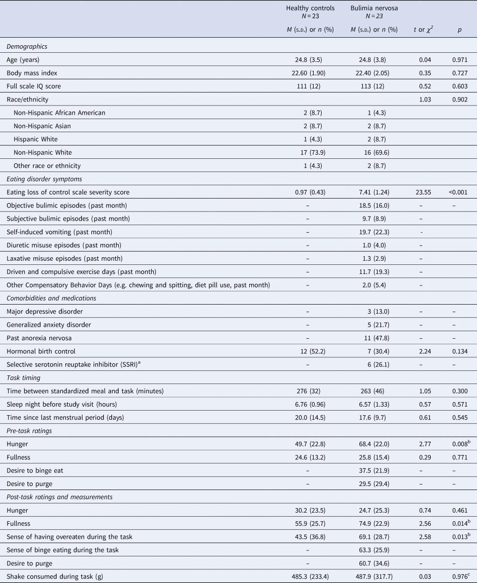
Forty-eight women completed study procedures. Two participants were excluded from all analyses due to task instruction noncompliance. Eating-task data for one BN participant and one HC were excluded due to technical difficulties. Final groups included in analyses (n = 23 per group) did not differ on age (range: 18–33), body mass index (BMI), FSIQ, hormonal birth control use, or state-related variables (Table 1).
Sample characteristics and state comparisons

a In the bulimia nervosa group, six women were taking SSRI medication [fluoxetine (n = 4), sertraline (n = 1), paroxetine (n = 1)]. In addition, three women with bulimia nervosa reported taking pro re nata (PRN) benzodiazepines [lorazepam (n = 1), clonazepam (n = 2)] in the last month but abstained from taking these medications in the week prior to scanning.
b Because groups did not differ on objective measures of intake (content of last meal, time since last meal, or amount of shake consumed during the task), we did not pursue additional analyses examining pre-task group differences in hunger ratings or post-task group differences in ratings of fullness or the sense of having overeaten.
c Mann–Whitney U Test p = 0.670.
Task performance
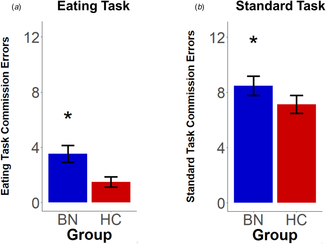
Compared with HC, women with BN made more commission errors on the eating (b = 0.69, s.e. = 0.28, t = 2.48, p = 0.013, R 2β = 0.069) and standard (b = 0.71, s.e. = 0.28, t = 2.49, p = 0.013, R 2β = 0.068) tasks (Fig. 2; see online Supplementary material for similar results of t tests).
Eating and standard go/no-go task performance in the full sample. The bulimia nervosa (BN) group made more errors of commission than did healthy controls (HC) on both the (a) eating go/no-go task and (b) standard go/no-go task. Error bars represent the standard error of the mean.

Full-sample group differences in prefrontal activation
In the full sample, no fNIRS Group × Condition interactions on either task passed whole-PFC FDR correction for multiple comparisons (online Supplementary Tables S4 and S5 present results at a less stringent threshold). Effect size maps indicate that group differences were larger on the eating than the standard task (online Supplementary Fig. S2).
Subgroup differences in prefrontal activation
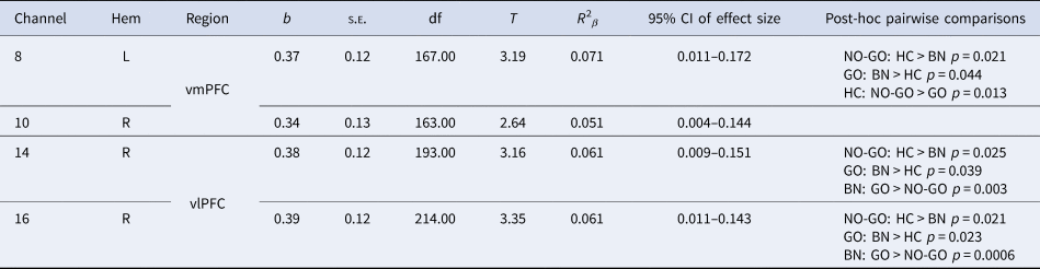
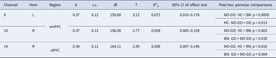
Exploratory analyses indicated that during eating-task inhibition, the BN subgroup defined by high ELOCS severity (n = 12) showed abnormally reduced activation compared with HC in the bilateral ventromedial prefrontal cortex (vmPFC) and right ventrolateral prefrontal cortex (vlPFC; p FDR < 0.05; Table 2, Fig. 3a). Notably, ELOCS severity scores were unrelated to rated feelings of binge eating during the task (p = 0.531), and there was only 59.1% overlap in subgroups defined by these scores and ratings; however, the subgroup defined by a strong sense of binge eating during the task (n = 12) showed similar reductions in medial and lateral prefrontal activation compared with HC (Table 3, Fig. 3b). Analyses comparing HC to the BN subgroup (n = 12) with the highest frequency of LOC eating episodes (moderately correlated with ELOCS Severity, z = 0.03, p = 0.041) revealed eating-task Group × Condition interactions in the same regions, but with smaller effect sizes that did not pass multiple comparisons correction (online Supplementary Table S6). On the standard task, no BN subgroup comparisons with HC passed multiple comparisons correction (online Supplementary Table S7).
Eating go/no-go task group × condition mixed-effects models comparing activation in healthy controls to participants with bulimia nervosa and severe LOC eating

Results are p < 0.05, false discovery rate corrected; Hem, hemisphere; vlPFC, ventrolateral prefrontal cortex; vmPFC, ventromedial prefrontal cortex; CI, confidence interval. The Satterthwaite method was used to estimate degrees of freedom.
Eating go/no-go task group × condition mixed-effects models comparing activation in healthy controls and participants with bulimia nervosa who felt strongly that they binge ate during the eating task

Results are p < 0.05, false discovery rate corrected; Hem, hemisphere; vlPFC, ventrolateral prefrontal cortex; vmPFC, ventromedial prefrontal cortex; CI, confidence interval. The Satterthwaite method was used to estimate degrees of freedom.
Prefrontal activation during the eating go/no-go task in bulimia nervosa subgroups compared to healthy controls. (a) Women with severe loss-of-control eating in the past month (n = 12) showed reduced activation in medial and lateral prefrontal cortex relative to healthy controls. (b) Women with the strongest sense of binge eating in the laboratory during the eating go/no-go task (n = 12) showed similar diminished activation. Maps depict t-statistics from cortical areas showing Group × Condition interactions in linear mixed-effects models (all p FDR < 0.05). Bar plots show levels of activation during GO and NO-GO blocks of the eating task for channels showing the largest interaction effect sizes in medial and lateral prefrontal cortex. Error bars represent the standard error of the mean. Image reconstruction was rendered infnirSoft (Ayaz, Reference Ayaz2010) using methodology described in Ayaz et al. (Reference Ayaz, Izzetoglu, Platek, Bunce, Izzetoglu, Pourrezaei and Onaral2006). vlPFC, ventrolateral prefrontal cortex; vmPFC, ventromedial prefrontal cortex.

Continuous associations with symptoms and performance
Exploratory analyses within the full BN sample indicated that higher ELOCS Severity, more frequent LOC eating episodes, and a stronger sense of binge eating during the eating go/no-go task were all associated with less NO-GO v. GO activation in right vlPFC (online Supplementary Table S8; Supplementary Fig. S3). We did not detect any Condition × ELOCS Severity or Condition × LOC eating frequency effects on the standard task (ps > 0.05, uncorrected).
Commission errors moderated group effects on NO-GO block activation in dorsomedial PFC and right lateral PFC on the eating task, and in most of the PFC on the standard task (online Supplementary Table S9). Poorer performance was associated with less activation of these regions in the BN group, but more activation of the same regions in HC (online Supplementary Fig. S4).
Sensitivity analyses
Results of sensitivity analyses are detailed in the Supplement. Eating-task results were similar or exaggerated after excluding women with comorbid MDD and GAD, those taking SSRIs, or those with past AN, suggesting that these factors likely did not contribute to the findings.
Discussion
Identifying brain alterations that underpin aberrant eating behavior is essential for translational research focused on the cognitive neuroscience of eating disorders (Steinglass & Foerde, Reference Steinglass and Foerde2018). This study used a novel go/no-go task and fNIRS to assess the ability to inhibit eating responses and its underlying neural correlates in women with and without BN. When women with BN who reported the most severely dysregulated eating in the real world attempted to inhibit their eating responses on the task, they insufficiently engaged prefrontal regions involved in goal-directed action control and decision-making. Less activation of these regions was similarly associated with poorer eating-task inhibition in BN. Consistent with prior behavioral results from tasks using neutral or food pictures in BN (Wu, Hartmann, Skunde, Herzog, & Friederich, Reference Wu, Hartmann, Skunde, Herzog and Friederich2013), observed effect sizes for both behavioral and neural alterations in BN were slightly larger for eating-specific inhibition than for button-pressing inhibition. The current findings provide evidence of the potential utility of fNIRS in detecting neural processes associated specifically with dysregulated eating. Study results also add to data suggesting the most pronounced prefrontal dysfunction in the most symptomatic individuals with BN, and support the notion that lateral and medial prefrontal cortices may play a key role in BN symptomatology.
Our behavioral results align with previous neuropsychological task findings (Wu et al., Reference Wu, Hartmann, Skunde, Herzog and Friederich2013) and indicate that women with BN made more commission errors than HC on general and food-specific go/no-go tasks. These data suggest an impaired ability to control both consummatory and more general behavioral responses. However, to our knowledge, our findings are the first to capture objectively quantifiable deficits in inhibitory control over eating behavior in BN.
In addition, participants with BN showed lateral and medial prefrontal cortical dysfunction that was dependent on the severity of dysregulated eating and inhibitory ability. Lateral PFC is involved specifically in the inhibition of unwanted behaviors (Milner, Reference Milner and Goldman-Rakic1995), cognitive regulation of craving (Kober et al., Reference Kober, Mende-Siedlecki, Kross, Weber, Mischel, Hart and Ochsner2010; Siep et al., Reference Siep, Roefs, Roebroeck, Havermans, Bonte and Jansen2012), and satiation (Del Parigi et al., Reference Del Parigi, Chen, Gautier, Salbe, Pratley, Ravussin and Tataranni2002). During eating-task inhibition, compared with HC, BN subgroups with the most severe sense of LOC over their eating in the past month and a stronger sense of binge eating during the eating go/no-go task showed medium-size reductions in right vlPFC response. Exploratory analyses in the full sample confirmed that reduced right vlPFC activation during eating-task inhibition was associated with LOC eating severity and frequency, the experience of binge eating during the task, and eating inhibition errors. These results are in line with consistently observed inverse associations of symptom severity with surface volume and cortical thickness of right vlPFC in adolescents and adults with BN (Berner et al., Reference Berner, Stefan, Lee, Wang, Terranova, Attia and Marsh2018; Cyr et al., Reference Cyr, Kopala-Sibley, Lee, Chen, Stefan, Fontaine and Marsh2017; Marsh et al., Reference Marsh, Stefan, Bansal, Hao, Walsh and Peterson2015; Westwater, Seidlitz, Diederen, Fischer, & Thompson, Reference Westwater, Seidlitz, Diederen, Fischer and Thompson2018). They also align with prior reports of decreased lateral PFC activation in adults with BED across all conditions of a food-picture go/no-go task during fNIRS (Rösch et al., Reference Rösch, Schmidt, Lührs, Ehlis, Hesse and Hilbert2021), and in adults with BN who were instructed to focus on feelings elicited by food v. non-food images during fMRI (Uher et al., Reference Uher, Murphy, Brammer, Dalgleish, Phillips, Ng and Treasure2004). Extending this previous research, our findings support a potential specific link between vlPFC dysfunction and dysregulated eating, both in and out of the laboratory.
Women with BN who had the most dysregulated eating also showed diminished activation compared with HC in the vmPFC. This region modulates decisions to consume food (Hare, Camerer, & Rangel, Reference Hare, Camerer and Rangel2009), and has been implicated in successful go/no-go task inhibition of button-pressing responses to food pictures in healthy adults (He et al., Reference He, Huang, Zhang, Turel, Ma and Bechara2019). In addition, vmPFC lesions are associated with persistent responding for food rewards despite their devaluation after satiation (Reber et al., Reference Reber, Feinstein, O'Doherty, Liljeholm, Adolphs and Tranel2017). As in vlPFC, greater cortical thickness reductions of the vmPFC have been linked to more frequent symptoms over time in adolescents with BN (Cyr et al., Reference Cyr, Kopala-Sibley, Lee, Chen, Stefan, Fontaine and Marsh2017). Thus, our results support theories that both medial and lateral PFC dysfunction contribute to dysregulated eating in BN (Bartholdy et al., Reference Bartholdy, O'Daly, Campbell, Banaschewski, Barker, Bokde and Stedman2019; Cyr et al., Reference Cyr, Kopala-Sibley, Lee, Chen, Stefan, Fontaine and Marsh2017).
In addition to their roles in behavioral control in the context of salient stimuli, lateral and medial PFC are also typically engaged during response inhibition, rapid adjustment of prepotent responses, and decision-making in the context of neutral stimuli (Bechara, Damasio, & Damasio, Reference Bechara, Damasio and Damasio2000; Chambers et al., Reference Chambers, Bellgrove, Stokes, Henderson, Garavan, Robertson and Mattingley2006; Fellows & Farah, Reference Fellows and Farah2007). However, on the standard go/no-go task, we did not detect any group differences in activation or neural associations with symptom severity. Instead, exploratory analyses indicated performance-moderated group differences in standard-task activation in lateral PFC and vmPFC. These performance-related results on the standard go/no-go task are consistent with prior findings that poorer Simon-Task performance is linked to less ventral and dorsolateral PFC activation in womens with BN (Marsh et al., Reference Marsh, Steinglass, Gerber, O'Leary, Walsh and Peterson2009). As such, lower prefrontal activation may contribute to the severity of difficulty engaging control over multiple behaviors in BN.
To date, only one other neuroimaging study has assessed food-specific and general inhibition in the same sample of participants with BN (Skunde et al., Reference Skunde, Walther, Simon, Wu, Bendszus, Herzog and Friederich2016). Contrary to the authors' hypotheses, previous behavioral meta-analytic findings (Wu et al., Reference Wu, Hartmann, Skunde, Herzog and Friederich2013), and our eating-specific findings, hypoactivation in sensorimotor areas on a go/no-go task was detected only when inhibiting responses to neutral, not food, pictures (Skunde et al., Reference Skunde, Walther, Simon, Wu, Bendszus, Herzog and Friederich2016). These discrepant results may be related to this prior study's lack of group differences in behavior, higher frequency of no-go trials, use of food pictures instead of food consumption, or intermixing of neutral and food-picture inhibition blocks.
However, consistent with results from this prior fMRI go/no-go study (Skunde et al., Reference Skunde, Walther, Simon, Wu, Bendszus, Herzog and Friederich2016), we detected corrected group differences only when women with the most severe symptoms were compared with controls. These high-severity BN subgroups may more closely match prior BN samples that showed decreased activation compared with controls during Simon Task response inhibition (Marsh et al., Reference Marsh, Steinglass, Gerber, O'Leary, Walsh and Peterson2009, Reference Marsh, Horga, Wang, Wang, Klahr, Berner and Peterson2011), but results from these women may not generalize to individuals with less severe forms of BN. Additional neuroimaging research using both food- and non-food specific inhibitory control task variants is needed. Nevertheless, extant findings (Marsh et al., Reference Marsh, Steinglass, Gerber, O'Leary, Walsh and Peterson2009, Reference Marsh, Horga, Wang, Wang, Klahr, Berner and Peterson2011; Skunde et al., Reference Skunde, Walther, Simon, Wu, Bendszus, Herzog and Friederich2016) and our results consistently support an association of control-related neural alterations with more severely dysregulated eating in BN.
The current study has several strengths. Unlike prior studies of BN that used the Simon Task, which measures primarily conflict and error-monitoring (Marsh et al., Reference Marsh, Steinglass, Gerber, O'Leary, Walsh and Peterson2009, Reference Marsh, Horga, Wang, Wang, Klahr, Berner and Peterson2011), this study combined neuroimaging with go/no-go tasks. These tasks require individuals to withhold behavior, which may be relevant to the inability to refrain from binge eating or purging. Our participants completed a novel eating task as well as a standard button-pressing task, which served as a measure of general response inhibition and a useful benchmark for the interpretation of our eating-task findings. Moreover, our use of an eating-specific, not just food-picture-specific, adaptation of a go/no-go task with a portable functional neuroimaging technology permitted objective, quantifiable measurement of attempts to control consummatory responses and their neural underpinnings.
Nevertheless, results should be interpreted in light of limitations that highlight directions for future research. Our samples were relatively small, participants were all adult females, and the BN group all self-induced vomiting, potentially limiting the generalizability of our results. The standard task was administered first to avoid potential carry-over arousal from the eating task, but this may have produced order effects. Although groups did not differ on the self-reported timing or content of their last meal before scanning, future studies should also include objective measures of pre-study metabolic state (e.g. blood-glucose measurement). We consistently detected altered activation of medial and lateral aspects of the PFC in women with more severely dysregulated eating; however, given the centimeter-level spatial resolution of fNIRS, and because we did not use digitization to co-register fNIRS data to an anatomical scan, we cannot draw inferences about precise locations of dysfunction within medial and lateral PFC. Given the regions covered by the fNIRS sensor used, we could not measure activation in more dorsal aspects of the PFC, which also play a role in behavioral control (Wriessnegger et al., Reference Wriessnegger, Bauernfeind, Schweitzer, Kober, Neuper and Mueller-Putz2012). In addition, because of sipping-response complexity, we could not conduct trial-by-trial, event-related analysis of correct responses.
Sample heterogeneity and fNIRS limitations may have impacted our ability to detect group differences during standard-task inhibition. For example, sensitivity analyses excluding women with past AN revealed reduced bilateral vlPFC activation in the BN-only subgroup compared with HC on the standard task. Although our sample was representative of the broader BN population in its inclusion of women with a history of AN (Bardone-Cone et al., Reference Bardone-Cone, Maldonado, Crosby, Mitchell, Wonderlich, Joiner and Le Grange2008), these exploratory findings suggest that this history may be linked to enhanced PFC function during non-food specific inhibitory control. Future work in larger samples is needed to better understand this result and to investigate whether our findings generalize to other eating disorder symptom trajectories and diagnoses. In addition, since the vlPFC and vmPFC are also involved in reward-related processes (Chudasama & Robbins, Reference Chudasama and Robbins2006), stronger effects on the eating task could be driven by a combination of control and reward alterations in BN. FNIRS does not permit assessment of subcortical, reward-related regions (e.g. ventral striatum), but studies integrating fNIRS, fMRI, less palatable foods, and other novel tasks that involve intermittent or earned access to food (Bodell & Keel, Reference Bodell and Keel2015; Goldschmidt et al., Reference Goldschmidt, Dickstein, MacNamara, Phan, O'Brien, Le Grange and Keedy2018; Racine, Horvath, Brassard, & Benning, Reference Racine, Horvath, Brassard and Benning2019) could help distinguish contributions of reward and control processes and circuits to the observed effects.
Our initial results support further investigation of treatment strategies that could enhance food-specific inhibition and medial and lateral PFC function in individuals with LOC eating (e.g. Manasse et al., Reference Manasse, Lampe, Gillikin, Payne‐Reichert, Zhang, Juarascio and Forman2020 Chami et al., Reference Chami, Cardi, Lawrence, MacDonald, Rowlands, Hodsoll and Treasure2020; Dunlop, Woodside, & Downar, Reference Dunlop, Woodside and Downar2016; Kekic et al., Reference Kekic, McClelland, Bartholdy, Boysen, Musiat, Dalton and Schmidt2017; Van den Eynde et al., Reference Van den Eynde, Claudino, Mogg, Horrell, Stahl, Ribeiro and Schmidt2010; Walpoth et al., Reference Walpoth, Hoertnagl, Mangweth-Matzek, Kemmler, Hinterhölzl, Conca and Hausmann2008). However, longitudinal imaging data from adolescents with BN indicate that reduced thickness of vmPFC and vlPFC persists despite symptom remission, suggesting that structural alterations in the areas implicated in the current study may be trait markers of BN symptoms (Cyr et al., Reference Cyr, Kopala-Sibley, Lee, Chen, Stefan, Fontaine and Marsh2017). Longitudinal research is needed to determine whether altered functioning of vmPFC and vlPFC during eating-specific inhibitory control in women with severe BN symptoms is trait- or state-like. Moreover, replication of our methods in a larger sample of individuals with severe BN is warranted. Ultimately, as fNIRS-measured activation robustly predicts outcome in patients with addiction (Huhn et al., Reference Huhn, Sweeney, Brooner, Kidorf, Tompkins, Ayaz and Dunn2019), our methods may be useful for predicting treatment response and relapse in patients with bulimic symptoms.
Conclusion
Assessing neural activation during attempts to inhibit food consumption represents an important first step toward elucidating the neurocognitive underpinnings of eating-disorder-specific behavior. Although prior studies suggest alterations in the function and structure of vmPFC and vlPFC in adults and adolescents with BN or BED and children with LOC eating (Balodis et al., Reference Balodis, Molina, Kober, Worhunsky, White, Sinha and Potenza2013; Berner et al., Reference Berner, Stefan, Lee, Wang, Terranova, Attia and Marsh2018; Cyr et al., Reference Cyr, Kopala-Sibley, Lee, Chen, Stefan, Fontaine and Marsh2017, Reference Cyr, Yang, Horga and Marsh2018; Goldschmidt et al., Reference Goldschmidt, Dickstein, MacNamara, Phan, O'Brien, Le Grange and Keedy2018; Marsh et al., Reference Marsh, Steinglass, Gerber, O'Leary, Walsh and Peterson2009, Reference Marsh, Horga, Wang, Wang, Klahr, Berner and Peterson2011, Reference Marsh, Stefan, Bansal, Hao, Walsh and Peterson2015; Rösch et al., Reference Rösch, Schmidt, Lührs, Ehlis, Hesse and Hilbert2021), our data directly implicate these regions in the severe dyscontrol of eating in adult BN. In addition, the current findings indicate that eating-specific neurocognitive tasks and portable neuroimaging may be useful tools for identifying the neural processes that contribute to the experience of LOC that characterizes binge eating.
Supplementary material
The supplementary material for this article can be found at https://doi.org/10.1017/S0033291722000198
Acknowledgements
Cardinal™ generously donated the straws for the eating go/no-go task, and Andrew Caspary developed and programmed the response coding software used for the eating go/no-go task. We would like to thank: Allison Tipton and Jennifer Gilbert for their assistance with recruitment and data collection and entry; Katya Rubia, Anthony Ruocco, and Achala Rodrigo for task design consultation; Shahrokh Golshan and Danielle Arigo for additional statistical consultation; and Jason Lavender, Takisha Corbett, and Zoe Mestre for their feedback on early versions of the manuscript. We referenced this study's methodological approach in a review paper (Berner et al., Reference Berner, Winter, Matheson, Benson and Lowe2017). Some of the results were presented at annual meetings of the Society for the Study of Ingestive Behavior and the Eating Disorders Research Society.
Author contributions
LAB conceived of and designed the study, developed the eating and standard go/no-go tasks, collected the data, conducted analyses, and led manuscript preparation. SRW assisted with data collection, analysis, and manuscript preparation. HA and MI provided consultation on task optimization and results interpretation. HA pre-processed the fNIRS data and assisted with the generation of neuroimaging figures. PAS provided extensive statistical consultation. RM provided consultation on study design, participant recruitment, and results interpretation. JN provided access to her lab space and equipment. AJM assisted with recruitment and data collection. MRL assisted with recruitment and provided consultation on results interpretation. All authors contributed to manuscript revision and approved the final version.
Financial support
This study and the preparation of this manuscript were supported by a National Institute of Mental Health fellowship (F31MH097406), an American Psychological Association Dissertation Research Award, and an Academy for Eating Disorders Student Research Grant awarded to LAB. The content is solely theresponsibility of the authors and does not necessarily represent the official views of the NationalInstitutes of Health.
Conflict of interest
HA and MI helped to develop the imaging technology used in the present research and were thus offered a minor share in its manufacturer (fNIRS Devices, LLC). MRL is a paid research consultant to the Renfrew Center for Eating Disorders. All other others declare no potential conflict of interest.
Ethical standards
The authors assert that all procedures contributing to this work comply with the ethical standards of the relevant national and institutional committees on human experimentation and with the Helsinki Declaration of 1975, as revised in 2008.