Introduction
Arising in the fourth or fifth gestational weeks, teratomas are congenital tumours derived from all three germ cell layers that typically occur in the midline.Reference Wolter, Siegele and Cunningham1 While these tumours are rarely malignant, cervical teratomas can be large enough to cause airway obstruction, polyhydramnios, pulmonary hypoplasia and other perinatal complications.Reference Laje, Johnson, Howell, Bebbington, Hedrick and Flake2 Obstruction of the upper airway in the neonate was frequently fatal prior to the development of ex utero intrapartum treatment (‘EXIT’ procedure) in the 1990s.Reference Mychaliska, Bealer, Graf, Rosen, Adzick and Harrison3
The ex utero intrapartum treatment procedure was initially developed to deliver fetuses that had undergone in utero tracheal clipping, in order to induce prenatal lung growth in the setting of a congenital diaphragmatic hernia. The procedure preserves uteroplacental blood flow and maternal–fetal gas exchange, thereby providing time for clip removal and intubation.Reference Mychaliska, Bealer, Graf, Rosen, Adzick and Harrison3 This method was adapted to fetuses with large obstructive cervical masses, allowing time for intubation via direct laryngoscopy and bronchoscopy or tracheostomy.Reference Liechty, Crombleholme, Flake, Morgan, Kurth and Hubbard4 However, in the setting of some cardiothoracic anomalies, ex utero intrapartum treatment to airway alone is not a viable solution. In these situations, reports of ex utero intrapartum treatment to extracorporeal membrane oxygenation (‘ECMO’) have displayed success in controlling the airway and cardiopulmonary circulation, allowing for definitive airway management and subsequent treatment.Reference Michel, Rosenberg and Polley5–Reference Matte, Connor, Toutenel, Gottlieb and Fynn-Thompson9
Extremely large obstructive neck masses not only compress the airway, making intubation difficult, but also block access to and distort the trachea, making timely tracheostomy very difficult. In addition, the carina may be pulled superiorly into the thoracic inlet, which compresses the lungs and can result in pulmonary hypoplasia.Reference Marwan and Crombleholme10,Reference Liechty, Hedrick, Hubbard, Johnson, Wilson and Ruchelli11 We present the case of a large obstructive cervical teratoma, wherein ex utero intrapartum treatment to airway alone would not have yielded an acceptable outcome. In order to circumvent the obstacles of this case, the ex utero intrapartum treatment to extracorporeal membrane oxygenation procedure was utilised, to enable establishment of the airway followed by definitive management of the teratoma.
Case report
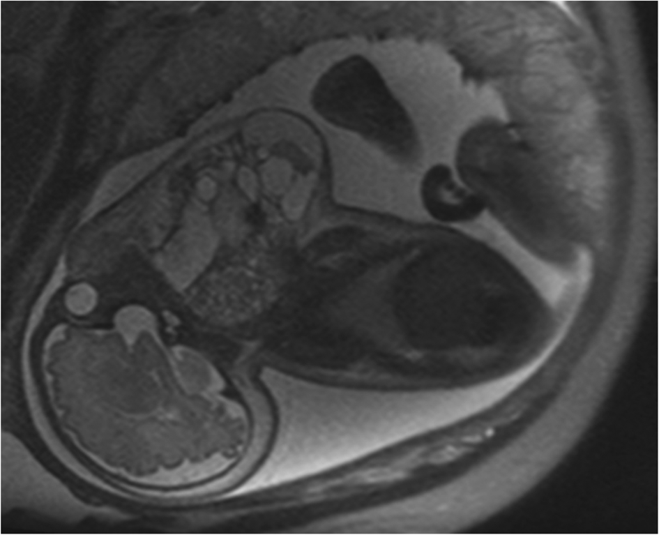
A 34-year-old, gravida 2 (2 pregnancies) para 1001 (1 full-term delivery, 0 pre-term, 0 abortions and 1 total living children) female presented to the paediatric otolaryngology clinic with polyhydramnios and fetal magnetic resonance imaging (MRI) findings concerning for a massive complex midline neck mass consistent with a teratoma (Figure 1).
A T2-weighted fetal magnetic resonance imaging scan conducted at 34 weeks and 5 days gestational age demonstrates polyhydramnios and a large midline complex mass, measuring 14 × 13 × 10 cm, consistent with giant cervicofacial teratoma.

Several factors suggested that ex utero intrapartum treatment to airway alone would not be sufficient for neonatal survival. These included the size of the mass, fetal MRI showing the mass immediately adjacent to the bony vertebrae, with no visualisation of the larynx or trachea, and concern for pulmonary hypoplasia.
Clinical suspicion was high that neither intubation via direct laryngoscopy nor timely tracheostomy would be feasible. At the very least, a prolonged ex utero intrapartum treatment procedure would be required, escalating the risk to both the mother and fetus. Furthermore, if significant pulmonary hypoplasia was present, there may not have been sufficient ventilation to sustain life even if the airway was secured. Extracorporeal membrane oxygenation was determined to be a possible solution.
After consultation with the paediatric cardiothoracic team, the decision was made to perform an ex utero intrapartum treatment procedure, with the possibility of ex utero intrapartum treatment to extracorporeal membrane oxygenation if an airway and/or adequate ventilation could not be obtained.
A prenatal ultrasound conducted several days prior to birth revealed the fetus to be in a vertex presentation. At 38 weeks’ gestation, the baby was delivered via caesarean section; however, over the course of several days, there had been conversion to a breech position, requiring the fetus to be completely delivered from the uterus. As a result, prolonged uteroplacental circulation was not possible, even though the placenta and cord were kept intact.
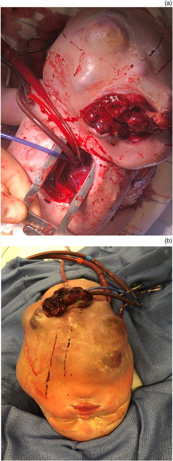
Immediately upon delivery, an attempt was made to establish a secure airway via rigid laryngoscopy and bronchoscopy. The compressive nature of the neck mass resulted in a lack of identifiable landmarks, prohibiting endotracheal intubation. Safe and expedient tracheostomy was not feasible, as uteroplacental circulation could only be maintained for 5 minutes. Once the umbilical pulse weakened, the decision was made to perform a median sternotomy to place the infant on central extracorporeal membrane oxygenation via the right atrium and aorta, as cervical access was not an option (Figure 2a).
(a) View of the median sternotomy and extracorporeal membrane oxygenation cannulae. The venous cannula is in the right atrium and the arterial cannula is in the aorta. Also pictured is an iatrogenic laceration to the inferior portion of the cervicofacial mass that occurred as a result of delivery. Cephalad is oriented towards the top right. (b) Infant on extracorporeal membrane oxygenation after transfer to the adjoining operating theatre. Cephalad is oriented towards the bottom.

The process of cannulating the child for extracorporeal membrane oxygenation took 8 minutes and the pulse oximetry readings did not drop below 70 per cent. The umbilical cord was divided, and the infant was brought to an adjoining operating theatre (Figure 2b).
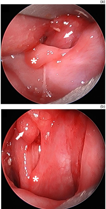
With improved patient positioning and the additional time afforded by extracorporeal membrane oxygenation, rigid laryngoscopy and bronchoscopy did demonstrate a compressed but intact larynx (Figure 3). The trachea and mainstem bronchi were also present, but were completely compressed and malacic. Endotracheal intubation was performed with a 3.5 mm uncuffed tube; however, there was no detectable end-tidal carbon dioxide (CO2) and ventilation was not adequate to sustain the neonate without extracorporeal membrane oxygenation. The child was then admitted to the neonatal intensive care unit on extracorporeal membrane oxygenation.
(a) Laryngoscopy performed during ex utero intrapartum treatment with no visualisation of the glottis; the uvula (asterisk) and epiglottis (arrow) are highlighted. (b) Laryngoscopy after extracorporeal membrane oxygenation displaying the glottis; the right arytenoid (asterisk) and epiglottis (arrow) are highlighted.

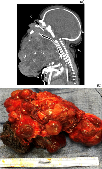
A computed tomography scan was obtained, to guide surgical planning (Figure 4a).
(a) Contrasted sagittal computed tomography scan on day of life 1, showing a complex midline cervicofacial mass measuring 15 × 14 × 11.3 cm. (b) Cervicofacial mass following resection, measuring 16.2 × 10.5 × 9.1 cm, with a weight of 585 g. Pathology revealed a grade 3 immature teratoma.

The infant developed consumptive coagulopathy as a result of the size and expansion of the mass on day of life 1. This necessitated urgent resection. The resected mass was determined to be a grade 3 immature teratoma, measuring 16.2 × 10.5 × 9.1 cm, with a weight of 585 g (Figure 4b).
The bilateral facial nerves, vagus nerves and hypoglossal nerves were preserved and functioning well post-operatively. There was a chylothorax that resolved with medium chain triglyceride tube feeding. Once the teratoma was removed, ventilatory pressure requirements decreased and end-tidal CO2 was detected. The child was subsequently weaned from extracorporeal membrane oxygenation on day of life 5, and the sternum was closed on day of life 7. A tracheostomy was performed after one week, and the child was discharged off the ventilator after five weeks. He is now 11 months of age, neurologically intact, and is tolerating oral feeding and a Passy-Muir valve, with plans for upcoming decannulation.
Discussion
Ex utero intrapartum treatment has dramatically minimised the fatality associated with large congenital neck masses.Reference Laje, Johnson, Howell, Bebbington, Hedrick and Flake2 The addition of extracorporeal membrane oxygenation to the treatment algorithm for neonates that cannot be ventilated or intubated can further reduce morbidity and mortality. The ex utero intrapartum treatment to extracorporeal membrane oxygenation procedure has been described previously in the setting of congenital cardiopulmonary anomalies.Reference Michel, Rosenberg and Polley5–Reference Shieh, Wilson, Sheils, Smithers, Kharasch and Becker8 It has also been described once in the extracorporeal membrane oxygenation literature for management of a cervical teratoma with intrathoracic extension;Reference Matte, Connor, Toutenel, Gottlieb and Fynn-Thompson9 however, likely due to the rarity of these presentations and the complexity of the operations involved, the ex utero intrapartum treatment to extracorporeal membrane oxygenation technique has not yet become widely considered in the setting of large obstructive cervical masses.
In the described case, successful immediate stabilisation of the airway via direct laryngoscopy and bronchoscopy with intubation was not achievable because of the characteristics of the mass and the resulting anatomical changes. In addition, tracheostomy was not possible in the time afforded by the ex utero intrapartum treatment procedure. While prolonged uteroplacental circulation of up to 93 minutes has been reported,Reference Laje, Johnson, Howell, Bebbington, Hedrick and Flake2 and has allowed for partial resection with tracheostomy and ex utero intrapartum treatment to resection in some cases, this was not possible in our case. The unexpected breech position of the fetus necessitated complete delivery from the uterus and shortened the total time of uteroplacental circulation. Performing an expedient ex utero intrapartum treatment to extracorporeal membrane oxygenation procedure saved the neonate's life and allowed time for subsequent intubation via rigid bronchoscopy while circumventing immature lungs, followed by controlled resection of the teratoma.
This case highlights several key considerations for physicians encountering similar clinical scenarios. First, extensive pre-operative planning is required to co-ordinate the succession of complex procedures needed, in order to yield the desired outcome. A multidisciplinary approach is necessary, and should include paediatric otolaryngologists, obstetricians, paediatric cardiothoracic surgeons, maternal and fetal anaesthesiologists, and neonatologists among others. Ideally, pre-operative simulation with all team members present should be performed, as was carried out in this case. Communication is paramount, and if airway access via ex utero intrapartum treatment alone cannot be obtained, extracorporeal membrane oxygenation should be instigated as soon as possible. Second, unexpected obstacles should be anticipated and incorporated into the delivery plan. The fetus’ breech position in this case significantly shortened the duration of uteroplacental circulation, making the decision to proceed to extracorporeal membrane oxygenation unavoidable. Third, consideration should always be given to the lung development of the fetus and the potential for inadequate ventilation, even if the airway is successfully secured. Extracorporeal membrane oxygenation bypasses the lungs and enables the neonate to survive until there is adequate ventilation capable of sustaining life.
The ex utero intrapartum treatment procedure does not come without significant risk, represented by increased susceptibility to maternal haemorrhage and haemodynamic instability due to the need for prolonged uterine relaxation.Reference Marwan and Crombleholme10 The addition of central extracorporeal membrane oxygenation elevates neonatal risk in the form of anticoagulation and morbidity associated with a sternotomy incision and extracorporeal membrane oxygenation support. In addition, future surgical procedures are complicated by increased bleeding associated with anticoagulation. The ex utero intrapartum treatment to extracorporeal membrane oxygenation procedure should be performed only as a lifesaving technique when all preceding interventions have failed. However, when employed in the correct circumstances, ex utero intrapartum treatment to extracorporeal membrane oxygenation provides the airway team with valuable time to secure the airway, bypass underdeveloped lungs and treat airway obstruction under a controlled setting.
• Cervical teratomas can cause neonatal airway obstruction, pulmonary hypoplasia and other complications
• Ex utero intrapartum treatment provides time to secure a difficult neonatal airway by maintaining a supply of oxygenated blood to the newborn
• However, this treatment to airway alone is not always sufficient to sustain life
• Extracorporeal membrane oxygenation has enabled survival of neonates with disease processes previously incompatible with life
• This paper describes a unique case of cervical teratoma managed by ex utero intrapartum treatment to extracorporeal membrane oxygenation
• The treatment resulted in neonatal survival, despite large mass size, inability to obtain a traditional airway and pulmonary hypoplasia
Conclusion
Ex utero intrapartum treatment procedures have allowed the securement of difficult neonatal airways, while maintaining a supply of oxygenated blood to the newborn. However, optimal outcomes are not always obtainable utilising ex utero intrapartum treatment alone. The arrival of extracorporeal membrane oxygenation has enabled the survival of neonates with disease processes previously incompatible with life. We present a unique case, and the first reported in the otolaryngology literature, where ex utero intrapartum treatment to extracorporeal membrane oxygenation allowed for bronchoscopy and endotracheal intubation followed by the controlled resection of a massively compressive cervical teratoma. The ex utero intrapartum treatment to extracorporeal membrane oxygenation option provides a crucial lifesaving alternative for select patients in the management of large obstructive congenital neck masses.
Competing interests
None declared