Introduction
Neurogenic tumours are common tumours of the nervous system, but they constitute a small fraction of neoplastic head and neck lesions. They include neurofibroma, schwannoma, neuroma, ganglioneuroma, ganglioneuroblastoma and neuroblastoma. Schwannoma is the most common solitary neurogenic tumour, followed by neurofibroma.Reference Wilson, McLaren, McIntyre, von Haacke and Maran1 Neurofibroma is a benign tumour that originates from the proliferation of various peripheral nerve constituents.Reference Ferner and O'Doherty2,Reference Rameh, Husseini, Tawil, Fuleihan and Hadi3 It is a heterogeneous group of disorders that involves the central nervous system and has cutaneous features. The most common locations are the extremities and the head and neck region. The tumours are circumscribed, benign and unencapsulated. Neurofibromatosis is a clinical diagnosis, whereas neurofibroma is a histopathological diagnosis. Plexiform and multiple localised neurofibromas are relatively more common in neurofibromatosis type 1 (NF1).Reference Wilson, McLaren, McIntyre, von Haacke and Maran1
Neurofibroma arising from the external nose is very rare.Reference Batsakis4 The first reports of external nose neurogenic tumour cases were by New and Devine in 1947,Reference New and Devine5 and Das Gupta et al. in 1969.Reference Das Gupta, Brasfield, Strong and Hajdu6 They considered neurofibroma and schwannoma as the same entity. The classical presentation is a slow-growing mass. It should be included as a differential diagnosis for painless asymptomatic masses of the external nose. The treatment of choice for all extracranial neurofibromas is surgery. The approach should keep the prominent location, nerve of origin, extent of excision and cosmesis in mind.
Malignant transformation is seen in 2–16 per cent of cases.Reference Wilson, McLaren, McIntyre, von Haacke and Maran1 Plexiform neurofibroma carries malignant potential, with an 8–13 per cent chance of malignant peripheral nerve sheath tumour development. The signs suggestive of transformation are increased tumour size, pain, hard consistency of the tumour and neurological deficits.Reference Ferner, Huson, Thomas, Moss, Willshaw and Evans7
Genetics
The NF1 gene is located on the long (q) arm of chromosome 17, at band 11.2 (17q11.2), and encodes for neurofibromin 1, which is a cytoplasmic protein. It activates ras-GTPase, which helps in cellular signal transduction; when activated, it leads to activation of other proteins necessary for cell growth and differentiation.Reference Upadhyaya and Cooper8 Biallelic inactivation of the NF1 tumour suppressor gene is the cause of neurofibroma. This is the result of either mutation or deletion of the NF1 gene.Reference Friedman, Pagon, Adam and Ardinger9 It is inherited in an autosomal dominant pattern that needs one altered gene for expression. NF1 has a highly penetrant nature and a high mutation changing rate. Any alteration in NF1 leads to neurocutaneous syndromes, and increases the risk of developing neurofibromas, plexiform neurofibroma or optic nerve gliomas.Reference Friedman, Pagon, Adam and Ardinger9 Mutational changes in NF1 genes can be detected by molecular cytogenetic testing. Genetic or molecular testing uses fluorescent in situ hybridisation, targeted at interphase and metaphase stages of development.10
Mutation of SPRED1 gene causes a mild neurofibromatosis-simulating NF1-like syndrome called Legius syndrome,Reference Spurlock, Bennett, Chuzhanova, Thomas, Jim and Side11 which lacks cutaneous manifestations. This gene is located on the long (q) arm of chromosome 15 (15q13.2). It encodes the spred-1 protein, which helps in regulating the ras and mitogen-activated protein kinase signalling pathway.
Although neurofibroma of the external nose is thought to have excellent survival prospects, it should be diagnosed and managed as soon as possible, to minimise functional and cosmetic compromise. This comprehensive review aimed to analyse clinical features, histological features, treatment options, outcomes and recurrences in neurofibroma patients.
Materials and methods
The PubMed and Embase databases, and Google Scholar search engine, were searched using the following medical subject heading (MeSH) terms: neurofibroma nose, neurogenic tumour and nose, neurofibroma external nose, neurofibroma dorsum of nose, plexiform neurofibroma of nose, diffuse neurofibroma of nose, solitary neurofibroma of nose, and neurofibroma nose and NF1. Inclusion criteria were all papers reporting neurofibroma of the external nose. Exclusion criteria were reports regarding schwannoma of the nose and nasal cavity, and non-English papers.
The pertinent papers were reviewed, along with the references, to ensure all related papers were included. Analysis of demographic features, presenting symptoms, clinical diagnosis methods, management techniques, diagnosis confirmation, treatments offered, recurrences, follow-up duration and revision surgery was performed.
Results
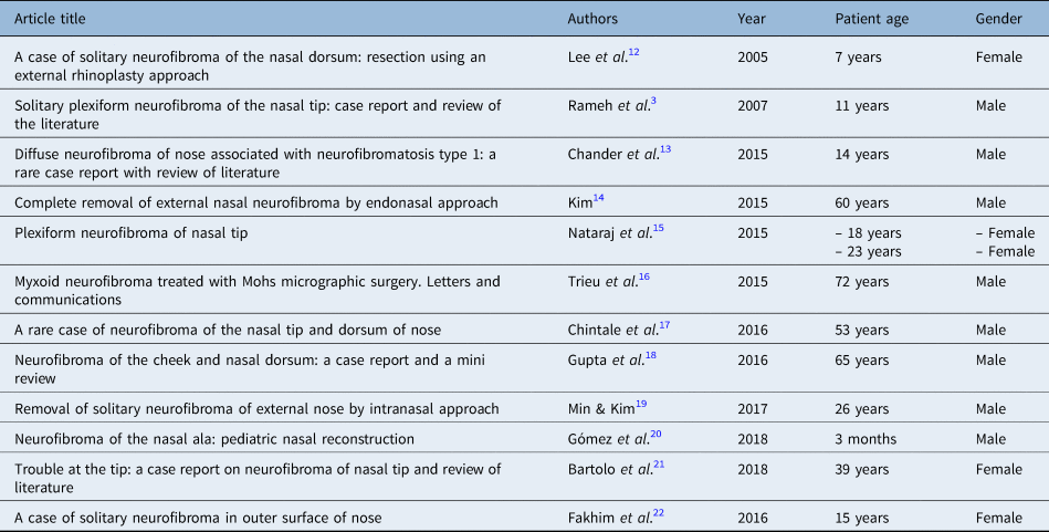
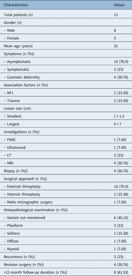
We found 12 articles that fulfilled the criteria. These included 11 case reports, and 1 case series including 2 patients (Table 1).Reference Rameh, Husseini, Tawil, Fuleihan and Hadi3,Reference Lee, Bae and Kim12–Reference Fakhim, Rezaeian, Feizi, Behzad and Piri22 No previous reviews or case series were identified. The mean age of presentation was 31 years (range, 0.25–72 years); 61.5 per cent of patients were male and 38.5 per cent were female (Table 2).
Updated review of neurofibroma of external nose cases

Characteristics of patients affected by neurofibroma of external nose

NF1 = neurofibromatosis type 1; FNAC = fine-needle aspiration cytology; CT = computed tomography; MRI = magnetic resonance imaging
The presentation of neurofibroma of the external nose was most often seen as an asymptomatic nasal mass or a non-specific symptom like nasal obstruction. Cosmesis was the primary concern for patients seeking medical advice.
The imaging techniques most commonly used were computed tomography (CT) and magnetic resonance imaging (MRI). Ultrasound and fine-needle aspiration cytology (FNAC) were performed in two cases, which suggested a superficial ill-defined hyperechoic lesion and a neurogenic tumour, respectively.
The diagnosis was most commonly reached on final histopathology. Association with NF1 was seen in 15.3 per cent of patients. Three patients (23 per cent) developed recurrence following their initial treatment. Table 2 lists all other important parameters identified in the literature review.
Discussion
Peripheral nerve sheath tumours are classified as benign or malignant. Benign peripheral nerve sheath tumours include neurofibroma and schwannoma. Malignant peripheral nerve sheath tumours include neurogenic sarcoma.Reference Batsakis4 Neurofibroma originates from a mixture of Schwann cells, fibroblasts, perineural cells and endoneurium. It can also be classified as a sporadic (solitary) lesion or a constituent of NF1 syndrome.Reference Kannan, Patil and Pattipati23 It can also present as multiple neurofibromas without von Recklinghausen disease.Reference Batsakis4,Reference Abell, Hart and Olson24 Ninety per cent of cases occur sporadically, while the rest are associated with NF1 or NF2.Reference Lassmann, Jurecka, Lassmann, Gebhart, Matras and Watzek25,Reference Skovronsky and Oberholtzer26 Both are caused by deletion of the NF1 gene. In sporadic cases, only involved cells show NF1 mutation, while in syndromic cases, germline mutation of NF1 is present. Neurofibromas show autosomal patterns resulting from spontaneous mutation or transfer of genes.Reference New and Devine5,Reference Das Gupta, Brasfield, Strong and Hajdu6 Among the 13 cases of neurofibroma of the external nose included in this review, 2 (15.3 per cent) had NF1. That makes a complete systemic examination essential, including café au lait spots and skin pigmentation, in cases of asymptomatic dorsal nasal masses.
Peripheral nerve sheath tumour was first described in 1908 by Verocay.Reference Verocay27 In the past, the distinction between neurofibroma and schwannoma was unclear, and these terms were used interchangeably.Reference New and Devine5,Reference Das Gupta, Brasfield, Strong and Hajdu6 Later, it was found that while schwannomas arise from the Schwann cells of the peripheral nerves, neurofibromas demonstrate heterogeneity in terms of origin, and showcase a combined proliferation of various elements of the peripheral nerves.Reference Ferner and O'Doherty2,Reference Berlucchi, Piazza, Blanzuoli, Battaglia and Nicolai28 Literature shows that 25–45 per cent of all neurofibromas are seen in the head and neck, and only 4 per cent are observed in the nasal cavity, mainly affecting the nasal septum.Reference Lee, Bae and Kim12,Reference Perzin, Panyu and Wechter29
Neurofibroma has an equal predilection for males and females, and often presents between the third and sixth decades of life.Reference Verocay27,Reference Gnepp30 Diffuse and plexiform neurofibromas occur mostly in children; plexiform neurofibromas rarely occur after five years of age.Reference Hernández-Martín and Duat-Rodríguez31 In this review, the male-to-female ratio was 8:5, and the age of patients ranged from 3 months to 72 years, with a mean of 31 years. Schwannomas are more common in the nose and paranasal sinuses in comparison to neurofibroma. In a review of 430 000 pathological specimens, there were only 6 cases of neurofibroma of the nose and paranasal sinuses, but none involved the nasal dorsum.Reference Perzin, Panyu and Wechter29
Neurofibroma of the external nose commonly arises from the first and second division of the trigeminal nerves. The olfactory nerve cannot give rise to neurofibroma, as it has no Schwann cells.Reference Batsakis4 In this review, the nerve of origin was mentioned in two cases as being the external nasal branch of the anterior ethmoidal nerve (ophthalmic nerve).Reference Kim14,Reference Min and Kim19 The nerves are embedded in these tumours; they cannot be separated during surgery and the nerves often have to be sacrificed. In contrast to this, schwannomas are truly encapsulated, and they push nerves aside rather than entrapping them.
The duration of symptoms varied from 20 days to several years. The most common clinical presentation was slow-growing, progressive, painless swelling.Reference Das Gupta, Brasfield, Strong and Hajdu6 The symptoms are usually non-specific, and the primary concern was cosmetic. Differential diagnoses to keep in mind are inflammatory lesions, lipomas, schwannomas, leiomyomas, granular cell tumours, adenomas and malignant tumours. Traumatic neuroma has to be ruled out in patients with a history of any trauma. In this series, two patients had a history of trauma.Reference Min and Kim19,Reference Fakhim, Rezaeian, Feizi, Behzad and Piri22
Pre-operative investigations
Before surgery, it is advisable to obtain radiology scans in the form of CT scan or MRI, to rule out any possibility of meningoencephaloceles or gliomas in cases of swelling over the nose.Reference Batsakis4 Radiologically, neurofibroma and schwannoma both show high signal on T2-weighted images; schwannoma shows a more defined capsule, but a definitive diagnosis cannot be reached.Reference Lemmerling, Moerman, Govaere, Praet, Kunnen and Vermeersch32 However, MRI is the investigation of choice, as it can differentiate between all three variants.Reference Hassell, Bancroft, Kransdorf, Peterson, Berquist and Murphey33 Seven cases in this review underwent radiology (30.7 per cent MRI, 23 per cent CT and 7.6 per cent ultrasound). Lee et al. performed CT, MRI and diethylene triamine penta-acetic acid cisternography in their patient to rule out meningocele.Reference Lee, Bae and Kim12 Others opted for MRI,Reference Rameh, Husseini, Tawil, Fuleihan and Hadi3,Reference Nataraj, Jagade, Parelkar, Hanawte, Singhal and Rengaraja15,Reference Fakhim, Rezaeian, Feizi, Behzad and Piri22 ultrasoundReference Chander, Surya Rao, Sekhar, Raja and Sridevi13 and contrast-enhanced CT.Reference Min and Kim19,Reference Fakhim, Rezaeian, Feizi, Behzad and Piri22 Gupta et al. performed FNAC in their report, which was suggestive of benign peripheral nerve sheath tumour.Reference Gupta, Kaur, Rai, Wadhwa and Singh18 Aspiration cytology has no role to play, as it does not differentiate neurofibroma from other non-epithelial tumours.Reference Perzin, Panyu and Wechter29 Ultrasound and CT scan findings can resemble lipoma or haemangioma.Reference Hassell, Bancroft, Kransdorf, Peterson, Berquist and Murphey33
The imaging appearance of neurofibroma at different sites varies. Diffuse neurofibroma grows like an infiltrative or plaque-like lesion, which involves the skin and subcutaneous tissues. There is prominent internal vascularity and marked enhancement. Most radiologists are well versed with the localised type of neurofibroma, and the radiological features are well documented. Because of the lesion's rarity, the characteristic plaques cannot be labelled as pathognomonic. The infiltrative growth pattern is non-specific, and can be seen in cellulitis, haemorrhage, haemangioma and angiolipoma.Reference Hassell, Bancroft, Kransdorf, Peterson, Berquist and Murphey33
On MRI, diffuse neurofibroma is isointense or mildly hyperintense in relation to muscle on T1-weighted images, and mild to markedly hyper-intensive in relation to muscle on T2-weighted images. Contrast-enhanced CT shows intense enhancement.Reference Hassell, Bancroft, Kransdorf, Peterson, Berquist and Murphey33 Four classical statistically significant MRI differences between malignant peripheral nerve sheath tumour and neurofibroma are: an increase in the largest axis diameter of the lesion, the presence of enhancement in the peripheries, an oedema-like zone in the periphery of the lesion, or an intra-tumoural cystic lesion. More than two features are suggestive of malignancy.Reference Wasa, Nishida, Tsukushi, Shido, Sugiura and Nakashima34
The advantages of ultrasound include its wide availability, the lack of exposure to ionising radiations, the shorter time required, its cost-effectiveness and early reporting.Reference Hassell, Bancroft, Kransdorf, Peterson, Berquist and Murphey33 It is limited by an inability to assess the extent and margin of the lesion. Chen et al. described the ultrasound features of neurofibroma in the subcutaneous zone as a hyperechoic mass surrounded by irregular hypoechoic structures.Reference Chen, Jia and Wang35 The deeper, infiltrative portion is diffusely hypoechoic, in contrast to subcutaneous tissue.Reference Hassell, Bancroft, Kransdorf, Peterson, Berquist and Murphey33
In this review, four patients (30.76 per cent) underwent biopsy before definitive surgery,Reference Rameh, Husseini, Tawil, Fuleihan and Hadi3,Reference Nataraj, Jagade, Parelkar, Hanawte, Singhal and Rengaraja15,Reference Chintale, Bhise, Kirdak and Shaikh17 and the results were inconclusive. Biopsy findings may indicate the benign neurogenic nature of the tumour; however, a definitive diagnosis cannot often be reached. Moreover, a biopsy can make the surgery difficult because of the adherence of skin and fibrosis at the biopsy site.
Histologically, a neurofibroma is non-encapsulated and poorly circumscribed, with ill-defined margins.Reference Campora, Radii and Campora36,Reference Glimer-Hill and Kline37 A neurofibroma consists of interlaced bundles of spindle cells with darkly stained nuclei, collagen bundles, and myxoid stroma with occasional mast cells and lymphocytes.Reference Miettinen, Weiss, Damjanov and Linder38 They tend to infiltrate the deeper structure.Reference Pablo and Diego39 They show immunostaining with S-100, neuron-specific enolase and Vimentin.Reference Lee, Bae and Kim12,Reference Perzin, Panyu and Wechter29,Reference Hillstrom, Zarbo and Jacobs40,Reference Macias, Rafael, Fernandes and Rosa41 However, schwannomas are more S-100 sensitive than neurofibroma.Reference Donnelly, Al-Sader and Blayney42 Meissner bodies are characteristic of diffuse neurofibroma.Reference Macias, Rafael, Fernandes and Rosa41 Three histopathological variants are localised, plexiform and diffuse.Reference Megahed43 The most common site for diffuse neurofibroma is the trunk, followed by the head and neck, and then extremities.Reference Ravi, Ram, Lindberg and Pandey44 In this review, variants were not mentioned for 46.15 per cent of patients.Reference Lee, Bae and Kim12–Reference Kim14,Reference Chintale, Bhise, Kirdak and Shaikh17,Reference Gupta, Kaur, Rai, Wadhwa and Singh18,Reference Gómez, Bernal and Martínez20 Three had plexiform,Reference Rameh, Husseini, Tawil, Fuleihan and Hadi3,Reference Nataraj, Jagade, Parelkar, Hanawte, Singhal and Rengaraja15 two had solitary,Reference Min and Kim19,Reference Fakhim, Rezaeian, Feizi, Behzad and Piri22 one had diffuseReference Bartolo, Spiteri and Ayoub21 and one had myxoidReference Trieu, Drosou, Schwartz and Goldberg16 variants. The cause of solitary neurofibroma is unknown, although MarocchioReference Marocchio, Oliveira, Pereira, Soares and Fleury45 and da Rosa et al.Reference da Rosa, Ribeiro, de Menezes, Pinheiro and Alves-Junior46 have reported it as a hamartomatous malformation or caused by trauma.
Surgical approaches and outcomes
The main goals of treatment are complete surgical excision of the tumour, with functional and cosmetic integrity. Close follow up may be necessary, as neurofibromas tend to infiltrate surrounding tissues and show potential for recurrence.Reference Lee, Bae and Kim12,Reference Fisher and Vuzevski47 They are chemoresistant and relatively less radiosensitive than other tumours, although neither chemotherapy or radiotherapy are preferred modalities for treating peripherally located neurofibromas. Neurofibromas carry a very low risk of malignant degeneration, thus precise surgery with cosmesis is a favoured crucial endpoint.
The most accepted surgical approach, considering the aesthetic and oncological aspects, is external rhinoplasty. In this review, 76.9 per cent of patients were treated by the external rhinoplasty approach. The intranasal approach is suggested if disease is unilateral, small and with clear, distinct margins. The advantages of external rhinoplasty are: wide exposure, safe and complete excision, and direct accessibility for any reconstructive flap if required.Reference Rameh, Husseini, Tawil, Fuleihan and Hadi3 However, an intranasal approach through marginal and intercartilaginous incisions is acceptable for small lesions.Reference Kim14,Reference Min and Kim19 Neurofibromas are more likely to recur if they are incompletely excised.Reference Perzin, Panyu and Wechter29,Reference Hirao, Gushiken, Imokawa, Kawai, Inaba and Tsukuda48
In a retrospective review of 39 NF1 patients who had 49 plexiform neurofibromas of the head and neck,Reference Wise, Cryer, Belasco, Jacobs and Elden49 11 tumours were small (i.e. less than 5 cm), and 38 were massive (i.e. more than 5 cm) and involved deep neck sites. Four out of 11 small neurofibromas and 18 out of 35 massive neurofibromas were operated on. There was 1 recurrence in the small neurofibroma group (25 per cent) and 18 recurrences in the massive tumour group (100 per cent), which was a statistically significant difference. The mean time to regrowth was 3.1 years. It was concluded that the size and site of plexiform neurofibroma are the most important predictors of outcome. Indications for surgery included an increase in neurological deficits, airway obstruction and disfigurement.
In another retrospective review of 23 patients who underwent surgery for low-grade malignant peripheral nerve sheath tumour or atypical neurofibroma,Reference Bernthal, Putnam, Jones, Viskochil and Randall50 the need for adjuvant therapy and surgical margins was reported; 78 per cent of cases were found to have microscopically positive margins. Both atypical neurofibroma and low-grade malignant peripheral nerve sheath tumour cases had a disease-specific survival rate of 100 per cent. Local recurrence was seen in 16.7 per cent of low-grade malignant peripheral nerve sheath tumour patients and 9.1 per cent of atypical neurofibroma patients; all cases were found to have microscopically positive surgical margins. The authors concluded that although 78 per cent of patients had positive surgical margins, none of the patients developed metastatic disease. The chances of local recurrence increase with positive margins.
Gupta et al. used Mustarde's local rotation advancement flap for a large neurofibroma of the cheek (4 × 5 cm) and nose (3 × 4 cm).Reference Gupta, Kaur, Rai, Wadhwa and Singh18 Involvement of the nasal tipReference Rameh, Husseini, Tawil, Fuleihan and Hadi3,Reference Nataraj, Jagade, Parelkar, Hanawte, Singhal and Rengaraja15,Reference Chintale, Bhise, Kirdak and Shaikh17 was identified in three patients in this review, for which the treatment remains more challenging because of its prominent position, thick and less flexible skin, more sebaceous glands, and consequent higher chance of recurrence.Reference Nataraj, Jagade, Parelkar, Hanawte, Singhal and Rengaraja15,Reference Hirao, Gushiken, Imokawa, Kawai, Inaba and Tsukuda48,Reference Cook, Rohrer, Cook, Migden, Nguyen and Mellette51–Reference Fattahi54 Bartolo et al. found that even after surgery, the nasal tip may stay bulky and need revision surgery.Reference Bartolo, Spiteri and Ayoub21
Gómez et al.Reference Gómez, Bernal and Martínez20 reported a resection of a left-sided nasal ala neurofibroma, which became complicated by necrosis of the ala, with collapse of the external nasal valve and stenosis of the nostril, which subsequently necessitated nasal reconstruction.Reference Bloom, Antunes and Becker55 This was performed in three stages, and nostril patency was achieved afterward. No recurrence was noted after four years.
Trieu et al.Reference Trieu, Drosou, Schwartz and Goldberg16 described a case in which different nasal subunits were involved and Mohs micrographic surgery was performed. Mohs micrographic surgery is a useful technique to treat difficult cases affecting different nasal subunits, considering the cosmetic and functional challenges. Recurrence was noted in 3 out of 13 patients (23 per cent), 2 of whom had NF1.Reference Chander, Surya Rao, Sekhar, Raja and Sridevi13,Reference Bartolo, Spiteri and Ayoub21 Trieu et al.Reference Trieu, Drosou, Schwartz and Goldberg16 also reported two recurrences in a patient, but this was not associated with NF1. We should keep watch for any recurrence.
Imatinib can decrease plexiform neurofibroma size. Interferon-alpha is a recent treatment modality that can be used as an adjunct in plexiform cases, but results are variable.Reference Staser, Yang and Clapp56,Reference Kebudi, Cakir and Gorgun57
Follow up of these patients is necessary because of the rare chance of malignant transformation of the neurofibroma. This transformation is more likely in neurofibroma compared with schwannoma cases. The likelihood of malignant transformation increases with the association of NF1 (2 per cent).Reference Wilson, McLaren, McIntyre, von Haacke and Maran1,10,Reference Marocchio, Oliveira, Pereira, Soares and Fleury45,Reference Weiss and Goldblum58 In this review, eight patients were followed up for more than 12 months, with no recurrence or malignant transformation. Neurofibroma should be ruled out in any cases of cutaneous swelling with NF1.Reference Chander, Surya Rao, Sekhar, Raja and Sridevi13 It may mimic dermatofibrosarcoma, exuberant granulation tissue or lipomatous neurofibroma. Neurofibroma patients should be followed up once every year.
Limitations
Neurofibroma of the external nose is uncommon. The articles available on this topic come from case reports and case series. The overall quality of the studies is poor. The follow-up duration is also different for all patients, limiting the data on long-term outcomes and recurrence rates. Because of the rarity of the disease entity, conducting a study on a large sample size will be difficult. This review is an effort to provide fundamental knowledge on neurofibroma of the external nose from an objective viewpoint.
Competing interests
None declared