INTRODUCTION
Most crustaceans are gonochoristic animals, and sexual dimorphism appears in characters such as the appendage of gonopore openings (Farmer, Reference Farmer1974; Sagi et al., Reference Sagi, Khlaila, Barki, Hulata and Karplus1996) and relative growth (e.g. Hartnoll, Reference Hartnoll and Wenner1985; Bauer & VanHoy, Reference Bauer and VanHoy1996; Nates & Felder, Reference Nates and Felder1999; Pardo et al., Reference Pardo, Fuentes, Olguín and Orensanz2009). However, specimens bearing both male and female primary or secondary characters are often seen, and these specimens are considered as intersex individuals (Sagi et al., Reference Sagi, Khlaila, Barki, Hulata and Karplus1996; Zou & Fingerman, Reference Zou and Fingerman2000; Rudolph, Reference Rudolph2002). Intersex incidences may be classified into at least two types. In the case of gynandromorph, half of the body has only male characters and the contralateral half consists of only female characters (Farmer, Reference Farmer1972; Johnson & Otto, Reference Johnson and Otto1981; Chace & Moore, Reference Chace and Moore1983; Taylor, Reference Taylor1986; Micheli, Reference Micheli1991). In the other case, intersex individuals are predominantly either male or female, but also possess characters of the opposite sex. For example, the gonopores of decapods open typically on the proximal parts (coxae) of the fifth pereiopods in the male, and coxae of the third pereiopods in the female, whereas intersex individuals have gonopores in both third and fifth pereiopods. Intersexuality of this type has been frequently reported in decapods: crayfish (Sagi et al., Reference Sagi, Khlaila, Barki, Hulata and Karplus1996, Reference Sagi, Manor, Segall, Davis and Khlaila2002; Rudolph et al., Reference Rudolph, Verdi and Tapia2001; Vazquez & López Greco, Reference Vazquez and López Greco2007); mud shrimps (Ngoc-Ho, Reference Ngoc-Ho2001; Pinn et al., Reference Pinn, Atkinson and Rogerson2001); hermit crabs (Turra, Reference Turra2004; Fantucci et al., Reference Fantucci, Biagi and Mantelatto2007); and freshwater crabs (Takahashi et al., Reference Takahashi, Araki, Nomura, Koga and Arizono2000; Ayaki et al., Reference Ayaki, Kawauchino, Nishimura, Ishibashi and Arizono2005). In the crayfish, Cherax quadricarinatus, 2–14% of the populations were intersex individuals having both male and female gonopores and the intersex individuals of this species always functioned as males, although they had ovaries in the pre-vitellogenic stage (Sagi et al., Reference Sagi, Khlaila, Barki, Hulata and Karplus1996, Reference Sagi, Manor, Segall, Davis and Khlaila2002). Similarly, most intersex decapods so far reported function as males even if they have vestigial female characters. Intersex females have also been reported in the crayfish, C. quadricarinatus (Vazquez & López Greco, Reference Vazquez and López Greco2007), the mud shrimp, Upogebia snelliusi (Sakai et al., Reference Sakai, Hirano and Saigusa2004), the ghost shrimp, Callianassa aquabaensis (Dworschak, Reference Dworschak2003) and the other groups of crustaceans (e.g. Ford et al., Reference Ford, Fernandes, Rider, Read, Robinson and Davis2004). On the other hand, Rudolph (Reference Rudolph2002) speculated that intersex individuals in the crayfish, Samastacus spinifrons, are in a transitional stage from male to female since most of them had gonoducts of both sexes irrespective of the pattern of gonopore arrangement.
The mud shrimp, Upogebia major inhabiting the mud tidal flats of Japan has distinct sexual dimorphism in its external structures (Oka, Reference Oka1941). Normal males lack a pair of pleopods on the first abdominal segment, but normal females possess these appendages. A pair of gonopores opens in the coxae of the fifth pereiopods in males, while of the third in females. Sexual dimorphism also appears in the dactylus connected to the propodus. Male dactyli exhibit a number of knobs on the outer surface and three distinguishable ridges on the inner surface, while female dactyli are relatively smooth on both surfaces. On the other hand, males have unique gonads with characteristics of both sexes that consist of ‘testis proper’ (true testis) and ‘ovarian part of testis’ (premature ovary) (Oka, Reference Oka1941; Kinoshita et al., Reference Kinoshita, Nakayama and Furota2003; Kang et al., Reference Kang, Nanri, Lee, Saito, Han, Hatakeyama and Saigusa2008). The major yolk protein precursor, vitellogenin is produced in males as well as females (Kang et al., Reference Kang, Nanri, Lee, Saito, Han, Hatakeyama and Saigusa2008). Upogebia major are neither protandrous nor protogynous hermaphrodites. The presence of intersex individuals distinguishable by the external morphology has not been reported in U. major. However, we have recently recognized that males possessing the first pleopods and females lacking them co-occurred in populations in the Seto-Inland Sea (Saigusa, personal observation).
To investigate if these specimens with morphological disorders are the intersex individuals, U. major was collected at nine sites in the Seto-Inland Sea, Japan and the types of morphological disorders were classified. The frequency of occurrence of the morphological disorders was compared among local populations. Allometric growth and gonad index were analysed to evaluate if these morphological disorders affect their secondary sexual characters. We finally discuss the possible causal factors of these incidences.
MATERIALS AND METHODS
Sampling of animals
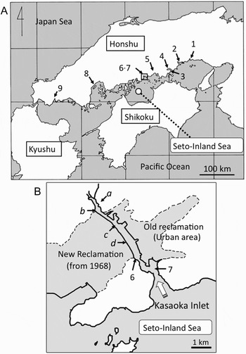
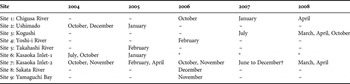
Nine sites (Sites 1–9 in Figure 1A) of tidal flats were selected for sampling of Upogebia major in the Seto-Inland Sea. Collections were made in the middle and lower intertidal zones at each site, where the burrow density was high. The substrate was dug to 50–70 cm deep using a hand-held shovel and the specimens were captured. Small individuals (total length < ~3 cm) were not collected. Months and years of sampling are summarized in Table 1. Specimens collected at Sites 1–8 were placed in seawater, transferred to the laboratory, and stored at –30°C, while specimens collected at Site 9 were fixed with 70% ethanol immediately after collection.
(A) Location of the nine sampling sites of Upogebia major in the Seto-Inland Sea, Japan (Sites 1–9); (B) sampling points in Kasaoka Inlet in detail (a to d, and 6 and 7). Most of the original mud tidal flat of Kasaoka Bay was lost by land reclamation started in 1968 (shown in grey). Anaerobic soft sediment thickly accumulated at Sites a to d, where no individuals were found.

Sampling sites of Upogebia major from 2004 to 2009 in Seto-Inland Sea.

–, no collection; *, population became locally extinct; †, every month from June to December.
Sampling was also made in six sites (Sites a–d and Sites 6 and 7) within Kasaoka Inlet (Figure 1B). Soft sediment accumulated thickly from Sites a–d, where no individuals inhabited. Upogebia major only inhabited Sites 6 and 7 in Kasaoka Inlet.
Inspection of the external morphology
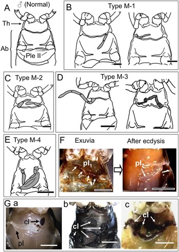
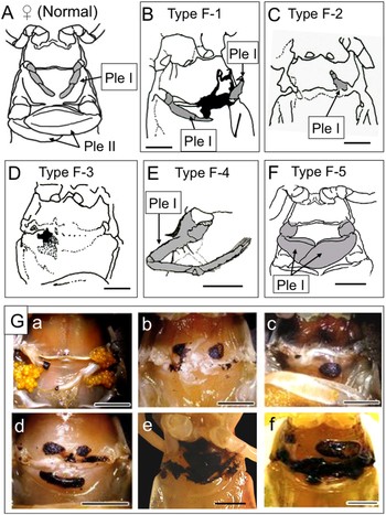
In the laboratory, the number, sex, morphology of the pleopods and cuticular lesions were inspected for in all specimens by the naked eye, or under a stereomicroscope if necessary. The abdomen of upogebiids consists of six segments. Normal males lack appendages (a pair of pleopods) on the first abdominal segment (Figure 2A), but normal females possess a pair of the first pleopods (Figure 3A) that are observed throughout the year regardless of the reproductive season. A pair of biramous leaf-like pleopods occurs on the second to fifth abdominal segments, and their morphology is very similar between males and females. The first pleopods of the female (Figure 3A) are morphologically simplified, but more embryos attach to the first pleopods than the second to fourth pleopods (no embryos attach to the fifth pleopods in Upogebia major). The types of morphological disorders were classified and the frequency of the occurrence in each collection site was counted.
Type classification of morphological disorders in males of Upogebia major. (A) Normal male: Th, thorax; Ab, abdomen; Ple II, second pleopod; (B) Type M-1 males (two instances); (C) Type M-2 male. Appendages occurring on the first abdominal segment are drawn in grey. Note that their morphology is similar to those normal females. Scale: 5 mm; (D) Type M-3 males (two instances); (E) Type M-4 male; (F) a pleopod remained after ecdysis. Left: exuvia of the right pleopod (pl1). Right: the pleopod (pl2) regenerated after ecdysis. Scale: 5 mm; (G) cuticular lesions (a–c) of the first abdominal segment (three incidences). Arrows (cl) show injuries on the exoskeleton. Scale: 5 mm.

Type classification of morphological disorders in females. (A) Normal female. Ple I and Ple II indicate the first and second pleopods, respectively; (B) Type F-1 female. Black area in the first abdominal segment indicates the cuticular lesions; (C) Type F-2 female. The right pleopod is lost, and a part of the left pleopod remains; (D) Type F-3 female. Black and dotted areas indicate cuticular lesion. Both pleopods are lost; (E) Type F-4 female possessing a pereiopod-like appendage; (F) Type F-5 female possessing leaf-like appendages; (G) cuticular lesions (a–f) of the first abdominal segment (six incidences). Scales are 5 mm.

A generalized linear model (GLM) with a Poisson error distribution and logarithm link function was applied to test the percentage frequency of individuals with morphological disorders between sites. Males and females were separately analysed. Any significant deviation of the percentage frequency from zero was tested by calculating the confidence interval (95%) of the percentage frequency assuming a normal distribution. Independence of the occurrences of the morphological disorders and cuticular lesions were also tested by a GLM with a Poisson error distribution and logarithm link function.
Allometric growth
The allometric relationship between the total length and propodus width is used as a definite index of secondary sexual characteristics because the propodus width becomes significantly different between males and females after sexual maturation (e.g. Felder & Lovett, Reference Felder and Lovett1989; Nates & Felder, Reference Nates and Felder1999). Total length and propodus width were measured in the specimens collected from each sampling site, and their allometric relationships were compared with those of normal individuals.
Some U. major specimens (<26% of the total samples) were associated with ectoparasitic symbionts. These specimens were included in the morphological inspection, but excluded from the data of allometric growth to distinguish whether the observed morphological disorders were directly caused by factors other than the direct influence of parasitism.
Gonad index
Males were dissected and the gonads were carefully extracted using fine forceps. The testis proper and the ovarian part of testis were weighed separately to the nearest 0.1 mg. Gonad index (GI) was estimated in the following manner: GI = {wet weight of gonads (testis proper + ovarian part of testis)/(gonad weight + body weight of each specimen)} × 100. GI was estimated for the males collected at Site 7 in 2007. Individuals infected with parasites were excluded from the data of GI due to the aforementioned reasons.
RESULTS
Males with morphological disorders
Four types of morphological disorders were recognized in males (Figure 2B, E). Males possessing a first pleopod similar to those of females on either side of the first abdominal segment were classified as Type M-1 (Figure 2B). The pleopods had one or two segments, but ovigerous setae did not occur even in the breeding season (January–April). Type M-2 males had a pair of pleopods similar to those of females on both sides of the first abdominal segment (Figure 2C). Ovigerous setae were not seen on either of these additional pleopods. These pleopods still remained after ecdysis (Figure 2F). On the other hand, the Type M-3 male had a pereiopod (walking leg)-like appendage on either side of the first abdominal segment (two instances are shown in Figure 2D). The structure of this appendage, however, was largely different from the five pairs of real walking legs. The Type M-4 male had a female-like pleopod and a leaf-like, biramous appendage, i.e. the characteristic of the second to fourth abdominal pleopods (Figure 2E). These males with morphological disorders in the first abdominal segment had other normal male characteristics, such as the position of gonopores and structure of dactyli.
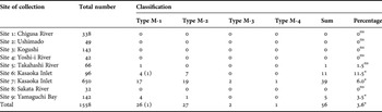
Frequency of the males possessing the first pleopods in each habitat is summarized in Table 2. The predominant type was Type M-2, followed by Type M-1. The other two types (Types M-3 and M-4) were very few. Kasaoka Inlet (Sites 6 and 7 in Figure 1B) had a higher frequency of males with morphological disorders than the other habitats in the Seto-Inland Sea: i.e. 11.5% in the inner part (Site 6) and 6.0% in the outer part (Site 7). The percentage of these males showed a statistically significant variation between the nine sites (GLM, P < 0.05). The 95% confidence interval of the percentage frequency at Sites 6, 7 and 9 significantly deviated from zero.
Number and percentage of males with morphological disorders.

Parentheses, number of males possessing cutitular lesions and morphological disorders at the same time.
*, significantly (P < 0.05) deviated from 0%; ns, not significant.
Type M-1, appendage similar to the pleopod of females on one side.
Type M-2, appendages similar to the pleopod of females on both sides.
Type M-3, appendage(s) similar to pereiopod on at least one side.
Type M-4, appendage(s) similar to abdominal pleopod on one side.
Females with morphological disorders
Most of the morphological disorders in females were deformities or loss of the first pleopods. Five types were recognized in relation to these incidences (Figure 3B–F). Type F-1 females possessed both the first pleopods, while at least one of them was short (<4 mm long) and/or morphologically incomplete. For example, as shown in Figure 3B, one pleopod was normal, but the other was incomplete and the tip was damaged. Type F-2 females lacked either pleopod on the first abdominal segment (Figure 3C). Type F-3 females lacked both of the first pleopods (Figure 3D). In contrast, the Type F-4 female had only one first pleopod with a pereiopod-like structure (Figure 3E). The setae on the distal margin of this pleopod were not ovigerous setae. Type F-5 was a female bearing a pair of pleopods with the leaf-like structures: i.e. the characteristic of the second to the fifth pleopods in both male and female (Figure 3F). All these individuals showed normal female characteristics other than the first abdominal segment, such as the position of gonopores and structure of dactyli.
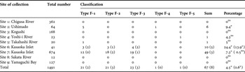
The occurrence of each type in females is summarized in Table 3. The most frequent type of disorders was Type F-3 followed by Type F-1 and Type F-2. These types were found mostly at Sites 6 and 7 (Kasaoka Inlet), and Type F-1 females were also found at Site 2 (Ushimado) and Site 5 (Takahashi River). Type F-4 and Type F-5 females were found in Site 2 (Ushimado) and Site 4 (Yoshi-i River), respectively. The Kasaoka Inlet population (Sites 6 and 7) had the higher frequencies of these incidences among the populations within the Seto-Inland Sea (24.4% and 7.3%, respectively). The percentage frequency of the morphological disorders showed statistically significant variations between the nine sites (GLM, P < 0.05). The 95% confidence interval of the percentage frequency at Sites 2, 6 and 7 significantly deviated from zero.
Number and percentage of females with morphological disorders.

Parentheses, number of females possessing cuticular lesions and morphological disorders at the same time.
*, significantly (P < 0.05) deviate from 0%; ns, not significant.
Type F-1, short and/or morphological incomplete pleopod(s) at least on one side.
Type F-2, lack of a single pleopod.
Type F-3, lack of both pleopods.
Type F-4, appendage similar to pereiopod on one side.
Type F-5, appendage similar to the second abdominal pleopod on one side.
Cuticular lesions in the first abdominal segment
Individuals with cuticular lesions were collected only at Kasaoka Inlet (Sites 6 and 7). Cuticular lesions were often found in the first abdominal segment in both males (Figure 2G) and females (Figure 3G). These appeared as a single or several black spots, when the abdominal segments were not deeply injured (e.g. Figures 2G-a & 3G-b). For example, in an ovigerous female (Figure 3G-a), the first abdominal segment was normal, but most of the right pleopod and the tip of the left pleopod were lost and black coloration remained on the pleopods. Most of the embryos had detached from the broken pleopods. Cuticular lesions appeared as bigger black spots, when the abdominal segment was more damaged. Multiple and wider black wounds would indicate more serious injuries (e.g. Figures 2G-b, c & 3G-d, e, f).
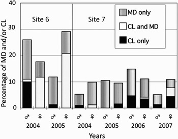
The frequency of cuticular lesions was usually lower than that of morphological disorders (Figure 4). In Site 6, it was often the case in females that cuticular lesions accompanied morphological disorders (i.e. 12% in 2004 and 21% in 2005), and the population at this site disappeared in 2006. Analysis using a GLM revealed that cuticular lesions and morphological disorders tended to occur simultaneously in females, but the occurrence of cuticular lesions and morphological disorders was independent in males; the interaction term of cuticular lesions and morphological disorders was significant (GLM, P < 0.05) only in females. Interestingly, the ratio of individuals with cuticular lesions increased from 2006 in Site 7 after the population in Site 6 became extinct.
Percentage of males and females with morphological disorders (MD) and/or cuticular lesions (CL) in the first abdominal segment. Investigations in Sites 6 and 7.

Allometric growth and gonad index
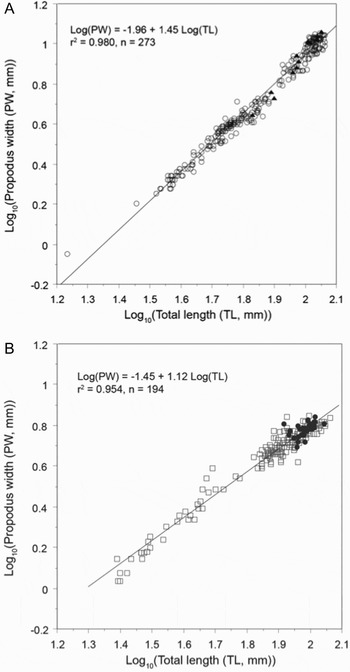
Allometric growth relationships between the log10-transformed values of total length and propodus width are shown in Figure 5. Coefficients of the regressions in the males (Figure 5A) and the females (Figure 5B) were significantly different from zero (P < 0.05), and the regressions of males and females were statistically different (P < 0.05). Both in males and females, the slope of the regression was statistically different from 1 (P < 0.05), suggesting positive allometric relationships. Minimum size of ovigerous females was 82.5 mm total length, indicating the approximate size of maturity. As shown in Figure 5A, plots of males with morphological disorders overlapped with the plots of normal males. Further statistical analysis showed that 11 out of 12 males with morphological disorders were included within the 95% confidence limit of the allometric relationship of the normal males. These results suggest that the presence of the first pleopods in males do not affect the secondary sexual characteristics.
Allometric growth relationship between log10-transformed value of total length (TL) and propodus width (PW). (A) Male. Specimens collected in 2007 were used for analysis. Open circles (○) indicate normal males, and solid triangles (▴) indicate males with morphological disorders; (B) female. Specimens collected in 2007 were used for analysis. Open squares (□) indicate normal females; solid circles (•) indicate normal ovigerous females. Specimens infected with parasites were not included in the allometric analysis.

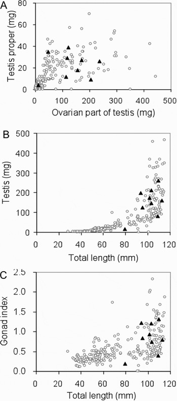
The wet weight of the ovarian part of testis was much higher than the testis proper (Figure 6A). The relative wet weight of testis proper against ovarian part of testis did not show a clear difference between the males with morphological disorders and normal males (Figure 6A). The weight of gonad (testis proper + ovarian part of testis) against total length was not different between males with the first pleopods and normal males (Figure 6B). Furthermore, the GI against total length was not different between males with the first pleopods and normal males (Figure 6C). These results suggest that the presence of the first pleopods is independent of the gonad development or other aspects of secondary sexual characters.
Comparison of the gonads between normal males and those with morphological disorders. Open circles (○) indicate normal males, and solid triangles (▴) indicate males with the first pleopods. (A) Relation between ovarian part of testis and testis proper; (B) relation between total length and gonad of the male; (C) relation between total length and gonad index.

DISCUSSION
Normal females of the thalassinidean decapod, Upogebia major possess a pair of pleopods on the first abdominal segment, while normal males lack them. However, males possessing the first pleopods occurred in populations in the Seto-Inland Sea, Japan. Upogebia major collected at nine sites showed morphological disorders in sexual dimorphic structures: i.e. additional pleopods in males, loss and abnormalities of pleopods in females, and cuticular lesions. Frequency of the occurrence of individuals with morphological disorders showed local variation. These morphological abnormalities seem to be independent of the gonadal development and not to affect the secondary sexual characters (Figures 5 & 6). The points of discussion are, whether the morphological disorders in males and females are evidence of de-masculinization and masculinisation, and what are the possible factors causing these morphological disorders and cuticular lesions.
Interpretation of morphological disorders in males and females
It has been reported that the secondary sexual characters of decapod crustaceans are controlled by hormones synthesized in the androgenic gland (e.g. Liu et al., Reference Liu, Cheung and Chu2008) indicating that hormonally, the primary state of sex is female and some male characters are subsequently induced. The additional first pleopods of Type M-1 and Type M-2 males (Figure 2B, C & F) were very similar in morphology and size to those of normal females (Figure 3A), although no ovigerous setae are present on these pleopods even in the breeding period. Males with these pleopods could be interpreted as de-masculinized individuals because apparent female-specific structures appeared by alteration of sexual differentiation, i.e. male characters are not induced. On the other hand, interpretation of the females of which first pleopods were lost and/or were abnormal (Types F-1, F-2 and F-3) is difficult, although they are apparently incomplete females. These incomplete females could be interpreted as masculinizing individuals since they show male characteristics, whereas a possibility of accidental loss or wound affected by parasites cannot be eliminated (discussed below). It is interesting that sexual dimorphism such as the position of gonopores and structures of dactyli are not affected in these cases (Figures 5 & 6). It is presumed that these sexual dimorphic structures are due to primary genetic sex determination, although the genetic mechanism of sex determination has not yet been ascertained in U. major. On the other hand, a pair of first pleopods in females is indispensable for reproduction to hold embryos until hatching, and this structure is a secondary female-specific character related to sexual maturation. We should carefully evaluate intersexuality considering whether the abnormal sexual dimorphic structures are caused by alteration of primary (genetic) sex determination or secondary sexual differentiation.
In contrast, a pereiopod (walking leg)-like appendage in Type M-3 males (Figure 2D) was largely different from the five pairs of real walking legs. A leaf-like, dichotomous appendage of the Type M-4 male (Figure 2E) was identical to the second to fourth abdominal pleopods. Similarly in the females, the Type F-4 female had a pereiopod-like first pleopod (Figure 3E), and the Type F-5 female had a leaf-like first pleopod (Figure 3F). These disorders seem to reflect alterations of segmental identity, i.e. the first abdominal segment to the posterior cephalothoracic segment (Types M-3 and F-4) and the first abdominal segment to the more posterior abdominal segment (Types M-4 and F-5). Segment identity in the posterior thorax and anterior abdomen is determined by functions of the Hox gene products, Ultrabithorax (Ubx) and abdominal-A (Abd-A) in arthropods (Hughes & Kaufman, Reference Hughes and Kaufman2002). Alteration of segment identity seen in these types of disorders might be a consequence of mutation occurring in the Hox gene. The occurrence of these phenotypes (Types M-3 and M-4 in Figure 2, and Types F-4 and F-5 in Figure 3) was very rare, and was not restricted to Sites 6 and 7 where morphological disorders in sexual dimorphism appeared at high frequency (Table 3). We conclude that these morphological disorders would neither reflect de-masculinization nor masculinization.
The ratio of intersexuality in crustaceans has been thought to be very low. LeBlanc (Reference LeBlanc2007) described that the frequency of intersexuality is very rare, normally less than 1% in the field. Intersex males accounted for 0.65% in Upogebia deltaura (Tunberg, Reference Tunberg1986). Intersex females were 1.1% in Upogebia stellata (Pinn et al., Reference Pinn, Atkinson and Rogerson2001). A local variation of intersexuality has been documented in an estuarine amphipod Echinogammarus marinus (Ford et al., Reference Ford, Fernandes, Rider, Read, Robinson and Davis2004). Intersex males possess rudimentary female brood plates and intersex females possess one or two genital papillae. The two reference sites had fewer intersex individuals (5–8%), but polluted habitats had more intersex specimens (14–15%). In the present study, the ratio of occurrence of individuals with morphological disorders was less than 3.5% for males in 7 sites other than Sites 6 and 7 (Table 2), while that of the females was less than 4.3% in 6 sites other than Sites 6, 7 and 2 (Table 3). In contrast, the ratio of morphological disorders was significantly high in Sites 6 and 7 (Kasaoka Inlet) for both males (11.5% in Site 6 and 6.0% in Site 7) and females (24.4% in Site 6 and 7.3% in Site 7). These results suggest that spontaneous occurrence of morphological disorders in sexually dimorphic structures is not high, and crustaceans seem to have a high sexual plasticity and to be sensitive to ambient environmental changes (see Ford, Reference Ford2008).
Possible factors inducing morphological disorders
Morphological abnormalities have so far been documented in a number of decapod crustaceans (LeBlanc, Reference LeBlanc2007), and potential causes of deformities may be genetic factors, environmental pollution including food quality, and parasitism (Simons & Jones, Reference Simons and Jones1981; Hancock et al., Reference Hancock, Hughes and Bunn1998; Mashiko, Reference Mashiko2000; Ford et al., Reference Ford, Fernandes, Rider, Read, Robinson and Davis2004; Béguer et al., Reference Béguer, Pasquaud, Noël, Girardin and Boët2008). Organic pollutants could produce morphological deformities in penaeid shrimps and fiddler crabs (Weis et al., Reference Weis, Gottlieb and Kwiatkowski1987; Weis & Kim, Reference Weis and Kim1988; Betancourt-Lozano et al., Reference Betancourt-Lozano, Baird, Sangha and Gonzalez-Farias2006). Heavy metals may also produce morphological abnormalities in the embryonic development (Itow et al., Reference Itow, Loveland and Botton1998).
The major trophic mode of upogebiids is filter feeding of fine particles including phytoplankton suspended in the water column (Dworschak, Reference Dworschak1987; Nickell & Atkinson, Reference Nickell and Atkinson1995; Yokoyama et al., Reference Yokoyama, Tamaki, Koyama, Ishihi, Shimoda and Harada2005). Highly eutrophicated tidal flats would contain much organic material as well as heavy metals and organotin compounds. Kasaoka Inlet (Sites 6 and 7) was formed by reclamation and the soft sediments accumulated on the seabed are readily re-suspended (Figure 1B). The accumulation and subsequent re-suspension on the habitat of U. major are probable causes of the physical and chemical deterioration of the burrow environment with the internal space of the burrows highly increasing the accumulation of fine particles.
In areas adjacent to the mouth of the Kasaoka Inlet (Figure 1B), concentrations of tributyltin (TBT) and triphenyltin (TPT) were 17 ± 5 µg/kg and 4 ± 3 µg/kg, but their concentrations were very high especially in the inner areas of Kasaoka Inlet, i.e. 24–90 µg/kg and 25–200 µg/kg (Fukue et al., Reference Fukue, Sato, Yanai, Nakamura, Yamasaki, Yong and Thomas2001). Exposure to TBT (concentration of 0.5–5 µg/l) induces deformities in regenerated claws and limbs of the fiddler crab Uca pugilator (Weis et al., Reference Weis, Gottlieb and Kwiatkowski1987; Weis & Kim, Reference Weis and Kim1988). The TBT concentrations in Kasaoka Inlet are higher compared to the abnormality-inducible concentrations for the fiddler crab. We assume that the contamination of pollutants such as heavy metals and organotin compounds are possible factors that induce morphological disorders (Figure 4), although we have not investigated the direct effects of these candidate factors.
Burrows of upogebiids are inhabited by a wide variety of animals, such as bivalves, annelids, copepods, shrimps, crabs and gobiid fish (Anker et al., Reference Anker, Jeng and Chan2001; Itani, Reference Itani and Tamaki2004; Nanri & Saigusa, unpublished observations). Two major parasites of U. major were Metabopyrus ovalis (isopod) attaching to the gill chamber and Peregrinamor ohshimai (bivalve) attaching to the ventral thorax (Kato & Itani, Reference Kato and Itani1995; Itani, Reference Itani and Tamaki2004). Itani (Reference Itani2001) observed that a small brachyuran, Sestrostoma sp. clung to the first abdominal segment of U. major and caused cuticular damages. We have also found the Sestrostoma sp. clinging to abdominal segments of U. major. It is highly probable that the black coloration of the lesions (Figure 3) is a defence response against cuticular damage caused by the ectoparasitic symbionts. It has been suggested that parasitism affects the endocrine system of crustaceans to cause de-masculinization (e.g. LeBlanc, Reference LeBlanc2007). In Kasaoka Inlet, parasitism may cause not only the direct effect (cuticular lesions), but also affect the endocrine system leading to the occurrence of morphological disorders in sexually dimorphic structures (Figure 4). Further investigation of the effects of parasites and the probable synergistic role of pollutants is required in areas with high frequency of morphological disorders.
ACKNOWLEDGEMENTS
This study was supported by grants of the Long-range Research Initiative (LRI) provided by the Japan Chemical Industry Association, Japan (2004–2008) and Ministry of Environment, Japan (2009).