Implications
The present study utilized two well-established in vivo chicken models for development, regression and remodeling of oviductal tissue to identify the RAS-related protein 1 (RASD1) gene as an important estrogen-responsive candidate gene responsible for growth, development, remodeling and maintenance of chicken oviduct. Results of this study also identified estrogen-responsive microRNA candidates involved in the post-transcriptional regulation of expression of RASD1 gene. Although specific biological mechanisms whereby RASD1 effects morphological and functional changes in the oviduct of chickens remain to be determined, expression of RASD1 is likely modulated by several microRNAs in response to various estrogen-mediated processes in the reproductive system of chickens.
Introduction
In chickens, the reproductive system is composed of one functional ovary and its associated oviduct. The chicken oviduct consists of four segments: the infundibulum (site of fertilization) proximal to the ovary; the magnum (secretion of components of egg white); the isthmus (formation of the soft shell membrane); and shell gland (calcification of the egg shell). The oviduct is the most dynamic organ undergoing diverse cellular and molecular changes in response to female sex steroids, which is pivotal to establishing an optimal microenvironment for transport of the egg and embryonic development over the course of the female reproductive cycle (Buhi et al., Reference Buhi, Alvarez and Kouba1997). As the primary ovarian-derived sex hormone, estrogen regulates various biological events involved in development and functions of the oviduct such as modulating contraction of its smooth musculature, formation of tubular glands, and secretions by its epithelial cells (Palmiter and Wrenn, Reference Palmiter and Wrenn1971; Monroe et al., Reference Monroe, Berger and Sanders2000; Monroe et al., Reference Monroe, Jin and Sanders2002; Louet et al., Reference Louet, LeMay and Mauvais-Jarvis2004; Hewitt et al., Reference Hewitt, Harrell and Korach2005; Hrabia et al., Reference Hrabia, Wilk and Rzasa2008). Expression of many estrogen-induced genes in the oviduct of chickens mediates key morphological, physiological and functional changes in the female reproductive tract.
Using microarray analysis, candidate genes that were significantly altered in chick oviducts in response to estrogen stimulation were identified (Song et al., Reference Song, Seo, Choi, Rengaraj, Kim, Lee, Kim, Yun, Jeong and Han2011). One of the candidate genes, dexamethasone (DEX)-induced RASD1 (also called dexras1) was up-regulated by estrogen in chick oviducts (Song et al., Reference Song, Seo, Choi, Rengaraj, Kim, Lee, Kim, Yun, Jeong and Han2011). The RASD1 gene was first identified as a DEX (a corticosteroid) inducible gene in the At-T20 mouse pituitary corticotroph cell line (Kemppainen and Behrend, Reference Kemppainen and Behrend1998). The mechanism involved in rapid up-regulation of RASD1 by glucocorticoids involved glucocorticoid response elements in the 3'-flanking region of the human Rasd1 gene (Kemppainen et al., Reference Kemppainen, Cox, Behrend, Brogan and Ammons2003). RAS-related protein 1 is expressed in brain at high concentrations, and heart, liver, kidney, pancreas and reproductive organs at lower concentrations (Kemppainen and Behrend, Reference Kemppainen and Behrend1998; Tu and Wu, Reference Tu and Wu1999; Fang et al., Reference Fang, Jaffrey, Sawa, Ye, Luo and Snyder2000; Bernal and Crespo, Reference Bernal and Crespo2006; Agretti et al., Reference Agretti, De Marco, Pinchera, Vitti, Bernal and Tonacchera2007). RAS-related protein 1 belongs to the Ras superfamily of small GTPases involved in various cellular processes such as a cell-signaling regulator of steroid hormone actions (Cismowski et al., Reference Cismowski, Ma, Ribas, Xie, Spruyt, Lizano, Lanier and Duzic2000). Expression of RASD1 gene is also modulated by β-estradiol, as well as glucocorticoid and DEX (Kemppainen and Behrend, Reference Kemppainen and Behrend1998; Fang et al., Reference Fang, Jaffrey, Sawa, Ye, Luo and Snyder2000; Graham et al., Reference Graham, Key, Kilpatrick and Dorin2001; Kim et al., Reference Kim, Cho, Kim, Lee, Yoon, Lee, Moon, Park, Hong, Na, Shin, Kwon, Song, Choi and Choi2017) and plays important roles in regulating various processes in the female reproductive organs (Kim et al., Reference Kim, Cho, Kim, Lee, Yoon, Lee, Moon, Park, Hong, Na, Shin, Kwon, Song, Choi and Choi2017). However, very little information is available concerning the responsiveness of the RASD1 gene to estrogen and its physiological function(s) in the avian reproductive system. Therefore, the objective of this study was to investigate the regulation of expression of RASD1 in the oviduct of chickens.
Because the chicken oviduct is a very reactive organ showing remarkable responsiveness to steroid hormones such as estrogen during egg formation and oviposition, it is an excellent research model for investigation of hormonal actions in developmental processes and functions (Dougherty and Sanders, Reference Dougherty and Sanders2005; Chousalkar and Roberts, Reference Chousalkar and Roberts2008). We tested the hypothesis that chicken RASD1 is an estrogen-regulated gene involved in the development and functions of the chicken oviduct during: (1) diethylstilbestrol (DES)-induced development of the chick oviduct; and (2) regression and estrogen-dependent regeneration of the oviduct of laying hens undergoing molting. To our knowledge, this is the first report on regulation of expression of the RASD1 gene during the DES-stimulated development the chick oviduct and regeneration of the oviduct of laying hens undergoing molting.
Material and methods
Experimental animals and animal care
The use of chickens for this study was approved by the Animal Care and Use Committee of Korea University. White Leghorn chickens were exposed to a regimen of 15 h light and 9 h dark with ad libitum access to feed and water, and managed according to standard poultry husbandry guidelines.
Tissue samples
Study one
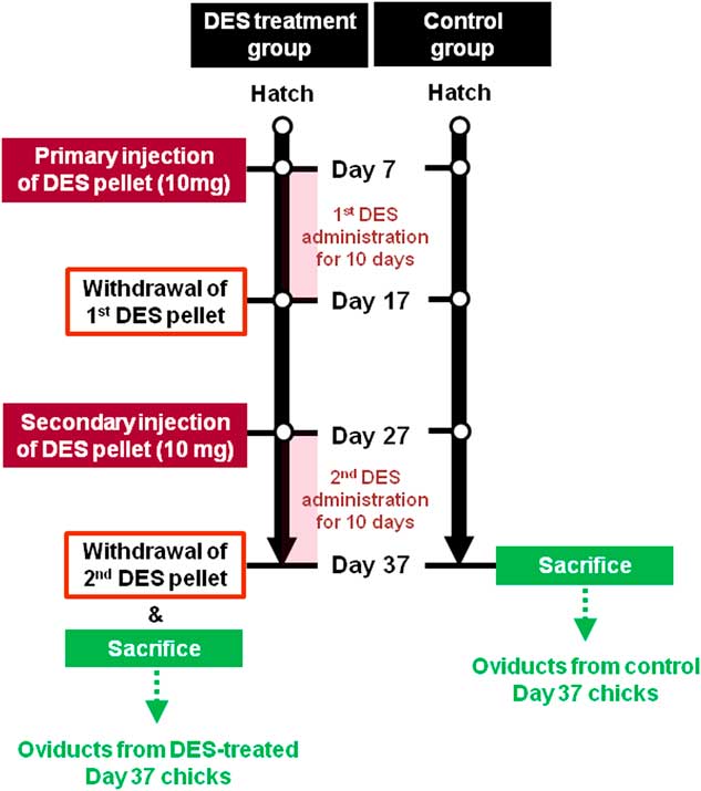
Diethylstilbestrol (a synthetic estrogen agonist) treatment and oviduct retrieval were conducted as reported previously (Song et al., Reference Song, Seo, Choi, Rengaraj, Kim, Lee, Kim, Yun, Jeong and Han2011). As shown in Figures 1 and 2, a 10-mg DES pellet was implanted into the abdominal region of 1-week-old female chicks to release the hormone for 10 days. Next, the DES pellet was removed from all chicks for 10 days, and then a 10 mg daily dose was administered for 10 additional days. Control chicks were raised normally without any treatment for 37 days after hatching. Five 37-day-old control chicks and five 37-day-old DES-treated chicks were euthanized using 60% to 70% carbon dioxide to provide samples of oviduct which was cut into 5- to 7-mm pieces and frozen in liquid nitrogen. The remaining tissues were cut into 10- to 15-mm pieces and fixed in fresh 4% paraformaldehyde in phosphate-buffered saline (PBS) (pH 7.4). After 24 h, fixed tissues were changed to 70% ethanol for 24 h and then dehydrated and embedded in Paraplast-Plus (Leica Microsystem, Wetzlar, Germany). Paraffin-embedded tissues were sectioned at 5 µm.
Diagrammatic illustration of the in vivo model for research on estrogen-dependent development of the chick oviduct. 1-week-old female chicks were implanted with a 10 mg diethylstilbestrol (DES) pellet in the abdominal region for 10 days, and then the DES pellet was removed for 10 days. The DES-treated chicks were next administered a 10 mg DES pellet for 10 additional days. Control chicks were raised normally without any treatment for 37 days after hatching.

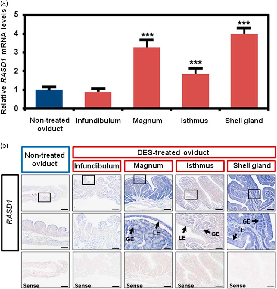
Differential expression and localization of RAS-related protein 1 (RASD1) messenger RNA (mRNA) in the chick oviduct in response to diethylstilbestrol (DES) stimulation. (a) Relative expression of RASD1 transcripts was analyzed by quantitative reverse transcription-PCR analyses using oviducts from DES-treated and control chicks on Day 37 post-hatching. These experiments were performed in triplicate and data normalized to GAPDH gene expression. Each bar is mean±SEM of three independent experiments. Statistically significant differences between non-treated oviduct and each segment of DES-treated oviduct: ***P<0.001. (b) Localization of RASD1 mRNA in oviducts of DES-treated and non-treated Day 37-chicks was determine using in situ hybridization analyses. GE=glandular epithelium; LE=luminal epithelium. Scale bar represents 200 µm (the first and third horizontal panels) and 50 µm (the second horizontal panels).

Study two
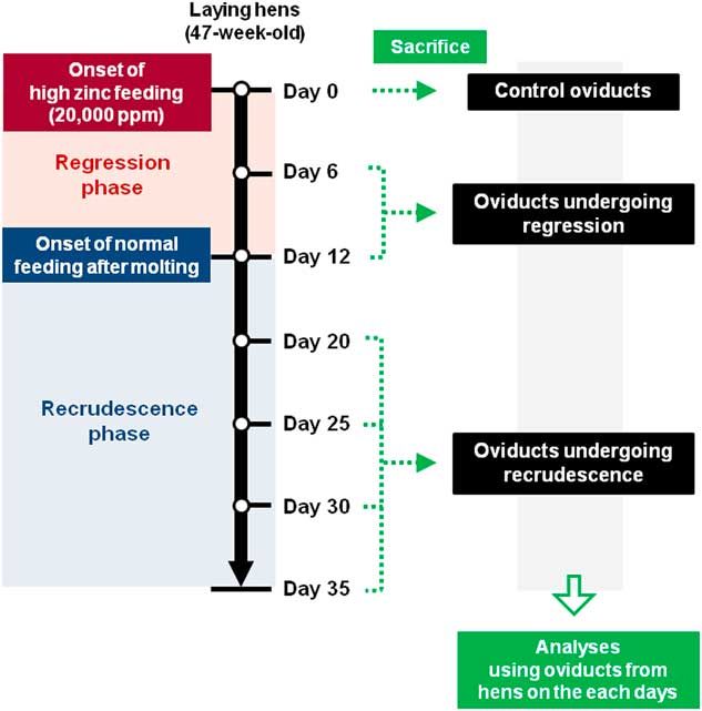
As shown in Figures 3 and 4, molting of laying hens was induced as described previously by adding 20 000 ppm zinc to the diet to effectively reduce feed-intake and induce molting (Creger and Scott, Reference Creger and Scott1977; Berry and Brake, Reference Berry and Brake1985). In brief, hens (n=5/time point) in each subgroup including the molting group (normal feeding group, 6 and 12 days after onset of zinc feeding) and the recrudescence group (20, 25, 30 or 35 days after onset of zinc feeding, and 8, 13, 18 or 23 days of normal feeding after cessation of egg production and removal from the high-zinc diet) were euthanized using 60% to 70% carbon dioxide before collecting the oviduct on each assigned day (Jeong et al., Reference Jeong, Lim, Ahn, Lim, Lee, Bae, Kim, Bazer and Song2013).
Diagrammatic illustration of the process for artificially induced molting to establish an in vivo model of regression and regeneration of the chicken oviduct. Molting was induced by feeding a diet containing high amounts of zinc, and laying hens in the molting group completely ceased egg production within 12 days after feeding the high-zinc diet. The laying hens in the recrudescence group were fed a normal commercial diet after complete cessation of egg laying (on 12 days after onset of zinc feeding). The days shown next to the arrow indicate days for feeding a high-zinc diet (6 and 12 days) and for feeding a normal diet after induction of complete molting (20, 25, 30 and 35 days after onset of feeding a high-zinc diet). Hens were euthanized on Day 0, 6, 12, 20, 25, 30 and 35 and the oviducts analyzed.

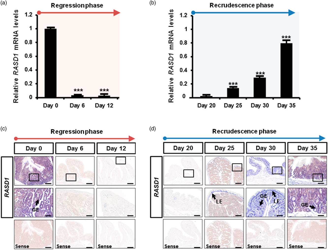
Differential expression and localization of RAS-related protein 1 (RASD1) messenger RNA (mRNA) in the oviduct during regression and recrudescence periods. (a) Relative expression of chicken RASD1 transcripts was analyzed by quantitative reverse transcription-PCR analyses using oviducts from hens fed a normal commercial diet (Day 0), high-zinc diet (Day 6 and Day 12) and (b) normal commercial diet after complete regression of the oviduct (Day 20, 25, 30 and 35). These experiments were performed in triplicate and data normalized to GAPDH gene expression. Each bar is mean±SEM of three independent experiments. Statistically significant differences compared with Day 0 (a Day 0 v. Day 6 or Day 12) or Day 20 (b: Day 20 v. Day 25, Day 30 or Day 35): ***P<0.001. (c) The localization of RASD1 mRNA in the oviduct of hens on different days of the regression and (d) recrudescence phases of molting were analyzed by in situ hybridization. GE=glandular epithelium; LE=luminal epithelium. Scale bar represents 200 µm (the first and third horizontal panels) and 50 µm (the second horizontal panels).

RNA isolation
Total RNA was isolated from frozen tissues using Trizol reagent (Invitrogen, Carlsbad, CA, USA) according to manufacturer’s recommendations. The quantity and quality of total RNA was determined by spectrometry and denaturing agarose gel electrophoresis, respectively.
Quantitative reverse transcription-PCR analysis
Complementary DNA (cDNA) was synthesized using total RNA extracted from each tissue and AccuPower® RT PreMix (Bioneer, Daejeon, Korea). Gene expression levels were measured using SYBR® Green (Sigma, St. Louis, MO, USA) and a StepOnePlus™ Real-Time PCR System (Applied Biosystems, Foster City, CA, USA). The GAPDH gene was simultaneously analyzed as a control and used for normalization for variation in loading. RASD1 and GAPDH were analyzed in triplicate. Based on the sequence of RASD1 messenger RNA (mRNA), the sense primer and antisense primer amplified specific product. For GAPDH, the sense primer (5'-ACA CAG AAG ACG GTG GAT GG-3') and antisense primer (5'-GGC AGG TCA GGT CAA CAA CA-3') amplified specific product. Using the standard curve method, we determined the level of expression of the RASD1 gene using the standard curves and C
T
values, and normalized them based on GAPDH expression. The PCR conditions were 95°C for 3 min, followed by 40 cycles at 95°C for 30 s, 60°C for 30 s and 72°C for 30 s using a melting curve program (increasing the temperature from 55°C to 95°C at a rate of 0.5°C/10 s) and continuous fluorescence measurement. ROX dye (Invitrogen) was used as a negative control for the fluorescence measurements. Sequence-specific products were identified by generating a melting curve in which the C
T
value represented the cycle number at which a fluorescent signal was statistically greater than background, and relative gene expression was quantified using the
 $$2^{{{\minus}\Delta \Delta C_{T} }} $$
method (Livak and Schmittgen, Reference Livak and Schmittgen2001).
In situ hybridization analysis
For hybridization probes, PCR products were generated from cDNAs with the primers used for reverse transcription-PCR analysis. The products were gel-extracted and cloned into TOPO® vector (Invitrogen). After verification of the sequences, plasmids containing the correct gene sequences were amplified with T7- and SP6-specific primers (T7:5'-TGT AAT ACG ACT CAC TAT AGG G-3'; SP6:5'-CTA TTT AGG TGA CAC TAT AGA AT-3') then digoxigenin (DIG)-labeled RNA probes were transcribed using a DIG RNA labeling kit (Roche Applied Science, Indianapolis, IN, USA). Tissues were collected and fixed in 4% paraformaldehyde, embedded in paraffin and sectioned at 5 μm on APES-treated (silanized) slides. The sections were then deparaffinized in xylene and rehydrated to diethylpyrocarbonate (DEPC)-treated water through a graded series of alcohol. The sections were treated with 1% Triton X-100 in PBS for 20 min and washed two times in DEPC-treated PBS. After washing in DEPC-treated PBS, they were digested with 5 μg/ml proteinase K (Sigma) in TE buffer (100 mM Tris-HCl, 50 mM ethylenediaminetetraacetic acid, pH 8.0) at 37°C. Paraffin-embedded tissue sections were incubated twice for 5 min each in DEPC-treated PBS and incubated in TEA buffer (0.1 M triethanolamine) containing 0.25% (v/v) acetic anhydride. The sections were next incubated in a prehybridization mixture containing 50% formamide and 4X standard saline citrate (SSC) for at least 10 min at room temperature. After prehybridization, the sections were incubated with a hybridization mixture containing 40% formamide, 4X SSC, 10% dextran sulfate sodium salt, 10 mM dithiothreitol, 1 mg/ml yeast transfer RNA, 1 mg/ml salmon sperm DNA, 0.02% Ficoll, 0.02% polyvinylpyrrolidone, 0.2 mg/ml RNase-free bovine serum albumin and denatured DIG-labeled cRNA probe overnight at 42°C in a humidified chamber. After hybridization, sections were washed for 15 min in 2X SSC at 37°C, 15 min in 1X SSC at 37°C, 30 min in NTE buffer (10 mM Tris, 500 mM NaCl and 1 mM ethylenediaminetetraacetic acid) at 37°C and 30 min in 0.1X SSC at 37°C. After blocking with a 2% normal sheep serum (Santa Cruz Biotechnology, Inc., Dallas, TX, USA), the sections were incubated overnight with sheep anti-DIG antibody conjugated to alkaline phosphatase (Roche). The signal was visualized by exposure to a solution containing 0.4 mM 5-bromo-4-chloro-3-indolyl phosphate, 0.4 mM nitroblue tetrazolium, and 2 mM levamisole (Sigma).
Quantitative reverse transcription-PCR analysis for microRNAs
MicroRNA (miRNA) first-strand cDNA was synthesized from total RNA from oviducts of DES-treated and non-treated chicks using a miRNA first-strand cDNA synthesis kit (Agilent Technologies, Santa Clara, CA, USA). The expression of miRNAs was measured using the High-Specificity miRNA QPCR Core Reagent Kit (Agilent Technologies) and a StepOnePlusTM Real-Time PCR System (Applied Biosystems) following guidelines from the vendor. For endogenous control, U6 small nuclear RNA (snRNA) was used to normalize variations in the amount of cDNA template across target samples. And, relative quantification of miRNAs was analyzed using the 2–ΔΔCT method.
Statistical analyses
Data for quantitative PCR were subjected to one-way ANOVA according to the GLM (PROC-GLM) of the SAS program (SAS Institute, Cary, NC, USA) and those were confirmed using the F-test and the Student’s t test to determine whether there was significant difference in gene expression in the chick oviduct in response to DES treatment, and the oviducts of laying hens during the molting and recrudescence periods. Differences with a probability value of P<0.05 were considered statistically significant. Data are presented as mean±SEM unless otherwise stated.
Results
Expression of RAS-related protein 1 messenger RNA increase in the developed chick oviduct by diethylstilbestrol stimulation
We first determined differential expression of RASD1 during the estrogen-induced development of the oviducts of chicks because the RASD1 gene was identified as a candidate gene involved in oviduct development as its expression was altered at least 2-fold in our previous study (Song et al., Reference Song, Seo, Choi, Rengaraj, Kim, Lee, Kim, Yun, Jeong and Han2011). In the present study, we determined the effects of DES on RASD1 expression in the chicken oviduct (Figure 1). Quantitative reverse transcription-PCR analysis indicated that DES increased RASD1 mRNA by 3.3- (P<0.001), 1.9- (P<0.001) and 4.0-fold (P<0.001) in the magnum, isthmus and shell gland, respectively as compared with non-treated oviducts of chicks on Day 37 post-hatching (Figure 2a). Consistent with those results, in situ hybridization analyses revealed that RASD1 mRNA expression was abundant in the luminal (LE) and glandular (GE) epithelia of the magnum and shell gland, but little or no RASD1 mRNA was detected in oviducts of control chicks or infundibulum of DES-treated chicks (Figure 2b).
Tissue and cell-specific expression of RAS-related protein 1 messenger RNA changes during regression and recrudescence of the oviduct of laying hens
Tissue- and cell-specific expression of RASD1 in the oviduct of chicks treated with DES suggested that regulation of regression and recrudescence of the oviduct during induced molting hens involved estrogen. We used our established in vivo model for inducing molting to determine tissue- and cell-specific expression of RASD1 on different days of regression and regeneration of the oviduct during the molting periods (Jeong et al., Reference Jeong, Lim, Ahn, Lim, Lee, Bae, Kim, Bazer and Song2013) (Figure 3). First, expression of RASD1 mRNA during regression phase (at Days 6 and 12) was decreased to 6% (P<0.001) and 3% (P<0.001) compared with Day 0 (control) values (Figure 4a). Then, with return of the hens to a normal diet, expression of RASD1 mRNA increased from Days 20 to 35 (recrudescence phase) (Figure 4b). The RASD1 mRNA was expressed abundantly in GE of the magnum portion of the oviduct which undergoes the most dramatic changes during reproductive tissue remodeling on Days 0, 30 and 35, but expression of RASD1 was low in LE of the oviduct on Days 0, 20 and 35, and minimal between Days 6 and 25 of the oviduct regression and recrudescence periods (Figures 4c and d).
Expression of candidate chicken microRNAs targeting changes in RAS-related protein 1 transcripts in response to diethylstilbestrol stimulation
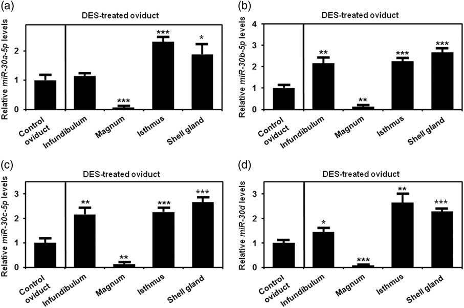
The possible involvement of miRNA-mediated post-transcriptional regulation of chicken RASD1 gene in the oviduct in response to hormonal action was determined for chick oviducts (DES-treated and non-treated chicks). Based on analysis of potential miRNA binding sites within the 3'-untranslated region of the chicken RASD1 gene using the miRNA target prediction database (miRDB; http://mirdb.org/miRDB/), we selected four candidate miRNAs targeting chicken RASD1 transcript (miR-30a-5p, miR-30b-5p, miR-30c-5p and miR-30d) with high target scores. As shown in Figure 5, expression of these four miRNAs decreased to below 3% (P<0.001) of values for control chicks in the magnum of oviducts from DES-treated chicks. On the other hand, relative expression levels of all four miRNAs increased in the isthmus and shell gland, whereas expression of miR-30b-5p, miR-30c-5p and miR-30d increased in the infundibulum.
Expression of candidate miRNAs targeting RAS-related protein 1 (RASD1) transcripts in the oviduct of chicks in response to diethylstilbestrol (DES) stimulation. Relative expression of (a) miR-30a-5p, (b) miR-30b-5p, (c) miR-30c-5p and (d) miR-30d was analyzed using quantitative reverse transcription-PCR analyses with miRNA first-strand complementary DNA templates from oviducts of DES-treated and non-treated chicks on Day 37 post-hatching. These experiments were performed in triplicate and normalized to control U6 snRNA expression. Statistically significant differences: ** P<0.01, ***P<0.001.

Discussion
Based on the global gene expression profile data from our previous study, showing up-regulation of RASD1 at least 2-fold in the estrogen-stimulated chick oviduct (Song et al., Reference Song, Seo, Choi, Rengaraj, Kim, Lee, Kim, Yun, Jeong and Han2011), we selected RASD1 as an estrogen-responsive gene which might be responsible for regulation of morphological and functional changes in the chick oviduct. RAS-related protein 1 is regulated by several steroid hormones (Brogan et al., Reference Brogan, Behrend and Kemppainen2001; Kemppainen et al., Reference Kemppainen, Cox, Behrend, Brogan and Ammons2003; Song et al., Reference Song, Seo, Choi, Rengaraj, Kim, Lee, Kim, Yun, Jeong and Han2011) and is a signaling protein for actions of steroid hormones in several cells and tissues (Fang et al., Reference Fang, Jaffrey, Sawa, Ye, Luo and Snyder2000; Brogan et al., Reference Brogan, Behrend and Kemppainen2001; Lellis-Santos et al., Reference Lellis-Santos, Sakamoto, Bromati, Nogueira, Leite, Yamanaka, Kinote, Anhe and Bordin2012). Although female reproductive organs are highly susceptible to female sex hormones, hormonal regulation of RASD1 expression and its physiological function(s) in the female reproductive system are poorly understood. In this study, using two well-established in vivo chicken models, we: (1) confirmed that estrogen stimulates tissue-and cell-specific expression of RASD1 gene and four candidate miRNAs targeting RASD1 transcripts in the chick oviduct; and (2) demonstrated temporal and cell-specific changes in expression of RASD1 transcripts in the oviducts of laying hens during as the oviduct underwent regression and regeneration during the molting and post-molting periods. These results provide novel insight into the responsiveness of RASD1 to estrogen and roles of estrogen-responsive miRNAs regulating cell-specific expression of the RASD1 gene in the oviduct of chickens.
RAS-related protein 1 is expressed in human and mouse reproductive organs including uterus, ovary, and testis, as well as brain, heart, liver and kidney, which suggests that it is a regulator of cellular signaling in males and females (Kemppainen and Behrend, Reference Kemppainen and Behrend1998; Fang et al., Reference Fang, Jaffrey, Sawa, Ye, Luo and Snyder2000; Kim et al., Reference Kim, Cho, Kim, Lee, Yoon, Lee, Moon, Park, Hong, Na, Shin, Kwon, Song, Choi and Choi2017). Although the RASD1 gene was first identified as a DEX (a corticosteroid) inducible gene, it is also responsive to estrogen (Brogan et al., Reference Brogan, Behrend and Kemppainen2001; Vaidyanathan et al., Reference Vaidyanathan, Cismowski, Wang, Vincent, Brown and Lanier2004; Kim et al., Reference Kim, Cho, Kim, Lee, Yoon, Lee, Moon, Park, Hong, Na, Shin, Kwon, Song, Choi and Choi2017). Expression of the Rasd1 gene increased in pituitary glands of mice in response to estrogen as well as glucocorticoids and in MCF-7 breast cancer cells treated with estrogen (Brogan et al., Reference Brogan, Behrend and Kemppainen2001; Vaidyanathan et al., Reference Vaidyanathan, Cismowski, Wang, Vincent, Brown and Lanier2004). Expression of Rasd1 mRNA is highly expressed in the endometrial epithelium of mice during the estrous cycle and in response to estradiol (Kim et al., Reference Kim, Cho, Kim, Lee, Yoon, Lee, Moon, Park, Hong, Na, Shin, Kwon, Song, Choi and Choi2017). Those observations are consistent with our data showing that DES induced RASD1 expression in chick oviducts undergoing dramatic morphological changes of reproductive organs including development of ovarian follicles, increases in size of the ovary and oviduct, development of tubular glands and differentiation of LE epithelial cells in oviduct (Song et al., Reference Song, Seo, Choi, Rengaraj, Kim, Lee, Kim, Yun, Jeong and Han2011). The estrogen-induced increase in RASD1 transcripts were predominantly in the LE and GE of the magnum and shell gland. In response to the two primary ovarian hormones, estrogen and progesterone, epithelial cells forming lumen and gland in oviducts undergo proliferation and differentiation. The induced expression and secretion of RASD1 in GE and LE activates intracellular signal transduction pathways implicated in oviduct growth and development. RAS-related protein 1 has been identified as a cell-signaling regulator in various cell-signaling pathways (Cismowski et al., Reference Cismowski, Ma, Ribas, Xie, Spruyt, Lizano, Lanier and Duzic2000). Rapid induction of target molecules and genes by estrogen action occurs through the action of the estrogen receptor coupled with multiple intracellular signaling molecules. Recently Kim et al. (Reference Kim, Cho, Kim, Lee, Yoon, Lee, Moon, Park, Hong, Na, Shin, Kwon, Song, Choi and Choi2017) reported that the rapid increase in Rasd1 expression in uteri of mice is regulated by estrogen-receptor dependent intracellular signaling pathways including both the ERK1/2 and P38 MAPK signaling cascades. In epithelial cells of oviduct, estrogen receptor 1α (ESR1) is localized to both ciliated and secretory epithelial cells, whereas ESR2 is largely confined to ciliated epithelial cells (Okada et al., Reference Okada, Ohta, Inoue, Hiroi, Muramatsu and Iguchi2003; Shao et al., Reference Shao, Egecioglu, Weijdegard, Kopchick, Fernandez-Rodriguez, Andersson and Billig2007). Although the molecular mechanisms guiding the estrogen-mediated expression and roles of RASD1 in the developing chick oviduct requires further investigation, the estrogen-mediated up-regulation of RASD1 gene in epithelial cells of the oviduct could involve activation of estrogen receptors coupled with intracellular signaling cascades identified in previous studies.
Actions of estrogen in the female reproductive organs affect morphological, physiological and functional changes through diverse regulatory cascades involving activation of transcription factors and expression of target genes. Dramatic remodeling of the reproductive system of laying hens during the molting process is coupled with physiological changes such as fluctuations in circulating concentrations of hormones (Berry, Reference Berry2003; Socha et al., Reference Socha, Sechman, Mika and Hrabia2017). In the present study, there were temporal and cell-specific changes in expression of the RASD1 gene during the regression and regeneration phases of the molting process. Expression of the RASD1 gene was differentially regulated in the magnum portion of the oviduct during remodeling of the reproductive tract of laying hens. Expression of the RASD1 gene decreased during the regression phase (from Days 0 to 12) and then increased during the regeneration of the regressed oviduct (from Days 20 to 35), particularly in the temporal patterns of expression of the RASD1 gene during molting and oviduct recrudescence were closely related to known changes in concentrations of estrogen in plasma of laying hens during the regression and recrudescence phases due to changes in ovarian steroidogenesis (Hoshino et al., Reference Hoshino, Suzuki, Kakegawa, Imai, Wakita, Kobayashi and Yamada1988; Jeong et al., Reference Jeong, Lim, Ahn, Lim, Lee, Bae, Kim, Bazer and Song2013). Taken together, changes in RASD1 expression are closely associated with developmental stages and tissue/cell type in oviducts of chickens regulated by estrogen and culminating in growth, development and remodeling of the oviduct in chickens.
Results of the present study are also the first to demonstrate estrogen-dependent changes of expression of candidate chicken miRNAs which inhibit post-transcriptional modifications of chicken RASD1 transcripts. As essential regulators of target gene expression, miRNAs regulates various biological actions including the development and functions of female reproductive organs (Nothnick, Reference Nothnick2012). MicroRNA-mediated post-transcriptional regulation is closely related with the changes in expression of target genes at the cellular level for dramatic morphological changes in the oviduct in response to estrogen. Expression of miRNAs can also be modulated by estrogen in reproductive tissues (Carletti and Christenson, Reference Carletti and Christenson2009; Klinge, Reference Klinge2012). In the present study, we selected four candidate miRNAs regulating RASD1 gene expression and demonstrated differential expression of those miRNAs by estrogen. DES decreased expression of miR-30a-5p, miR-30b-5p, miR-30c-5p and miR-30d in the magnum of chick oviducts whereas DES decreased expression of the RASD1 gene in magnum portion of the chick oviduct. Thus, the miRNAs are estrogen-responsive molecules and the estrogen-induced down-regulation of those miRNAs allowed for increased expression of the RASD1 gene in the chicken oviduct.
Acknowledgments
This research was supported by a Korea University Grant. The authors have declared no conflict of interest.