Arrhythmias are common in patients admitted to the intensive care unit.Reference Goodman, Shirov and Weissman 1 – Reference Tarditi and Hollenberg 3 There is extensive adult literature demonstrating that disturbances in cardiac rhythm are frequently seen on continuous electrocardiographic monitoring postoperatively, in sepsis, and in patients with cardiac disease.Reference Reinelt, Karth, Geppert and Heinz 2 , Reference Goldman 4 – Reference Walsh, Tang, Wijewardena, Yarham, Boyle and Gaunt 7 In addition, arrhythmias have been associated with systemic problems brought on by hypoxaemia, hypercarbia, electrolyte disturbances, acid–base abnormalities, and multi-organ system failure, all problems that are frequently encountered in the intensive care unit.Reference Goldman 4 , Reference Kirkpatrick, Heilbrunn and Sankaran 5 , Reference Hoffman, Wernovsky and Wieand 8 , Reference Polanczyk, Goldman, Marcantonio, Orav and Lee 9 In adults, arrhythmias have been reported in 6–13% of non-cardiac postoperative patients, demonstrating an association between hospital length of stay and mortality.Reference Goodman, Shirov and Weissman 1 , Reference Goldman 4 , Reference Kirkpatrick, Heilbrunn and Sankaran 5 , Reference Polanczyk, Goldman, Marcantonio, Orav and Lee 9 – Reference Brathwaite and Weissman 11
The adult intensive care patient, however, is quite distinct from the paediatric intensive care patient (paediatric intensive care unit). The physiology, diseases commonly seen, and absence of ischaemic heart disease make the paediatric population a markedly different group. Although studies have looked at arrhythmias in critically ill adults, there are little data regarding the rates and types of arrhythmias in critically ill paediatric patients. The primary aim of this study was to determine the rates and types of cardiac rhythm disturbances in children admitted to the paediatric intensive care unit without a primary cardiac diagnosis. Secondary aims were to determine diagnoses or clinical variables that are predictors for arrhythmias in critically ill children – for example, respiratory failure and use of inotropes – and determine whether the development of an arrhythmia is associated with severity of illness and paediatric intensive care length of stay.
Methods
Study design
After approval by the institutional review board, a prospective observational review of continuous cardiac telemetry on paediatric intensive care patients at the Children’s Hospital at Montefiore was performed from March to June 2012. All medical and surgical patients less than or equal to 21 years of age admitted to the paediatric intensive care unit during this time were included and followed up daily from admission through discharge from the intensive care unit. Patients with a history of congenital or acquired primary cardiac disease, those admitted for a primary cardiac arrhythmia, and postoperative cardiac surgical patients were excluded from the analysis.
Data collection
All patients admitted to the paediatric intensive care unit were placed on continuous electrocardiographic monitoring with arrhythmia alarms and full-disclosure telemetry (Philips system L.00.20, Andover, Massachusetts, United States of America). Telemetry was programmed with pre-defined alarm settings for arrhythmias, based in part upon patient age. Those who met the inclusion criteria constituted the study sample. Cardiac telemetry was reviewed every 24 hours by one of the investigating paediatric cardiologists, and all final diagnoses were confirmed by a paediatric cardiac electrophysiologist. Telemetry was reviewed to analyse arrhythmia alarms and heart rate trends. In addition, demographic and clinical variables were recorded within the first hour of admission and then daily until paediatric intensive care unit discharge. These variables included the following: age (years), gestational age at birth (weeks) if there was a history of prematurity, gender, race, height (cm), weight (kg), body surface area (m2), length of stay in the intensive care unit (days), diagnosis, and co-morbidities such as any genetic, metabolic, pulmonary, renal, gastrointestinal, endocrine, neurological, immunological, haematologic or oncologic disease, or multi-system organ failure. The patient’s mode of respiratory support – that is, mode of mechanical ventilation – presence of respiratory failure, use of albuterol or aminophylline, use of vasoactive or inotrope medications, use of inhaled nitric oxide, diagnosis of sepsis or trauma, presence of invasive catheters, procedure performed in the case of postoperative patients, anaemia, acid–base and electrolyte abnormalities, results of thyroid function testing, and results of toxicology screen, if performed, were also recorded. The type of arrhythmia and any therapeutic interventions were also recorded. The cause and details for all mortalities were documented. A Pediatric Index of Mortality-2 score was calculated for each patient within the first hour of admission. This is a standard score used by the paediatric intensive care team to estimate mortality risk from data available at admission, to predict patient outcome, and also to describe how ill the child is upon presentation.Reference Slater, Shann and Pearson 12 A Vasoactive-Inotrope Score was calculated on admission for any patient requiring a vasoactive or inotrope. This score has been used as a marker of illness severity by quantifying the degree of cardiovascular support required.Reference Gaies, Gurney and Yen 13
Definition of tachyarrhythmias and bradyarrhythmias
All cardiac rhythm disturbances for each patient were recorded during the study period. All cardiac rhythm abnormalities were catalogued, including premature atrial contractions, premature ventricular contractions, non-sinus rhythms, bradyarrhythmias – excluding sinus bradycardia – and tachyarrhythmias – excluding sinus tachycardia. Tachyarrhythmias were identified as supraventricular tachycardia, ventricular tachycardia, an arrhythmia causing haemodynamic compromise, or one for which an intervention was performed by the intensive care unit team. Bradyarrhythmias were defined as episodes of second-degree atrioventricular block (Mobitz Type I or Type II), third-degree atrioventricular block, or sinus pauses that were either greater than 3 seconds or greater than two to three times the baseline cycle length. In addition, an arrhythmia was characterised as non-sustained if it terminated in <30 seconds, and as sustained if it persisted for ⩾30 seconds.
Statistical analysis
Statistical analyses included χ2 or Fisher’s exact test for categorical variables. Continuous variables that were not normally distributed were compared with the Mann–Whitney U test. Variables found to be statistically significant in bivariate analyses were used as co-variates in a multivariate logistic regression model to evaluate independent associations with arrhythmias. Values are reported as either median [IQR] for non-normally distributed or as mean±standard deviation for normally distributed continuous variables, and as a number and percentage for dichotomous variables. A p-value <0.05 in univariate analysis was determined to be significant and was then entered into the multivariate model. Multivariable logistic models were constructed with aminophylline use, central venous access, age, sex, and intubation (yes/no) to estimate adjusted odds ratios with 95% confidence intervals (95% confidence interval) for the primary outcome of any arrhythmia. Models with tachyarrhythmias as outcome were unadjusted, as there were too few outcomes for multivariable analyses. Hosmer-Lemeshow tests were used to assess logistic model fit. All analyses were performed using SPSS statistical software (Version 20; IBM, Armonk, New York, United States of America). A p-value <0.05 was considered statistically significant.
Results
Study population
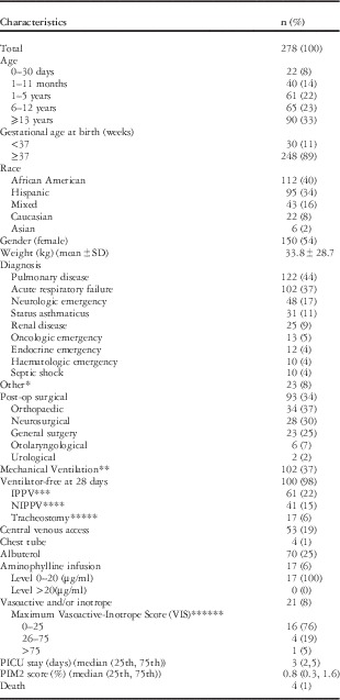
There were 278 consecutive patients who met the inclusion criteria and were included during the 4-month study period. The characteristics of the study population are shown in Table 1. A total of 53 patients (19%) had at least one arrhythmia during their paediatric intensive care stay, and six patients (2%) had one arrhythmia each that was identified as a tachyarrhythmia.
Characteristics of the study population (n=278).

PICU=paediatric intensive care unit
Values are n (%), mean±standard deviation, or median (25th, 75th percentiles)
* Other: trauma (n=9), toxicology (n=6), autoimmune (n=5), metabolic (n=2), acute hepatic failure (n=1)
** Mechanical ventilation: a total of 102 patients required either IPPV or NIPPV at any time during ICU stay, of which 100 patients were ventilator-free at 28 days
*** IPPV: invasive positive pressure ventilation. This number reflects therapy at any time during ICU stay
**** NIPPV: non-invasive positive pressure ventilation. This number reflects therapy at any time during ICU stay
***** Tracheostomy: This number reflects therapy at any time during ICU stay
****** Maximum Vasoactive-Inotrope Score (VIS)=[dopamine dose (μg/kg/minute)+dobutamine dose (μg/kg/minute)+100×epinephrine dose (μg/kg/minute)]+(10×milrinone dose (μg/kg/minute))+(10,000×vasopressin dose (U/kg/minute))+(100×norepinephrine dose (μg/kg/minute)) (34)
Rates and types of arrhythmias
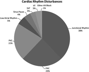
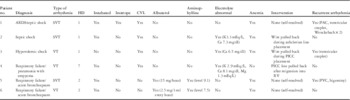
A total of 97 arrhythmias occurred in 53 patients (19%). The arrhythmias are shown in Figure 1. The most commonly encountered rhythm disturbances were as follows: junctional rhythm (n=37), premature atrial contractions (n=23), and premature ventricular contractions (n=21). Once a patient experienced an initial arrhythmia, they often experienced another, with 25 patients (47%) having more than one arrhythmia recorded during admission. Most patients experienced an arrhythmia within days 1–4 of admission (45/53 [85%]), with the majority noted on days 1 and 2 (32/53 [60%]). A total of six patients experienced one tachyarrhythmia each. Of these six tachyarrhythmias, five occurred during the first 2 days of the patient’s stay in the intensive care unit. There were three patients with supraventricular tachycardia – two non-sustained and one sustained – and three patients with non-sustained ventricular tachycardia among the six patients with tachyarrhythmias. Of these six arrhythmias, four were related to placement or migration of central venous lines. The remaining two patients were receiving aminophylline infusions at the time of arrhythmia. No pharmacological intervention was performed for termination of any arrhythmia. The details of these six patients are given in Table 2.
Cardiac rhythm disturbances. PACs=premature atrial contractions; PVCs=premature ventricular contractions; SVT=supraventricular tachycardia; VT=ventricular tachycardia.

Patients with tachyarrhythmias.

ARDS=acute respiratory distress syndrome; CVL=central venous lines, HD=hospital day; PAC=premature atrial contraction; PICC=peripherally inserted central catheter; PVC=premature ventricular contraction; RV=right ventricle; SVT=supraventricular tachycardia; VT=ventricular tachycardia
Among the six patients with tachyarrhythmias, four experienced rhythm disturbances during placement or migration of central venous lines. The first patient was a 13-year-old girl with acute respiratory distress syndrome undergoing cannulation of the internal jugular vein for veno-venous extracorporeal membrane oxygenation, who experienced sustained supraventricular tachycardia at a rate of 225 beats/minute. The arrhythmia terminated spontaneously immediately before administration of IV adenosine. The second patient was a 7-year-old girl with septic shock who had a 5-beat run of supraventricular at 300 beats/minute during placement of a subclavian central line. The third patient was a 20-year-old girl with Crohn’s disease and hypovolaemic shock who had a 6-beat run of ventricular tachycardia at 150 beats/minute that occurred during placement of a peripherally inserted central catheter. Finally, the fourth patient was an 11-year-old girl with respiratory failure due to pneumonia with empyema who had an 11-beat run of ventricular tachycardia at a peak rate of 215 beats/minute. After ventricular tachycardia was noted on telemetry, a chest x-ray revealed migration of her peripherally inserted central catheter into the right ventricle.
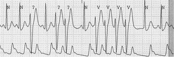
The remaining two patients with tachyarrhythmias were both receiving aminophylline infusions at the time of their arrhythmia. The first patient was an 18-month-old girl with severe bronchospasm and acute respiratory failure due to viral bronchiolitis. She experienced a non-sustained run of supraventricular tachycardia at 300 beats/minute while on an aminophylline infusion of 1 mg/kg/hour. The second patient was an 11-month-old boy, also with severe bronchospasm and acute respiratory failure due to viral bronchiolitis. He experienced a 4-beat run of ventricular tachycardia at 250 beats/minute while on an aminophylline infusion of 0.9 mg/kg/hour (Fig 2). Both patients were given an aminophylline bolus of 6 mg/kg before infusion. Neither patient had an aminophylline level that reached toxic range at the time of rhythm disturbance, with levels of 9.1 and 7.5, respectively (therapeutic range 10–20 mcg/ml). Of note, a total of 17 patients were started on aminophylline infusions during admission, none of whom had toxic serum drug levels or associated electrolyte abnormalities.
Telemetry finding in an 11-month-old boy with bronchospasm and acute respiratory failure due to viral bronchiolitis. The tracing demonstrates sinus rhythm with a premature ventricular contraction (PVC), a ventricular couplet, and a 4-beat run of VT at a rate of 250 bpm.

Predictors for any rhythm disturbance
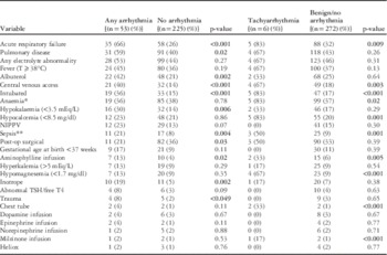
The first bivariate analysis was used to compare those patients experiencing any rhythm disturbance (n=53) with the remainder of the population (n=225). The details of this analysis are shown in Table 3. Variables associated with any rhythm disturbance included acute respiratory failure, pulmonary disease, electrolyte abnormalities, fever (T⩾38°C), use of albuterol, and central venous access. In the multivariable analysis model adjusting for age, sex, intubation, and sepsis, central venous access (odds ratio 3.1; 95% confidence interval 1.3–7.2) and use of aminophylline (odds ratio 5.1; 95% confidence interval 1.7–14.9) were independent predictors for the development of any arrhythmia (p<0.01) (Table 4).
Clinical variables in the study population.

NIPPV=non-invasive positive pressure ventilation; TSH=thyroid stimulating hormone
Values are presented as numbers (%). p-values were calculated with χ2 or Fisher’s exact test as appropriate. The left half of the table demonstrates the first bivariate analysis comparing patients with any rhythm disturbance with those with no arrhythmias. The right half of the table demonstrates the second bivariate analysis comparing patients with a tachyarrhythmia with the remainder of the patient population. Bold values indicates statistical significance for the variables.
* Anaemia is defined as haemoglobin <2 SD from the mean for age
** Sepsis is defined as a systemic inflammatory response syndrome secondary to infection, as documented by microbiology cultures or evidenced clinically by signs of infection (35)
Multivariate model for predictors of any arrhythmia.

* Odds ratios (95% CI) estimated from logistic models. Multivariate models included central venous access, aminophylline infusion, age, sex, intubation, and sepsis
Predictors for a tachyarrhythmia
An additional bivariate analysis was used to compare patients who developed a tachyarrhythmia (n=6) with the rest of the patients (n=272). The details of this analysis are also shown in Table 3. In this bivariate analysis, variables associated with a tachyarrhythmia included acute respiratory failure, central venous access, sepsis, anaemia, hypocalcaemia (<8.5 mg/dl), hypomagnesaemia (<1.7 mg/dl), use of aminophylline, higher mean maximum Vasoactive-Inotrope Score [(4.6±16.9 versus 0.79±5.8), and higher median Pediatric Index of Mortality-2 score [1.2 (0.6, 4.1) versus 0.8 (0.3, 1.4) (p<0.05)]. Central venous access and use of aminophylline were statistically significant predictors for the development of all arrhythmias in multivariate analysis and a tachyarrhythmia in univariate analysis.
Clinical outcomes
Those patients observed to have an arrhythmia on telemetry, whether or not a tachyarrhythmia, were also noted to have a more complicated course in the intensive care unit. This included longer median duration (days) of mechanical ventilation [3 (0, 10) versus 0 (0, 1), p<0.001] and longer median stay (days) in the intensive care unit [7 (4, 13) versus 2 (1.5, 4), p<0.001] (Table 5).
Patient outcomes based on the presence or absence of arrhythmia.

PICU=paediatric intensive care unit
Values are median (25th, 75th percentiles) with p-values calculated using the Mann–Whitney U test, or mean±standard deviation with p-values calculated using the independent sample t-test
Complications
During the study period, there were four deaths (1%) in the study cohort, none of which were associated with a primary arrhythmia. Each patient who died had a “Do Not Resuscitate” order at the time of death, and the rhythm just before the time of death was not included as an arrhythmia for analysis.
Discussion
In this prospective, observational study, we found that cardiac rhythm disturbances occurred in 19% of children admitted to the paediatric intensive care unit. Although rhythm disturbances were not uncommon, tachyarrhythmias occurred infrequently, in only 2% of admissions over a 4-month period, and there were no significant bradyarrhythmias. Among patients noted to have any type of arrhythmia, the course was found to be associated with a longer duration of mechanical ventilation and longer length of stay in the intensive care unit. In addition, central venous access and aminophylline infusions were independently associated with the occurrence of tachyarrhythmias.
The aetiology of arrhythmias in patients admitted to the intensive care unit is likely multifactorial. Hypoxaemia, hypercarbia, sepsis, and inflammation, all common entities during acute illness, have been identified as contributing factors to rhythm disturbances.Reference Hoffman, Wernovsky and Wieand 8 , Reference Goodman, Weiss and Weissman 18 In addition, continuous infusions of inotropes, such as dopamine, are known to be arrhythmogenic.Reference Hadjizacharia, O’Keeffe and Brown 14 Patients in the intensive care unit may have a significant stress response and increased catecholamine levels, which lead to elevated sympathetic tone.Reference Hadjizacharia, O’Keeffe and Brown 14 In adults, there are data indicating that atrial fibrillation may be triggered and maintained by such sympathetic and inflammatory mechanisms.Reference Chung, Martin and Sprecher 15 Similar inflammatory responses to those seen after cardiopulmonary bypass, with activation of the complement cascade and a systemic inflammatory response, have been demonstrated after major non-cardiothoracic surgeries.Reference Bobbio, Caporale and Internullo 16 , Reference Lin, Calvano and Lowry 17 Maximal inflammatory responses occur within the first 4 days after surgery, the time during which arrhythmias are most commonly seen.Reference Walsh, Tang, Wijewardena, Yarham, Boyle and Gaunt 7 These results correlated with our findings, in that 85% of the patients who experienced an arrhythmia did so within the first 4 days of admission.
Despite the multitude of adult studies on this topic, there are relatively few studies looking at arrhythmias in critically ill children. Badrawi et al reported the rates of arrhythmias in more than 400 patients in a neonatal intensive care unit.Reference Badrawi, Hegazy, Tokovic, Lotfy, Mahmoud and Aly 19 “Benign” arrhythmias were noted in 8.5% of the observed patients and “non-benign” arrhythmias in 1.5%. Larger chest circumferences, higher baseline heart rate, and lower oxygen saturations were shown to have a statistically significant association with non-benign arrhythmias.Reference Badrawi, Hegazy, Tokovic, Lotfy, Mahmoud and Aly 19 , Reference Dubin 20 A recent report by Rosman et al documented that, although arrhythmia alarms were common in the paediatric intensive care unit, the vast majority were either artefact or extreme heart rate values for age.Reference Rosman, Blaufox, Menco, Trope and Seiden 21 There are several studies assessing arrhythmias in critically ill children in the cardiac intensive care unit and after surgery for congenital heart disease. During the initial 24-hour postoperative period after an arterial switch procedure for transposition of the great vessels with intact ventricular septum at the Children’s Hospital Boston, Rhodes et al reported atrial ectopy in 78% and ventricular ectopy in 69% of patients.Reference Rhodes, Wernovsky and Keane 22 Telemetry monitoring in nearly 800 patients at the Children’s Hospital of Philadelphia by Hoffman et al showed 29% of patients to have at least one arrhythmia during their intensive care stay, the most common being non-sustained ventricular tachycardia in 18% of patients.Reference Hoffman, Wernovsky and Wieand 8 Ceresnak et al noted that up to 20% of patients may be paced in the postoperative period after major congenital heart surgery due to a cardiac rhythm disturbance.Reference Ceresnak, Pass and Starc 23
In this study, one predictor for the development of arrhythmias was placement or migration of a central venous line. Several authors have reported on the occurrence of arrhythmias during central line placement, and have concluded that they are self-limited and almost always without haemodynamic compromise.Reference da Silva and Waisberg 24 – Reference Stuart, Shikora and Akerman 26 Although the paediatric literature on this topic is limited, there has been an overall reported rate of 30% in children, with atrial arrhythmias comprising only 3%.Reference da Silva and Waisberg 24 In addition, there is a reported frequency of central line dislocation in ~2.5% of patients.Reference da Silva and Waisberg 24 It has been noted that peripherally inserted central catheters are more likely to become displaced compared with other long-term vascular access devices, which correlates to the one patient in our study whose central line migration was a peripherally inserted central catheter.Reference Vesely 27
The other statistically significant predictor for arrhythmias in this study was the use of intravenous aminophylline. Aminophylline is a xanthine derivative that functions as both a non-selective phosphodiesterase inhibitor and a non-selective adenosine receptor antagonist. Phosphodiesterase inhibition leads to increased tissue concentrations of cyclic adenine monophosphate, which in turn promotes catecholamine stimulation and release of epinephrine from adrenal medulla cells. In addition, the ability of aminophylline to inhibit adenosine-mediated renal vasoconstriction continues to be of clinical interest, and may increase the drug’s use in an effort to improve renal dysfunction in the intensive care unit. There have been several studies linking the use of aminophylline to tachycardia and other rhythm disturbances, but relatively few studies looking at the arrhythmogenic effects of aminophylline in critically ill children.Reference Hendeles, Bighley, Richardson, Hepler and Carmichael 28 – Reference Axelrod 31 Of the 17 patients started on an aminophylline drip, two patients experienced tachyarrhythmias. It is important to note that patients on aminophylline infusions did not have toxic serum drug levels and there were no associated electrolyte abnormalities, and thus it is reasonable to conclude that the arrhythmias were sequelae of the drug’s use. It is important to note that, although it was a predictor for the development of arrhythmias in this patient population, it never resulted in an event that required urgent clinical intervention or cessation of an aminophylline infusion.
The ability to recognise and interpret arrhythmias is essential in the paediatric intensive care unit. The determination of clinical triggers and the decision whether intervention is required, particularly for tachyarrhythmias, are of critical importance. This prospective observational study demonstrates associations between arrhythmias and several clinical variables, including placement of central venous lines and the use of aminophylline. With the growing use of aminophylline in critically ill children with renal insufficiency, further study of the association of aminophylline with cardiac rhythm disturbances and arrhythmogenicity may be warranted.Reference Axelrod 31 The association identified between arrhythmias and longer intensive care unit length of stay and requirement for ventilation suggests that arrhythmias may be markers of disease severity and illness in children in the paediatric intensive care unit.
Limitations
Although there was a thorough review of telemetry on a daily basis by the cardiology team, analysis was limited by the data stored in the telemetry system. As a result, it is possible that arrhythmias occurred that were not recorded as an arrhythmia alarm. The study was also observational, and laboratory testing for electrolyte or other abnormalities was performed on a case-by-case basis by the intensive care team and was not based on any particular protocol as part of this investigation. In addition, patients were not always followed on continuous cardiac monitoring before or after admission to the intensive care unit, and it was not known whether the observed patients had previous underlying rhythm disturbances. Given the nature of the study design, we were only able to identify associations between the exposure variables and the outcomes of interest, but were unable to establish causality. Future research would be helpful to assess the impact, if any, of arrhythmias on patient outcomes.
Conclusion
Cardiac rhythm disturbances were routinely observed in paediatric intensive care patients with non-cardiac diagnoses. In this study, arrhythmias occurred in 19% of patients, although tachyarrhythmias were rarely observed (2%). In this cohort, arrhythmias were associated with a more complicated course, including increased number of days requiring mechanical ventilation and increased length of stay in the intensive care unit, but did not have any association with mortality. In addition, central venous access and use of aminophylline were identified as two independently associated clinical factors that may be associated with the development of any arrhythmia, as well as tachyarrhythmias.
Acknowledgements
Financial Support
This research received no specific grant from any funding agency, commercial, or not-for-profit sectors.
Conflicts of Interest
None.