Introduction
A mammalian birth occurring due to somatic cell nuclear transfer (SCNT) was a remarkable demonstration of developmental plasticity (Wilmut et al., Reference Wilmut, Schnieke, McWhir, Kind and Campbell1997). Although the molecular basis of nuclear reprogramming after SCNT remains largely unknown, changes in chromatin configuration (Kikyo et al., Reference Kikyo, Wade, Guschin, Ge and Wolffe2000; Bordignon et al., Reference Bordignon, Clarke and Smith2001; Alberio et al., Reference Alberio, Johnson, Stick and Campbell2005) and epigenetic modifications, such as DNA methylation (Dean et al., Reference Dean, Santos, Stojkovic, Zakhartchenko, Walter, Wolf and Reik2001) and various modifications to histone tails (Santos et al., Reference Santos, Zakhartchenko, Stojkovic, Peters, Jenuwein, Wolf, Reik and Dean2003), are probably involved.
Epigenetics is defined as changes in gene transcription through modulation of chromatin, which are brought about by changes other than those in DNA sequence (Allis et al., Reference Allis, Jenuwein, Reinberg, Allis, Jenuwein, Reinberg and Caparros2007). In mammals, DNA methylation and post-translational modifications to histone tails are major epigenetic events. Histones (H2A, H2B, H3 and H4) are proteins essential to chromatin structure and they play an important role in regulating gene expression by covalently modifying amino acid residues through acetylation, methylation, phosphorylation and ubiquitination (Luger et al., Reference Luger, Rechsteiner, Flaus, Waye and Richmond1997; Turner, Reference Turner2000; Strahl & Allis, Reference Strahl and Allis2000; Bird, Reference Bird2002). Acetylated H3 at lysine 9 or 14 (H3K9/14ac) is associated with an active chromatin configuration (Rice & Allis, Reference Rice and Allis2001) and transcription (Fuks et al., Reference Fuks, Hurd, Wolf, Nan, Bird and Kouzarides2003), while methylated H3K9 (H3K9me) is associated with a repressed chromatin state (Fischle et al., Reference Fischle, Wang, Jacobs, Kim, Allis and Khorasanizadeh2003; Lachner et al., Reference Lachner, O'Sullivan and Jenuwein2003). Furthermore, acetylation of H3K9 is inversely correlated with DNA methylation and is associated with regulation of gene activation (Wu et al., Reference Wu, Wang, Potter, Liu, Smith, Wu, Huang and Plass2007). Therefore, abnormal epigenetic reprogramming of a transferred somatic cell nucleus, resulting in altered expression of important developmental genes to ultimately result in abnormalities and fetal losses, would not be unexpected (Bourc'his et al., Reference Bourc'his, Le Bourhis, Patin, Niveleau, Comizzoli, Renard and Viegas-Pequignot2001; Kang et al., Reference Kang, Koo, Park, Choi, Chung, Lee and Han2001, Reference Kang, Park, Koo, Choi, Kim, Lee and Han2002; Santos et al., Reference Santos, Zakhartchenko, Stojkovic, Peters, Jenuwein, Wolf, Reik and Dean2003; Wee et al., Reference Wee, Koo, Song, Kim, Kang, Moon, Kang, Lee and Han2006; Shao et al., Reference Shao, Ding, Gong and Xiao2008).
Changes in DNA methylation and histone modifications of cultured embryos can be induced by environmental factors, such in vitro culture (Doherty, Reference Doherty, Mann, Tremblay, Bartolomei and Schultz2000; Young et al., Reference Young, Fernandes, McEvoy, Butterwith, Gutierrez, Carolan, Broadbent, Robinson, Wilmut and Sinclair2001; Li et al., Reference Li, Vu, Ulaner, Littman, Ling, Chen, Hu, Behr, Giudice and Hoffman2005; Fauque et al., Reference Fauque, Jouannet, Lesaffre, Ripoche, Dandolo, Vaiman and Jammes2007). For instance, the epigenetic status of donor cells is related to the cell cycle, cell culture passage number and chemical exposure (Enright et al., Reference Enright, Jeong, Yang and Tian2003a,Reference Enright, Kubota, Yang and Tianb; Ke et al., Reference Ke, Davidson, Chen, Kluz and Costa2006). The long-term culture of bovine fibroblasts increased histone acetylation levels of H3K18 (Enright et al., Reference Enright, Jeong, Yang and Tian2003a) and blastocyst development and pregnancy rates were enhanced by using donor cells with high levels of histone acetylation of H3K9 (Yang et al., Reference Yang, Yang, Beaujean, Niu, He, Xie, Tang, Wang, Zhou and Ji2006, Reference Yang, Hao, Kessler, Brem, Wolf and Zakhartchenko2007). Moreover, cryopreservation has been recently shown to alter histone acetylation levels of H3K9 in donor cells and it indirectly affected the in vivo viability of cloned cat embryos (Gómez et al., Reference Gómez, Pope, Kutner, Ricks, Lyons, Ruhe, Dumas, Lyons, López, Dresser and Reiser2008). Similarly, epigenetic modifications may occur in cryopreserved bovine fibroblasts and such alterations may affect in vitro development of the reconstructed bovine embryos. Therefore, the objectives of the present study were to: (1) measure acetylation levels of H3K9 in cultured and cryopreserved bovine fibroblasts; and to (2) determine the influence of the epigenetic status of donor cells on the in vitro development of reconstructed embryos.
Materials and Methods
Experimental design
In Experiment 1, fibroblasts from each of the three cultures (C1, C2 and C3) frozen at PC were thawed and passaged one (P1), two (P2) or 10 (P10) times. Relative acetylation levels of H3K9 were measured at each of the three passages in each of the three cell cultures that were: (1) cultured for 3 days after cells reached 100% confluence and analysed after dissociation (cultured cells); or (2) frozen after 3 days of culture at 100% confluence, stored in liquid nitrogen (LN2) and thawed just before analysis (frozen/thawed cells). The relative acetylation levels were measured in three replicates per treatment.
In Experiment 2, fibroblasts from each of the three cell cultures (C1, C2 and C3) and at each of the three passages (P1, P2 and P10) were cultured for 3 days after cells reached 100% confluence and either (1) used immediately for HMC (cultured cells); or (2) frozen/stored in LN2 and thawed just before HMC (frozen/thawed cells). Embryo reconstruction was performed as described below. Cleavage, development to the blastocyst stage and total cell numbers in blastocysts were evaluated on day 8 of culture. The number of embryos in each treatment was derived from two to six replicates.
Chemical reagents
Unless otherwise stated, all chemicals were purchased from Sigma-Aldrich Chemical Co. Any use of trade, product, or firm names is for descriptive purposes only and does not imply endorsement by the U.S. Government.
Isolation, culture and cryopreservation of fibroblasts
Fibroblast cultures (1, 2 and 3) were generated from ear biopsies (Uni-Punch, Premier Medicals Products) from three adult Holstein cows (Bos taurus). Skin biopsies were washed in Ca2+- and Mg2+-free Dulbecco's phosphate-buffered saline (DPBS, Gibco) and minced into 1 mm2 pieces. Minced tissues were plated individually in 25 cm2 culture flasks (Nunc) containing 5 ml of DMEM supplemented with 10% (v/v) fetal bovine serum (FBS; HyClone), 6.4 mM glutamine, 0.1 mM sodium pyruvate and 50 µg/ml gentamicin at 38.5 °C in 5% CO2/air. After 7 to 10 days of incubation, monolayer outgrowths with fibroblast-like morphology at primary culture (PC) were disaggregated with 2.5 mg/ml of pronase and re-suspended in cooled (4 °C) CryoStor solution (CS10™, BioLife Solutions). Cell suspensions in 500 µl of CryoStor were aliquoted into cooled (4 °C) cryovials (1.2 ml, Nalgene) loaded into a cryo-chamber (Mr Frosty, Nalgene) and placed into a –80 °C freezer for 16–18 h before storage in liquid nitrogen (LN2).
For further passages, fibroblasts that had been frozen at PC were thawed and plated (200,000 live cells) into flat-sided tissue culture tubes (5.5 cm2, Nunc). Once 100% confluence was reached, culture medium was replaced daily and cells were cultured for another 3 days before dissociation for an additional passage until cryopreserved at P1, P2 or P10. For thawing, cryovials were removed from LN2 and warmed for 10 s in air at 22 °C before being submerged into a water bath at 37 °C or at 40 °C for 1 to 2 min. The cryoprotectant was removed by sequential dilution with DMEM medium.
Analysis of histone acetylation levels
The relative levels of histone acetylation of frozen/thawed or cultured cells were assessed by flow cytometry using minor modifications of Enright et al. (Reference Enright, Jeong, Yang and Tian2003a). Cell pellets from frozen/thawed and cultured cells from each of the three cell cultures at each of the three passages were re-suspended in cold ‘saline GM’ (Boquest et al., Reference Boquest, Day and Prather1999) and fixed in 100% ethanol at 4 °C for at least 12 h. Cells were then washed once with PBS containing 5 mM EDTA and incubated with a 1:100 dilution of the primary rabbit anti-acetyl-histone 3 lysine 9 antibody (H3K9; Upstate Cell Signaling Solutions) for 30 min at room temperature. After incubation, cells were washed twice in PBS + 5% FBS and incubated with secondary anti-rabbit goat IgG conjugated with fluorescein isothiocyanate (1:50 dilution; Chemicon International) for 30 min at 38.5 °C. Cells were counterstained with 30 µg/ml of propidium iodide (PI) and 0.3 mg/ml of RNase was added before filtering the cells through nylon mesh (30 µm pore size, Small Parts, Inc.). Appropriate flow cytometric controls included for auto-fluorescence (was replace the primary antibody with sheep immunoglobulin G) and nonspecific binding by secondary antibody (the primary antibody was omitted) were analysed. Data from ~10,000 cells per sample in triplicate were acquired with a FACS Calibur flow cytometer (Becton Dickinson [BD] Immunocytometry Systems) and data were analysed with CellQuest software (BD). The relative levels of histone acetylation were detected by the intensity of green fluorescence displayed and calculated using geometric means.
Embryo production by SCNT handmade cloning method
The handmade cloning method (HMC) was conducted according to Vajta et al. (Reference Vajta, Lewis, Hyttel, Thouas and Trounson2001; Reference Vajta, Maddox-Hyttel, Skou, Tecirlioglu, Peura, Lai, Murphy, Prather, Kragh and Callesen2005), with minor modifications. All manipulations were performed on a heated stage at 39 °C. Bovine oocytes were purchased from a commercial supplier (BioMed Inc.) and were delivered to the Audubon Center for Research of Endangered Species (ACRES) in New Orleans, USA by overnight express transport at 39 °C with suspension in in vitro maturation (IVM) medium. Briefly, at 20 h after onset of IVM, 100–150 cumulus oocyte complexes (COCs) were vortexed for 3 min in HEPES-buffered TCM199 (T) containing 1 mg/ml of hyaluronidase (T0; 0 = % of FBS). The oocytes were cultured in 0.5 µg/ml demecolcine to induce assisted enucleation. After 2 h of culture, the zona pellucida of denuded oocytes was removed by 2 to 5 min exposure to T0 containing 2.5 mg/ml of pronase. By using a stereomicroscope (×20), zona-free oocytes with extrusion cones or with a polar body were placed in a 30 µl droplet of T20 (20 = % of FBS) under mineral oil (Sage, BioPharma) and manually bisected with an ultra sharp splitting blade (AB Technology). All demi-oocytes were stained with 25 µg/ml Hoechst 33342 in T2 (2 = % of FBS) for 5 min and subsequent selection of half-oocytes without chromatin (cytoplasts) was performed by epifluorescence microscopy. Exposure to ultraviolet light was restricted to 1 s. Cytoplasts were then cultured in a 4-well dish with 400 µl of modified synthetic oviduct fluid medium supplemented with 2.7 mM myoinositol, 10 µl/ml essential (BMEM 50×) and non-essential amino acids (MEM 100×) and 5% FBS (Medio SOFaa; Holm et al., Reference Holm, Booth, Schmidt, Greve and Callesen1999) at 39 °C before being fused individually to a somatic cell.
For embryo reconstruction, one cytoplast was incubated briefly in 1 mg/ml phytohemagglutinin before being rolled gently over a single, presumably synchronized (G0/G1) fibroblast (cultured or frozen/thawed) located in a drop of T2 medium supplemented with 0.5% of bovine serum albumin (BSA; fatty acid free). Following attachment, the cytoplast-somatic cell pair and another single cytoplast were transferred into a 60 mm petri dish containing 5 ml of fusion medium (0.3 M mannitol, 0.1 mM Mg2+ and 1 mg/ml polyvinyl alcohol) and positioned between two stainless-steel electrodes attached to micromanipulators (LF-101; Nepa Gene). Fusion was induced by applying a 2 s AC pre-pulse of 10 V, 1 Mhz, followed by two 35-µs DC pulses of 25 V at intervals of 2 s. Triplets (cytoplast–cell–cytoplast) were washed and cultured in 5 µl droplets of SOFaaci medium under mineral oil and after 30 min, fusion was evaluated visually. Activation of triplets was performed 2–3 h after fusion (28 h after onset of IVM) by incubation in 5 µM Ca ionophore A23187 (in 400 µl T2) for 5 min at 38.5 °C and then in 5-µl droplets of SOFaa medium supplemented with 2 mM 6-dimethylaminopurine (6-DMAP) at 38.5 °C in 5% CO2 and 95% air under mineral oil for 4 h. Following activation, reconstructed triplets were cultured individually in 400 µl of SOFaa medium in a well-of-the-well (WOW) system (Vajta et al., Reference Vajta, Peura, Holm, Paldi, Greve, Trounson and Callesen2000) under mineral oil in a humidified atmosphere of 5% CO2, 5% O2 and 90% N2 at 38.5 °C. On day 8 of culture, development to the blastocyst stage and total cell numbers were evaluated.
Embryo assessment
Blastocyst cell number, both total and inner cell mass/trophectoderm cell (ICM/TPD) ratio were determined by using a protocol previously described by Wells (Reference Wells2000), with minor modifications. Blastocysts were incubated in 1 mg/ml Hoechst 33342 stain solution for 1 h at 38 °C. After incubation, blastocysts were exposed to 0.04% Triton-X100 in PBS containing 25 µg/ml PI for 45–60 s. Stained embryos were placed in a drop of mounting medium (Fluorguard Antifade Reagent™) on a clean glass microscope slide and cells were observed by epifluorescence microscopy. Photographic images were taken (D70, Olympus) and total cell numbers in the ICM and TPD were counted (Image Pro Plus software, version 5.0.1; Media Cybernetics). The ratio of ICM cells to TPD cells per embryo was determined by counting the number of blue (ICM) and red (TPD) cells stained by Hoechst and PI, respectively.
Statistical analysis
The geometric means of acetylation in fibroblasts in the treatment groups were analysed by three-way ANOVA (Experiment 1) and Tukey's method was used to discriminate means differences. The numbers of embryos that cleaved or developed to blastocyst stage were analysed by chi-squared test and the numbers of nuclei in blastocysts, ICM and TPD were analysed by three-way ANOVA (Experiment 2). Possible correlations between acetylation levels in donor cells and percentages of embryos at blastocyst stage were investigated using the Pearson correlation test. Statistics were performed by using SigmaStat version 3.1.1 (Systat Software, Inc.). The level of significance was set at p < 0.05.
Results
Experiment 1
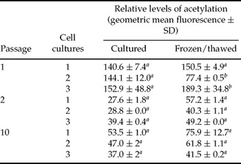
Data on the relative levels in acetylation of H3K9 in three bovine fibroblast cell cultures (C1, C2 and C3) at three passages (P1, P2 and P10) and two cell treatments (cultured vs. frozen/thawed cells) are presented in Table 1. Flow cytometric analyses revealed that the relative levels of histone acetylation of H3K9 were affected by the cell passage, with cells at P1 displaying higher intensity (142.5 ± 39.3) than those at P2 or P10 (40.4 ± 10.9 vs. 52.9 ± 14.1 respectively; p < 0.001). Overall, cryopreservation of donor cells did not alter levels of histone acetylation in each culture, except that frozen/thawed cells of culture 2 at P1 were lower (77.4 ± 0.5) and frozen/thawed cells of culture 3 at P1 were higher (189.3 ± 34.8; Table 1).
Flow cytometrically determined relative levels of acetyl H3K9 from three bovine fibroblast cultures at three passages and two cell treatment groups (cultured vs. frozen/thawed cells).

a,bDifferent superscripts within each cell treatment groups depict significant differences (p < 0.05). The relative acetylation level was measured in three replicates per treatment and ~10,000 cells in each replicate.
Experiment 2
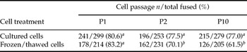
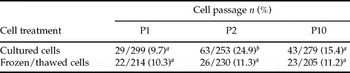
Because the genetic source (cows or culture; C1, C2 and C3) did not influence cleavage or development to the blastocyst stage, the data from the three cell cultures within the same cell passage and cell treatment were pooled (Tables 2 and 3).
Microscopically determined cleavage rates of cloned bovine embryos reconstructed with cultured or frozen/thawed donor cells after three cell passage times.

a,bDifferent superscripts between columns and rows depict significant differences (p < 0.05).
Development to the blastocyst stage (day 8) of cloned bovine embryos reconstructed with cultured or frozen/thawed donor cells at three different cell passages.

a,bDifferent superscripts between columns depict significant differences (p < 0.05).
Cleavage of reconstructed embryos was affected by cell treatment and cell passage. When embryos were reconstructed with cultured cells at P2 and P10, cleavage percentage were higher (77.5% and 77.0%, respectively) compared with frozen/thawed cells at P2 and P10 (70.1% and 61.5%, respectively; p < 0.05; Table 2). However, the cleavage rate of embryos reconstructed with frozen/thawed cells at P1 (83.2%) was similar to the rate observed with embryos reconstructed with cultured cells at P1 (80.6%; Table 2). Cell passage interacted with the treatment in embryos reconstructed with frozen/thawed cells at P1, showing a higher cleavage rate (83.2%) than those reconstructed with frozen/thawed cells at P2 and P10 (70.1% and 61.5%, respectively; p < 0.05; Table 2).
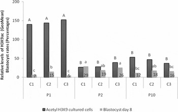
Development to the blastocyst stage was not influenced by the cell treatment or genetic source of the donor nucleus (Table 3). However in passage 2, cultured cells showed greater blastocyst development (24.9%) compared with other experimental groups (9.7% to 15.4 %, Table 3). In addition, an inverse correlation was observed between the relative levels of histone acetylation in donor cells and development to the blastocyst stage (r = −0.55; p = 0.01). Embryos reconstructed with cultured donor cells at P2 (Table 1) that had the lowest histone acetylation levels (C1 = 27.6 ± 1.8; C2 = 28.8 ± 0.0 and C3 = 39.4 ± 0.4) produced the highest rates of development to the blastocyst stage (C1 = 29.4%; C2 = 19.2% and C3 = 26.1%; Fig. 1).
Relative levels of acetyl H3K9 in bovine fibroblast cultures at three passages (P1, P2 and P10) and development to blastocyst stage (8 days) after reconstruction with three fibroblast cultures. A, B Black bars with different superscripts are significantly different in relative levels of acetyl H3K9 (p < 0.05).
a,b,cGrey bars with different superscripts are significantly different to development to blastocyst stage (p < 0.05). Numbers in gray bars are percentages of development to blastocyst stage.

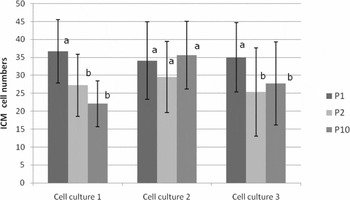
The total cell number and TPD cells in bovine cloned blastocysts were influenced by the genetic source of the donor nucleus, and the numbers of ICM cells by the cell passage. The ratios of ICM/TPD were only influenced by the cell treatment. Blastocysts derived from embryos reconstructed with cell culture 2 had higher TPD cells (61.9 ± 21.9) and total cell numbers (95.2 ± 29.2) than that of blastocysts derived from cell cultures 1 and 3 (TPD = 48.0 ± 21.7 vs. 52.2 ± 22.6; respectively; and the total cell numbers were 75.9 ± 31.1 and 80.2 ± 30.0, respectively; p < 0.05; Table 4). The numbers of ICM cells in blastocysts derived from cell cultures 1 and 3 at P1 were higher (36.7 ± 8.8 and 35.0 ± 6.4; respectively) than at P2 (27.2 ± 10.8 and 25.3 ± 9.4; respectively) and P10 (22.1 ± 9.7 and 27.7 ± 11.6 respectively; p < 0.05; Fig. 2). Blastocysts derived from embryos reconstructed with cell culture 2 at P10 had more cells in their ICM (35.6 ± 12.3) than in embryos reconstructed with cell culture 1 and 3 at P10 (22.1 ± 9.7 and 27.7 ± 11.6 respectively; p < 0.05; Fig. 2).
Total cell numbers of blastocysts (day 8) reconstructed with three donor cell cultures.

a,bDifferent superscripts in the same column depict statistical difference (p < 0.05).
ICM cell numbers in bovine blastocysts reconstructed with three donor cell cultures at three cell passages (P1, P2 and P10). Different superscripts within the same cell culture depict statistical difference (p < 0.05).

ICM/TPD ratios in P10 were higher in embryos reconstructed with cultured cells (C2 = 1:1.9 ± 0.1 and C3 = 1:1.9 ± 0.2) compared with the ICM/TPD ratios of embryos reconstructed with frozen/thawed cells (C2 = 1:1.6 ± 0.1 and C3 = 1:1.0 ± 0.3; p < 0.05). However, ICM/TPD ratios in P10 were lower in embryos reconstructed with cultured cells in C1 (1:1.4 ± 0.2) compared with the ratio in frozen/thawed cells in C1 (1:1.9 ± 0.2).
A positive correlation was observed between the relative levels of histone acetylation in donor cells and ICM cell numbers in the blastocyst stage (r = 0.52; p = 0.02).
Discussion
In the present study, we found that cryopreservation did not alter the overall acetylation levels of H3K9 in bovine fibroblasts analysed immediately after thawing (frozen/thawed) compared with fibroblasts cultured for a period of time after thawing. However, reduced cleavage rates were noted in embryos reconstructed with fibroblasts used immediately after thawing. In addition, increasing the times the culture was passaged reduced the relative acetylation levels of H3K9 in fibroblasts and, in turn, this reduction appeared to increase percentages of embryos that developed into blastocysts. Nonetheless, there was no effect of cell treatment on the total cell and ICM numbers, or on the ICM/TPD ratios. However, the genetic source did influence the total number of cells and the trophectoderm cell numbers, as well as the cell passage on the ICM cell numbers.
Several factors associated with in vitro culture may alter histone acetylation levels of cultured cells (Antequera et al., Reference Antequera, Boyes and Bird1990; Pnueli et al., Reference Pnueli, Edry, Cohen and Kassir2004; Yang et al., Reference Yang, Yang, Beaujean, Niu, He, Xie, Tang, Wang, Zhou and Ji2006; Ke et al., Reference Ke, Davidson, Chen, Kluz and Costa2006; Allegrucci, Reference Allegrucci, Way's., Denning, Priddle, Mummery, Ward-van Oostwaard, Andrews, Stojkovic, Smith, Parkin, Jones, Warren, Yu, Brena, Plass and Young2007; Noer, Reference Noer2008). In fact, Enright et al. (Reference Enright, Jeong, Yang and Tian2003a) reported that prolonged in vitro culture increased acetylation levels of H3K18 and H4K8 in bovine fibroblasts cultured from P5 to P15. In the present study, we also observed an influence of passages in culture on the relative levels of histone acetylation. But, contrary to the results presented by Enright et al. (Reference Enright, Jeong, Yang and Tian2003a), we observed a reduction of acetylation levels in H3K9 at P2 and P10 compared with that observed at P1. Although we cannot make direct comparisons between our results and those of Enright et al. (Reference Enright, Jeong, Yang and Tian2003a), it is possible that the culture conditions and genotype of fibroblast cells between the two studies were not exactly alike and this could account for differences in acetylation levels.
Acetylation function in H3K9, H3K18 and H4K8 has been associated with transcriptional activation; however, acetylation in H3K9 is regulated by different transcriptional co-activators (Spencer et al., Reference Spencer, Jenster, Burcin, Allis, Zhou, Mizzen, McKenna, Onate, Tsai, Tsai and O'Malley1997; Schiltz et al., Reference Schiltz, Mizzen, Vassilev, Cook, Allis and Nakatani1999). Therefore, it is possible that acetylation of H3K9 in bovine fibroblasts is regulated by different transcription co-activators than those that regulate acetylation in H3K18 and H4K8. In turn, activation of these co-activators during in vitro culture may result in a different acetylation pattern in H3K9. Future studies designed to elucidate which transcriptional co-activators acetylate specific sites on histone in bovine fibroblasts will help in understanding how in vitro culture affects epigenetic status.
Low temperature and cryopreservation have been considered as additional factors that modify acetylation patterns in histones. In fact, plant cells exposed to low temperatures during vernalization exhibited a reduction in acetylation levels of lysine 29 and 27 on histone 3 (H3K9/27; Amasino, Reference Amasino2004) and cryopreservation of strawberry shoot-tips induced a significant change in global DNA methylation status. Moreover, the relative levels of acetylation of lysine 9 in histone 3 (H3K9) in sand cat fibroblast cells were significantly decreased after cryopreservation (Gómez et al., Reference Gómez, Pope, Kutner, Ricks, Lyons, Ruhe, Dumas, Lyons, López, Dresser and Reiser2008). Nonetheless, in the present study, cryopreservation did not affect relative acetylation levels of H3K9 in bovine fibroblasts. In fact, acetylation levels were similar between frozen/thawed and cultured fibroblasts. A likely explanation for the differences between our results and those reported by others is that there may be inherent variation among species and cell culture sources.
Acetylation levels of donor cells have been correlated with the success of SCNT. Donor cells from different mammalian species with high acetylation levels produced a higher percentage of embryos developed to the blastocyst stage (Yang et al., Reference Yang, Yang, Beaujean, Niu, He, Xie, Tang, Wang, Zhou and Ji2006; Zakhartchenko et al., Reference Zakhartchenko, Yang, Hao and Wolf2007) and established higher pregnancy rates (Zakhartchenko et al., Reference Zakhartchenko, Yang, Hao and Wolf2007). In this study, fibroblasts with the lowest acetylation levels produced higher development to the blastocyst stage. In previous studies, donor cells were collected from fetal or neonates, while in our study donor cells were collected from adults. Therefore, not only the age of the cell, but also in vitro culture conditions and genotype of the cells may have influenced acetylation and development.
Cloning efficiency is affected by certain characteristics of the donor cells such as genetic source, type of cell, passage of culture and cell line (Kato et al., Reference Kato, Tani and Tsunoda2000; Roh et al., Reference Roh, Shim, Hwang and Yoon2000; Vignon et al., Reference Vignon, Le Bourhis, Laloy, Lavergne, Servely, Richard, Renard and Heyman2003; Powell et al., Reference Powell, Talbot, Wells, Kerr, Pursel and Wall2004; Mastromonaco et al., Reference Mastromonaco, Perrault, Betts and King2006; Al-Rostum et al., Reference Al-Rostum, Bhojwani, Poehland, Becker, Viergutz, Brunner and Kanitz2007; Beyhan et al., Reference Beyhan, Forsberg, Eilertsen, Kent-First and First2007; Poehland et al., Reference Poehland, Al-Rostum, Becker, Viergutz, Brunner, Kanitz and Bhojwani2007). Cryopreservation of donor cells also affects in vitro development of cloned embryos. Bovine and sand cat cloned embryos reconstructed with frozen/thawed cells showed lower cleavage rates than embryos reconstructed with donor cells that have been cultured for a period of time before SCNT (Kishi et al., Reference Kishi, Itagaki, Takakura, Sudo and Teranishi2003; Gómez et al., Reference Gómez, Pope, Kutner, Ricks, Lyons, Ruhe, Dumas, Lyons, López, Dresser and Reiser2008). Similarly, in the present study, cleavage rates of bovine cloned embryos reconstructed with frozen/thawed cells at P2 or P10 were lower than that of embryos reconstructed with cultured cells at the same passage. In contrast, a previous report indicated that cryopreservation did not affect cleavage rates of bovine embryos reconstructed with frozen/thawed cells in comparison to embryos reconstructed with cultured cells (Hayes et al., Reference Hayes, Rodriguez, Gonzalez, Falcon, Aguilar and Castro2005). Although the reasons for the contradictory results are not clear, the lower cleavage rate of embryos reconstructed with frozen/thawed cells possibly may be a consequence of cryopreservation-induced changes. It has been reported that apoptotic cells that have an intact plasma membrane can fuse with recipient cytoplasts and embryos reconstructed with donor cells undergoing apoptosis have a lower incidence of cleavage (Gómez et al., Reference Gómez, Pope, Kutner, Ricks, Lyons, Ruhe, Dumas, Lyons, López, Dresser and Reiser2008). Although we did not evaluate the incidence of apoptosis in donor cells, it is possible that some embryos were reconstructed with donor cells that were undergoing apoptosis. In previous studies, we observed that 1 to 5% of bovine fibroblasts frozen using protocols identical to those in the present study were apoptotic, despite having intact cell membranes. A lower cleavage rate was observed in embryos reconstructed with frozen/thawed cells (77%) as compared with that of embryos reconstructed with cultured cells (81%; Chacón et al., Reference Chacón, Gómez, Jenkins, Leibo, Wirtu, Dresser and Pope2009). Therefore, further studies are needed to confirm whether apoptosis in donor cells is responsible for the lower cleavage rate observed in Experiment 2.
The total cell numbers and the proportions of ICM and TPD cells in blastocysts have been used as an indicator of the quality of embryos produced in vitro (Thompson, Reference Thompson1997; Van Soom et al., Reference Van Soom, Ysebaert and de Kruif1997; Fleming et al., Reference Fleming, Kwong, Porter, Ursell, Fesenko, Wilkins, Miller, Watkins and Eckert2004). Embryo growth depends on both the rate of cell proliferation and cell death and these rates are directly influenced by embryo culture conditions (Young et al., Reference Young, Sinclair and Wilmut1998). In fact, the ‘large offspring syndrome’ in cattle has been associated with lower numbers of ICM cells and total cells, thus favouring trophectoderm growth (Walker et al., Reference Walker, Hartwich and Seamark1996). Aberrations in the ICM/TPD ratio have been associated with the cloning process and not with in vitro culture (Koo et al., Reference Koo, Kang, Choi, Park, Kim, Oh, Son, Park, Lee and Han2002) because higher ICM and lower TE cell numbers in cloned embryos may be responsible for insufficient placental development (Koo et al., Reference Koo, Kang, Choi, Park, Kim, Oh, Son, Park, Lee and Han2002; Ross et al., Reference Ross, Beyhan and Cibelli2007). In addition, reduced total and ICM cell numbers in cloned embryos compared with IVF embryos was correlated with abnormal Oct4 expression (Boiani et al., Reference Boiani, Eckardt, Leu, Scholer and McLaughlin2003).
We found that embryos reconstructed with fibroblasts from cell culture 2 had higher development to the blastocyst stage and these blastocysts had higher numbers of total, ICM and TPD cells, indicating a clear influence of the genotype. Moreover, embryos reconstructed with cells that were positively influenced by the genetic source (cell culture 2) and that had the lowest acetylation levels (P2) produced the highest number of embryos that developed to the blastocyst stage, indicating that both the genetic source and the acetylation levels of donor cells influenced embryo development. Although, we did not observe an influence of the acetylation levels on the total cell numbers of derived blastocysts, we observed that the relative acetylation levels of donor cells also influenced the numbers of ICM cells, where lower acetylation produced lower cell numbers. These results indicated that development to the blastocyst stage and the ICM cell numbers in blastocysts are affected by both the genetic source and acetylation levels of donor cells. The higher cell numbers, lower acetylation levels and higher development to blastocyst stage of cells from culture 2 at P2 suggest that, of the cell lines and passages that we examined, these parameters could be the most important combination for producing potentially viable embryos. Further studies that evaluate the in vivo viability of the different groups of embryos will determine the possible association between relative acetylation levels of H3K9 in donor cells, cryopreservation effect and number of ICM cells in blastocysts.
Acknowledgements
This work was supported by Audubon Center for Research of Endangered Species (ACRES), USGS and, partially by a grant from the ACRES and LSU system collaborative projects. Colombian National University and Colciencias financed a fellowship and part of the residency of Liliana Chacón while she was conducting the research in New Orleans, Louisiana, USA.