INTRODUCTION
Cellular death types and involved pathways in unicellular parasites have great relevance to the understanding of the development and evolution of infections (Besteiro et al. Reference Besteiro, Williams, Morrison, Coombs and Mottram2006). Programmed cell death (PCD) is a regulated process allowing organisms to eliminate cells, avoiding an inflammatory response (Hengartner, Reference Hengartner2000). PCD, including apoptosis and autophagy, is well characterized in higher eukaryotes. Apoptosis (type I PCD) is a regulated mechanism of self-killing, involved in many biological processes, that is characterized in mammalian cells by cell volume reduction, extracellular phosphatidylserine exposure, caspase activation, internucleosomal DNA fragmentation, blebbing of the plasma membrane, formation of apoptotic bodies and loss of mitochondrial membrane potential with cytochrome c release into the cytoplasm (Menna-Barreto et al. Reference Menna-Barreto, Salomão, Dantas, Santa-Rita, Soares, Barbosa and de Castro2009a). Trypanosoma cruzi (T. cruzi) was the first unicellular organism in which apoptosis was described (Ameisen et al. Reference Ameisen, Idzioerk, Billaut-Mulot, Tissier, Potentier and Ouaissi1995). Autophagy (type II PCD) is the major mechanism used by eukaryotic cells for bulk degradation of proteins and organelles and is essential for maintaining cellular homeostasis and for cellular development during differentiation, metamorphosis, and aging (Alvarez et al. Reference Alvarez, Kosec, Sant'Anna, Turk, Cazzulo and Turk2008a). Type II PCD is characterized by the formation of autophagosomes, portions of cytoplasm engulfed in double-membrane vesicles, cytoskeletal preservation and absence of inflammatory response (Levine and Yuan, Reference Levine and Yuan2005). Considering that T. cruzi differentiation inside the human host is mediated by autophagy it has been proposed that the inhibition of this process could be an alternative to control the infection (Alvarez et al. Reference Alvarez, Kosec, Sant'Anna, Turk, Cazzulo and Turk2008b). In protozoa, PCD may play a role in the regulation of parasite number in biological vectors and in definitive host tissues, in the maintenance of clonality, as a mechanism of immunomodulation and of parasite differentiation (Welburn et al. Reference Welburn, Barcinski and Williams1997; Freire-de-Lima et al. Reference Freire-de-Lima, Nascimento, Soares, Bozza, Castro-Faria-Neto, de Mello, DosReis and Lopes2000; Alvarez et al. Reference Alvarez, Kosec, Sant'Anna, Turk, Cazzulo and Turk2008a). On the other hand, necrosis is defined as a non-apoptotic cell death process, displaying some aspects of programmed cellular suicide. It is characterized by dramatic alterations in mitochondria, ATP depletion, generation of reactive oxygen species, loss of calcium homeostasis, cytoplasmic vacuolization and, finally, breakdown of the plasma membrane with an inflammatory response (Zong and Thompson, Reference Zong and Thompson2006). Different techniques have been employed in the study of T. cruzi cell death, i.e. destructive techniques such as flow cytometry, fluorescence microscopy, Western blot, agarose-gel electrophoresis, and ultrastructural analysis or less destructive ones like protease inhibition analysis (Alvarez et al. Reference Alvarez, Kosec, Sant'Anna, Turk, Cazzulo and Turk2008a, Reference Alvarez, Kosec, Sant'Anna, Turk, Cazzulo and Turkb; Jiménez et al. Reference Jimenez, Paredes, Sosa and Galanti2008; Menna-Barreto et al. Reference Menna-Barreto, Salomão, Dantas, Santa-Rita, Soares, Barbosa and de Castro2009b; Irigoín et al. Reference Irigoín, Inada, Fernandes, Piacenza, Gadelha, Vercesi and Radi2009).
Nuclear magnetic resonance (NMR) spectroscopy is a non-invasive method that can be used to study the metabolism of cells and tissues as well as to monitor the effects of drugs both in vitro and in vivo providing dynamic information for the study of biological systems (Griffiths et al. Reference Griffiths and Glickson2000; Boiani et al. Reference Boiani, Aguirre, González, Cerecetto, Chidichimo, Cazzulo, Bertinaria and Guglielmo2008; Caterina et al. Reference Caterina, Perillo, Boiani, Pezaroglo, Cerecetto, González and Salerno2008). The phenomenon of mobile lipid accumulation following the induction of either apoptosis or cytostasis could be studied by NMR because of its capability to visualize these lipids (visible mobile lipids, VMLs) as a more appropriate tool to denote some types of cellular death. Several lipid-associated signals are visible in 1H NMR spectroscopy, the most important ones being the methylene (–CH2–) signal near to 1·3 ppm, and the methyl (–CH3) signal at 0·9 ppm. Increased methylene resonances in 1H NMR spectra resulting from the accumulation of VMLs correlate with the onset of apoptosis in a number of drug-treated cell models (Blankenberg et al. Reference Blankenberg, Storrs, Naumovski, Goralski and Spielman1996) reflected in an increase in the CH2/CH3-ratio (Mikhailenko et al. Reference Mikhailenko, Philchenkov and Zavelevich2005), and in some cases increment in the signals from polyunsaturated fatty acids (PUFA) at 2·8 and 5·4 ppm (Hakumäki et al. Reference Hakumäki, Poptani, Sandmair, Ylä-Herttuala and Kauppinen1999). Other modified signals are those from the choline-containing compounds (choline –Cho-, phosphatidylcholine –PTC-, phosphocholine –PC-, as well as taurine and myo-inositol) near to 3·2 ppm which have been associated with apoptosis and cell loss (Milkevitch et al. Reference Milkevitch, Shim, Pilatus, Pickup, Wehrle, Samid, Poptani, Glickson and Delikatny2005). However, changes in these signals appear not to be universal, at least in the early stages of apoptosis (Lehtimäki et al. Reference Lehtimäki, Valonen, Griffin, Väisänen, Gröhn, Kettunen, Vepsäläinen, Ylä-Herttuala, Nicholson and Kauppinen2003). The biogenesis, biochemical nature and subcellular localization of the lipids that originate these VML in NMR are still under debate; however, the 1H NMR spectroscopy has been accepted as a reliable tool to elucidate lipid biochemistry mechanisms involved in eukaryotic cells and tissues (Hakumäki and Kauppinen, Reference Hakumäki and Kauppinen2000; Quintero et al. Reference Quintero, Cabañas and Arús2007).
The aim of this study was to determine whether the 1H NMR-VML technique is able to distinguish different epimastigote-T. cruzi death processes. Our results show that the CH2/CH3 ratio and changes in the region of the methyl-choline moiety, -N+(CH3)3, but not modifications in the PUFA signals, could be used to distinguish between the types of T. cruzi death pathways applying this non-invasive technique.
MATERIALS AND METHODS
Chemicals
Chemicals were purchased from Sigma-Aldrich unless otherwise indicated. Nifurtimox was purchased from Bayer.
Cell cultures
For 1H NMR studies, T. cruzi epimastigote forms (Y strain) were cultured at 28°C for 5–7 days (exponential phase of growth) under aerobiosis in axenic BHI-tryptose milieu (33 g/l brain-heart infusion, 3 g/l tryptose, 0·02 g/l hemin, 0·3 g/L D-(+)-glucose, supplemented with 10% (v/v) calf serum, 200 000 units/l penicillin and 0·2 g/l streptomycin). For TUNEL and propidium iodide- staining studies, T. cruzi epimastigote forms (Y strain) were cultured at 28°C for 5–7 days (exponential phase of growth) under aerobiosis in axenic Diamond milieu (12·5 g/l yeast extract, 12·5 g/l tryptose, 12·5 g/l tryptone, 106 mM NaCl, 29 mM H2K2PO4, 23 mM HK2PO4, pH 7·2, 7·5 mM hemin supplemented with 10% (v/v) calf serum, 75 units/ml penicillin and 75 mg/l streptomycin).
Trypanosoma cruzi treatments
Growth in axenic culture
Cultures of Y strain epimastigotes in the exponential growth phase (5×10⁶ parasites/ml) were washed with PBS, re-suspended in BHI-tryptose milieu and then subjected to culture for 14 days. Samples (10 μl) were fixed in 90 μl of formalin (3·8%, w/v) and the number of epimastigotes was determined by counting in a Neubauer chamber. For 1H NMR studies-processes see section 2.6.
H2O2 or nifurtimox
Cultures of Y strain epimastigotes in the exponential growth phase (150×106 parasites/ml) were incubated with 2·6–530 μM (per 106 parasites/ml) freshly prepared H2O2 (for 30 min at 28°C) or with 2 μM nifurtimox (per 106 parasites/ml, for 48 h at 28°C). Treatment was stopped by centrifugation at 3000 g and the sediment was re-suspended in PBS. T. cruzi epimastigotes under the same conditions but without treatment were included as controls. For 1H NMR studies-processes see below.
Starving of cells
Cultures of Y strain epimastigotes in the exponential growth phase (150×106 parasites/ml) were washed twice with PBS, re-suspended in PBS at 50×106 cells/ml, and incubated for 24 h at 28°C. Treatment was stopped by centrifugation at 3000 g and the sediment re-suspended in PBS. T. cruzi epimastigotes at the same conditions but without treatment were included as controls. For 1H NMR studies-processes see section below.
TUNEL assays
Parasites after H2O2 or nifurtimox treatments were collected by centrifugation at 3000 g, washed twice in PBS and re-suspended in the same buffer. Once placed on a slide and dried at room temperature, the cells were fixed with methanol (70%) and washed in PBS. After permeabilization with 0·2% Triton X-100, the cells were incubated with a reaction mix containing dUTP-FITC (fluorescein isothiocyanate). Nuclei were counterstained with DAPI (4′,6-diamidine-2-phenylindol) (1 mg/ml). Fluorescence was observed using Nikon Eclipse E400 microscopy and pictures were captured with a Nikon Coolpix 4500 digital camera. Results were quantified counting 200 cells in duplicate from 3 independent experiments. Apoptotic index was obtained from the ratio between TUNEL positive cells and total cells.
Ultrastructural analysis
Starved parasites were processed for transmission electron microscopy. After washing 3 times in PBS, the parasites were fixed in 2·5% glutaraldehyde (40 min/room temperature) and post-fixed in a solution containing 1% OsO4, 0·8% potassium ferricyanide and 2·5 mM CaCl2 (30 min/room temperature). Afterwards the cells were dehydrated in an ascending alcohol series following by acetone and embedded in Epoxy-resin (Araldita Durcupan, FLUKA). Thin sections, 0·5 μm, were stained with methyleneboraxic blue (1%) and examined in a Nikon Eclipse E200 microscope. Ultrathin sections were stained with uranyl acetate and lead citrate during 10 min and examined in a Jeol JEM 1010 transmission microscope operated at 80 kV.
NMR experiments
Cell sample preparation
Treated or un-treated (control) cells were harvested and centrifuged for 10 min at 3000 g. The pellet was washed 3 times in PBS, re-suspended in PBS (500 μL), transferred to a 5 mm NMR tube (ALDRICH, USA) and D2O (90 μL) was added. The mixture was homogenized prior to acquire the spectrum.
NMR spectra acquisition
1H NMR experiments were recorded at 20°C in a Bruker Avance DPX-400 spectrometer, operating at 400·132 MHz, with a 5 mm broadband inverse geometry probe. The acquisition parameters included: 90° pulse (zgpr, avance-version v 1.7.10.2, 1D sequence with f1 presaturation), 128 scans, and spectral width of 14·983 ppm. The acquisition time was 1·3664 s. Signal intensities were calculated by performing appropriate baseline corrections and then integrating the area under each of the resonances using MestRe-C NMR software (http://mestrelab.com/). Spectra were analysed using the Topspin 1.3 software package.
The integrated regions were: 1. for CH2: 1·20–1·35 ppm; 2. for CH3: 0·80–0·90 ppm. The visualized regions were: 3. for Cho: 3·10–3·30 ppm; 4. for PUFA: 2·80 and 5·40 ppm.
Statistical analysis
Values are expressed as means±s.e.m. of at least 3 independent experiments. Statistical comparisons were performed with unpaired Student's t-tests by using OriginPro 8 software. P<0·05 was considered statistically significant.
RESULTS
Detection of apoptosis in the stationary phase of Trypanosoma cruzi epimastigote growth
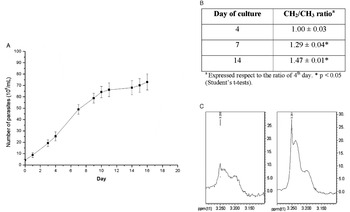
In a previous study, it was shown that natural death of T. cruzi epimastigotes occurs by PCD at the beginning of the stationary phase of growth in axenic cultures or under nutrient deprivation (Jimenez et al. Reference Jimenez, Paredes, Sosa and Galanti2008). Consequently, we studied the modifications in the VMLs profiles, using 1H NMR spectroscopy, in cultures of T. cruzi epimastigotes maintained in axenic milieu for 16 days (Fig. 1A). The best indicator of apoptosis evolution was the increment in the CH2/CH3-ratio as a function of the time of incubation. Thus, this ratio increased 29% from day 4 to day 7 of culture (Fig. 1B) reaching 47% at the stationary phase (14 days of culture). These results are in agreement with the apoptotic index previously obtained at the same times of incubation using the TUNEL technique (Jimenez et al. Reference Jimenez, Paredes, Sosa and Galanti2008). In the PUFA-signal regions, 2·8 and 5·4 ppm, we did not detect any relevant modifications; however, the ‘choline region’ clearly changed as a function of time (Fig. 1C). Though the ‘choline region’ changes were not as notorious as after treatment with H2O2 (see next section), the appearance of new peaks in this region could be indicative of the transformation of PTC into PC, Cho, or lysophosphatidylcholine. Those are well-known phenomena that play an important role in the membrane perturbations related to the apoptotic cascade (Hakumäki and Kauppinen, Reference Hakumäki and Kauppinen2000; Lutz, Reference Lutz2006).
Changes in the VML evaluated by 1H NMR spectroscopy for Trypanosoma cruzi epimastigotes (Y strain) in axenic milieu as a function of time. (A) Growth curve. (B) CH2/CH3 ratios for VMLs. (C) Choline region: parasites on the 4th (left) and 7th (right) days of incubation.

Analysis of the effects of H2O2 on Trypanosoma cruzi epimastigotes by 1H NMR
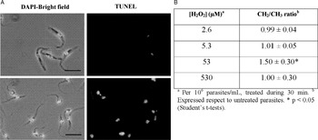
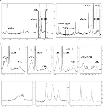
As previously described (Piacenza et al. Reference Piacenza, Peluffo and Radi2001; Deolindo et al. Reference Deolindo, Teixeira-Ferreira, DaMatta and Alves2010; Vilchez Larrea et al. Reference Vilchez Larrea, Alonso, Schlesinger, Torres, Flawiá and Fernández Villamil2011) hydrogen peroxide (H2O2) was able to induce different types of T. cruzi epimastigote cell death in a dose-dependent manner. Indeed, incubation of 150×106 parasites/ml for 30 min with H2O2 (30–85 μM per 106 parasites/ml) clearly provoked PCD as shown by the TUNEL assay (Fig. 2A). However, when the H2O2 concentration was increased up to near to 530 μM, a necrotic process took place as evidenced by light microscopy, i.e. trypan blue staining, mobility, and morphology (data not shown). These T. cruzi treatments encouraged us to investigate the VMLs modification by 1H NMR spectroscopy. Consequently, we studied the VMLs changes at different H2O2 concentrations. Applying H2O2 concentrations lower than those required to induce apoptosis, i.e. 2·6 or 5·3 μM, the CH2/CH3 ratios were similar to the values for untreated-parasites (Fig. 2B, Fig. 3B). Similarly, when the conditions were necrotic, i.e. 530 μM H2O2, the ratio was 1·0; however, when the conditions where apoptotic the CH2/CH3 ratio increased to 50%.
Effect of H2O2 on Trypanosoma cruzi epimastigotes. (A) T. cruzi epimastigotes (12×106/ml) were incubated with PBS (upper panels) or with 500 μM H2O2 (42 μM H2O2 per 106 parasites/ml) (lower panels) for 30 min at 28°C. Bars: 10 μm. (B) CH2/CH3 ratios for VMLs.

Trypanosoma cruzi epimastigotes were treated with different H2O2 concentrations and examined by 1H NMR. (A) Control (PBS, left) and H2O2 (right) 53 μM/106 parasites/ml (30 min). (B) CH2/CH3 regions for VMLs, left to right: untreated parasites (control, PBS), H2O2–parasites incubated during 30 min with 2·6, 53 and 530 μM/106 parasites/ml, respectively. (C) Choline region, left to right: untreated parasites (control, PBS, left), parasites exposed 30 min to 53 (centre), and 530 H2O2μM/106 parasites/ml (right).

In addition to the relevant changes on CH2/CH3 ratios, from VMLs, other significant modifications were observed after the different H2O2-treatments (Fig. 3A). Interestingly, in these 1H NMR experiments the whole parasites were associated with free alanine, ethanol, and glycine (for glycine signal see Fig. 6B–D) metabolites, confirmed by standards additions, which were not eliminated after exhaustive washing. The most important change was in the ‘choline region’. Under apoptotic the conditions of 53 μM of H2O2/106 parasites/ml for 30 min, new peaks were evident in this region (Fig. 3C) probably indicative of the presence of PTC and related compounds, i.e. lysophosphatidylcholine, associated with the phospholipase A2 activity during the apoptotic cascade (Hakumäki and Kauppinen, Reference Hakumäki and Kauppinen2000; Lutz, Reference Lutz2006). In contrast, the PUFA-regions showed a weak signal in the region of 2·8 ppm (Fig. 3A), but not at 5·4 ppm (data not shown), although with lower significance than the other MLVs. Under necrotic conditions, 530 μM of H2O2/106 parasites/ml for 30 min, the profile in the ‘choline region’ was similar to the profile under apoptotic conditions, with little modifications in the relative signals intensities (Fig. 3C) while the PUFA-peaks were undetectable.
1H NMR-VML spectroscopy analysis of Trypanosoma cruzi epimastigotes treated with nifurtimox
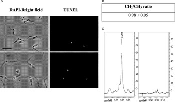
First we tested by TUNEL assay (Fig. 4A) and by light microscopy, i.e. trypan blue staining, mobility, and morphology (data not shown), whether nifurtimox, one of the drugs used for Chagas disease treatment (Cerecetto and González, Reference Cerecetto and González2010), produced necrotic death on T. cruzi epimastigotes. Then, we investigated the changes of VMLs by 1H NMR when T. cruzi was treated with nifurtimox under necrotic conditions, i.e. 2 μM (per 106 parasites/ml) during 48 h at 28°C. Results were similar to those observed when epimastigotes were treated with H2O2 at 530 μM (necrotic conditions, Fig. 2B); the T. cruzi CH2/CH3 ratio (Fig. 4B) and PUFA-signals were not modified by nifurtimox (not shown). Meanwhile, the ‘choline region’ suffered a clear modification showing that the peak of PTC disappeared as a result of the development of the necrotic process (Fig. 4C).
Effect of nifurtimox on Trypanosoma cruzi epimastigotes. (A) T. cruzi epimastigotes (12×106/ml) were incubated with PBS (upper panels) or with 8·3 μM nifurtimox (per 106 parasites/ml) (lower panels) for 24 h at 28°C. Scale bars: 10 μm. NMR-VML results for T. cruzi treatment with nifurtimox at necrotic conditions (2μM/106 parasites/ml, during 48 h at 28°C). (B) CH2/CH3 ratio. (C) Choline region: control (left) and nifurtimox-treated (right) parasites.

1H NMR-VML spectroscopy analysis of Trypanosoma cruzi epimastigotes under autophagic conditions
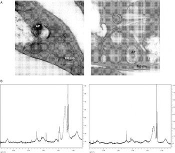
T. cruzi epimastigotes were starved for 24 h in PBS. When autophagy was reached, as detected by transmission electron microscopy (Fig. 5A), the cells were analysed by 1H NMR. Surprisingly, no significant difference in the CH2/CH3 ratios was observed between untreated and starving-parasites, 1·0±0·1 and 0·7±0·2, respectively. Similarly, no differences were either observed between both conditions in the Cho and PUFA regions (Fig. 5B).
Trypanosoma cruzi epimastigotes under autophagic conditions. (A) Transmission electron micrographs of parasites exposed for 24 h to PBS. AP, autophagosome. (B) 1H NMR spectra of whole parasites, control- (left) and starving-parasites (right).

DISCUSSION
Our interest focused on studying the use of a non-invasive technique, such as 1H NMR spectroscopy to study programmed cell death in T. cruzi as a target for drug control. This process was carefully studied in higher eukaryotes considering its relevance at the time of choosing a drug candidate (Blankenberg et al. Reference Blankenberg, Storrs, Naumovski, Goralski and Spielman1996; Hakumäki and Kauppinen, Reference Hakumäki and Kauppinen2000; Mikhailenko et al. Reference Mikhailenko, Philchenkov and Zavelevich2005; Milkevitch et al. Reference Milkevitch, Shim, Pilatus, Pickup, Wehrle, Samid, Poptani, Glickson and Delikatny2005).
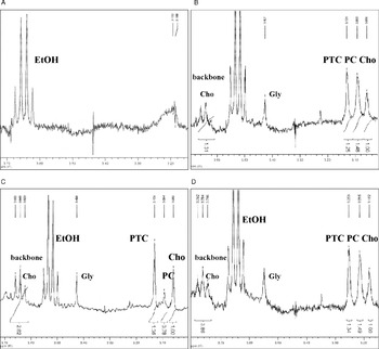
Our results, analysing VMLs, showed that the CH2/CH3 ratio increased under typical apoptotic conditions for epimastigotes of T. cruzi. These enhancements in the methylene signals are, also in the case of apoptotic eukaryotic cells, demonstrating the significance of the membrane turnover perturbations (Hakumäki and Kauppinen, Reference Hakumäki and Kauppinen2000). Furthermore, an increment in the choline-containing compound peaks was detected by 1H NMR under these conditions (Fig. 6). The choline region shows a disclosure in the signals with an enhancement of signals near to 3·18 and 3·21 ppm that could be assigned to Cho and PC, respectively (Bollard et al. Reference Bollard, Xu, Purcell, Griffin, Quirk, Holmes and Nicholson2002). The PTC signal was confirmed by PTC-standard addition (Fig. 6D). The appearance of peaks for PTC, PC, and Cho in apoptotic conditions (Fig. 6B) convinced us to assume that they are signals from membrane-lipids. This observation was also apparent in other eukaryotic apoptotic cells (Milkevitch et al. Reference Milkevitch, Shim, Pilatus, Pickup, Wehrle, Samid, Poptani, Glickson and Delikatny2005; Kettunen and Brindle, Reference Kettunen and Brindle2005). All these facts are in agreement with information related to lipid modifications upon induction of apoptosis (Al-Saffar et al. Reference Al-Saffar, Titley, Robertson, Clarke, Jackson, Leach and Ronen2002; Jimenez et al. Reference Jimenez, Paredes, Sosa and Galanti2008; Vilchez Larrea et al. Reference Vilchez Larrea, Alonso, Schlesinger, Torres, Flawiá and Fernández Villamil2011). However, under necrotic conditions (Fig. 6C), the appearance of the same signals (PTC, PC, and Cho) was more defined than in the apoptotic situation, i.e. narrow and broad, respectively (see also these appearances in Fig. 3C). It is possible that during necrosis and at the end stage of apoptosis, the cell membrane disruption and leakage of Cho-containing metabolites into the extracellular fluid explaining the presence of well-defined methyl-choline resonances, N+(CH3)3, were from associated membrane metabolites (Griffin et al. Reference Griffin, Mann, Scott, Shoulders and Nicholson2001).
‘Choline region’. (A) Control PBS-untreated parasites. (B) H2O2 (53 μM/106 parasites/ml, 0·5 h)-treated parasites. (C) H2O2 (530 μM/106 parasites/ml, 0·5 h)-treated parasites. (D) The same conditions as in (B) + PTC.

In order to study necrosis in T. cruzi, treatments with high concentrations of hydrogen peroxide or nifurtimox were applied. In both conditions, no modifications in the CH2/CH3 ratio and in PUFA-signals were observed; however dissimilar results were obtained for choline-containing compounds, particularly an increase in backbone Cho near to 3·8 ppm (Bollard et al. Reference Bollard, Xu, Purcell, Griffin, Quirk, Holmes and Nicholson2002) (Fig. 6C). These data suggest possible differences in the death-process induced by each necrotic agent. In trypanosomatids, a higher proportion of PUFAs than in their eukaryote mammalian host has been described, i.e. oleate and linoleate constitute around 60% of the total fatty acids in T. cruzi epimastigote and trypomastigote cellular forms (Florin-Christensen et al. 1997), suggesting an essential role in membrane fluidity to adapt the parasite to the environmental changes in its different hosts (Alloatti et al. Reference Alloatti, Testero and Uttaro2009; Alloatti and Uttaro, Reference Alloatti and Uttaro2011). In spite of these descriptions, in our experiments PUFAs from T. cruzi epimastigotes seemed to be non-VML in 1H NMR maybe as a result of the effects of the large dipolar couplings produced between lipids in cell membranes (Siminovitch et al. Reference Siminovitch, Ruocco, Olejniczak, Das Gupta and Griffin1988).
Data reported in the present study imply that 1H NMR of whole-T. cruzi in autophagy did not show any differential aspect in the VMLs. Although epimastigotes contains reservosomes (Cunha-e-Silva et al. Reference Cunha-e-Silva, Sant'Anna, Pereira, Porto-Carreiro, Jeovanio and de Souza2006) that concentrate lipids, these seemed to be not visible in the NMR spectroscopy. These invisible lipids could be immersed in vacuola that are immobilized in the cytoplasm and not exposed to relaxing protons (Hakumäki and Kauppinen, Reference Hakumäki and Kauppinen2000).
Our work, summarized in Table 1, may contribute to the use of a new non-invasive technique to analyse T. cruzi death pathways. As a consequence, studies on biological or biophysical aspects on T. cruzi plasma membrane and/or on the effect of natural drugs, as well as of synthetic compounds on this parasite would be easily conducted. To the best of our knowledge, there has been no previous report on the use of this technique for the study of cell death processes in trypanosomatid organisms.
Summary of results

a With respect to untreated parasites.
b n. m., no detected modifications.
ACKNOWLEDGMENTS
The authors are grateful to RIDIMEDCHAG-CYTED for the scholarship awarded to D.B. This work was supported by PEDECIBA (Uruguay), by FONDECYT 1090124 (to N.G.), by FONDECYT 1110053 (to G.C.) and by CONICYT-PBCT Anillo ACT 112 ‘Research in the Design of Pharmacological and Immunological Strategies for the Control of Parasitic and Neoplasic Aggressions’, Chile. D.B. is a fellow of ANII.