Introduction
There are currently no guidelines or recommendations for simultaneous cochlear implantation during vestibular schwannoma surgery. This is because of the difficulty of predicting the post-operative hearing results. In the literature, only a few studies on this topic are available. There are many case reports and a few case series, but it is hard to compare them as they use different audiological evaluation techniques.Reference West, Sass and Cayé-Thomasen1,Reference Bartindale, Tadokoro and Kircher2
Once the vestibular schwannoma has been removed, the option of cochlear implantation is generally evaluated according to not yet standardised parameters, as there is no definitive test capable of predicting the future hearing result. Some authors assess cochlear nerve function using audiological tests like electrical auditory brainstem response testing,Reference Lassaletta, Polak, Huesers, Díaz-Gómez, Calvino and Varela-Nieto3 while others rely on the morphological appearance of the cochlear nerve to predict a positive result after cochlear implantation.Reference Sanna, Medina, Macak, Rossi, Sozzi and Prasad4,Reference Wick, Butler, Yeager, Kallogjeri, Durakovic and McJunkin5
Another interesting question is whether the degree of pre-operative sensorineural hearing loss, because of the presence of a schwannoma, should be considered a negative prognostic factor for post-operative cochlear nerve function and for the audiological outcome after cochlear implantation. Moreover, it would be interesting to understand whether a pre-operative hearing loss due to the schwannoma might improve with tumour excision and simultaneous cochlear implantation, as direct cochlear nerve stimulation may be a possible source of functional hearing recovery. Other factors contributing to cochlear nerve damage and a possible worse audiological outcome should also be considered.
We carried out a retrospective study on patients with vestibular schwannoma who underwent surgery via a translabyrinthine or transcanal infrapromontorial approach and simultaneous cochlear implantation. It primarily evaluated the possibility of predicting the result of simultaneous cochlear implantation during schwannoma removal via an endoscopic morphological study of the cochlear nerve after vestibular schwannoma removal. A secondary endpoint of the study was to identify other possible predictors of post-operative hearing outcome, such as pre-operative hearing function, vestibular schwannoma dimensions and location (particularly involvement of the internal auditory canal fundus), and the tumour dissection technique in relation to the cochlear nerve (en bloc or piecemeal tumour removal).
Materials and methods
This study included vestibular schwannoma patients who underwent schwannoma removal via a translabyrinthine or transcanal infrapromontorial approach and simultaneous cochlear implantation. All procedures were carried out by one experienced surgeon. We selected patients with sporadic schwannomas and with schwannomas related to neurofibromatosis type 2. In the latter patients, we carried out cochlear implantation (hearing rehabilitation of the affected ear) because these individuals were likely to develop bilateral schwannomas. Conversely, in cases of sporadic schwannomas, the resection and cochlear implantation were carried out if the contralateral pre-surgery hearing was not serviceable, or the contralateral ear was unstable (i.e. chronic otitis media), when schwannoma resection was indicated and the lesion was small.
Vestibular schwannoma dissection and nerve evaluation
In both surgical approaches (translabyrinthine or transcanal infrapromontorial), once the dura of the internal auditory canal (from the fundus to the porus) and that of the posterior cranial fossa had been exposed, an incision was made along the internal auditory canal dura, and another was made perpendicular to the former on the posterior cranial fossa dura, to gain access to the pontocerebellar angle. Then the schwannoma was isolated and some neurosurgical patties were used to protect the neurovascular structures around the tumour, avoiding the use of a bipolar instrument so as not to cauterise the terminal branches of the internal auditory artery. Eventually, the vestibular schwannoma was gently removed, using dissectors and sharp tools, to preserve the integrity of the cochlear nerve and its vessels.
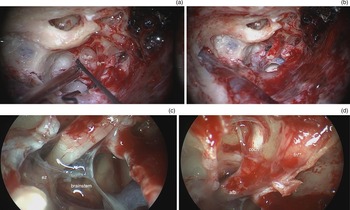
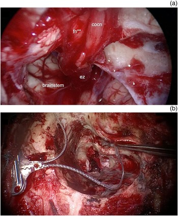
Once the tumour had been removed, the morphology of the nerve was evaluated. A 0°, 4 mm diameter, 15 cm length endoscope was introduced into the surgical cavity to assess the whole course of the cochlear nerve, from the entry zone to the fundus. Three parameters were considered: the integrity of the nerve fibres along the entire course; the whitish colour of the nerve itself after saline solution wash; and the presence of vascularity on the nerve surface (Figure 1). If all the three criteria were met, cochlear implantation was performed (Figure 2); in cases of total or partial interruption of the nerve, or when intraneural haematomas or bleeding with loss of vascularity were observed, the cochlear implantation was not carried out. Therefore, only patients with nerve integrity, with a light colour nerve and with preserved nerve vascularisation were selected for this study.
Surgical dissection, left ear. (a) Dissection procedure during schwannoma removal. (b) Microscopic view of the final surgical cavity. (c) Endoscopic magnification showing the transected vestibular nerves and the entry zone of the acoustic–facial bundle into the brainstem. (d) Endoscopic magnification displaying the preserved cochlear and facial nerves with their colour and vascularity. rw = round window; fn* = mastoid segment of the facial nerve; tum = vestibular schwannoma; fn** = meatal segment of the facial nerve; afb = acoustic–facial bundle; ez = entry zone; cocn = cochlear nerve

Endoscopic magnification and cochlear implantation, right ear. (a) Endoscopic view of the preserved cochlear and facial nerves, and of the entry zone in the brainstem. (b) Cochlear implant positioning after the vestibular schwannoma has been resected. cocn = cochlear nerve; fn** = meatal segment of the facial nerve; ez = entry zone

Patient grouping
We defined the patients who showed auditory responses during the follow-up period as responders, while those who did not show any auditory responses were defined as non-responders. The following variables were considered in the abovementioned two groups.
Pre-operative hearing function
Pre-operative pure tone average (PTA) for bone conduction and air conduction was calculated for 0.5, 1, 2 and 4 kHz frequencies. The patients were subsequently subdivided into two groups: those with pre-operative serviceable hearing and those with pre-operative non-serviceable hearing function. Serviceable hearing is defined as a PTA of 50 dB HL or lower, with a speech discrimination score of 50 per cent or higher.
Tumour dimension
Tumour dimension was measured by means of pre-operative magnetic resonance imaging (MRI).
Tumour location
The patients were also classified according to vestibular schwannoma extension to the internal auditory canal fundus. This involvement was evaluated through pre-operative MRI and through intra-operative videos.
Type of dissection
Another feature considered was the difficulty level of the surgical dissection of the tumour. It was usually removed en bloc; in cases of difficult dissection because of tumour adherence to nerves of the acoustic–facial bundle, dissection was carried out in a piecemeal fashion.
Activation and fitting of cochlear implant
The cochlear implant was activated one month after surgery, and patients subsequently underwent periodic checks. Pure tone audiometry was carried out using the cochlear implant, and PTAs were calculated for the 0.5, 1, 2 and 4 kHz frequencies. Post-operative hearing performance in both closed-set (vowel identification) and open-set (disyllabic word recognition, sentence recognition, common phrase comprehension) formats was analysed with a monitored live voice through the sound field at a level of 70 dB HL sound pressure level.
Statistical analysis
A univariate analysis of the data was conducted. The t-test was used for normally distributed continuous variables, while the chi-square test was used for categorical variables. Statistical significance was set for p-values of less than 0.05.
Results
A total of 16 patients (7 females and 9 males) were included in our study, with a mean age of 58 years. They were deemed as implantable following the abovementioned endoscopic analysis of cochlear nerve integrity. The average follow-up period was 13 months. One patient suffered from neurofibromatosis type 2 while 15 had a sporadic schwannoma. For tumour removal and cochlear implant positioning, 13 patients underwent a translabyrinthine technique, while in only 3 cases a transcanal infrapromontorial approach was carried out.
Pre-operative hearing function
Regarding pre-operative hearing function, mean PTA was 48 dB HL for bone conduction and 56 dB HL for air conduction. Seven of our patients had pre-operative serviceable hearing and nine had non-serviceable hearing, prior to surgery.
Tumour dimension
The mean schwannoma dimension was 10.8 mm, and the lesions ranged in size from 3.8 mm to 16 mm. The only neurofibromatosis type 2 patient in our case series had a 6.5 mm schwannoma in the treated ear and a 2.4 mm lesion in the contralateral ear.
Tumour location
Pre-operative gadolinium-enhanced MRI and intra-operative videos showed that 10 out of 16 vestibular schwannomas involved the fundus of the internal auditory canal.
Type of dissection
In 12 cases, the dissection was performed in an en bloc fashion, whereas in 4 cases a piecemeal dissection was necessary.
Responders and non-responders
The results for each variable are reported below, according to our subdivision of patients into responders and non-responders to cochlear implantation.
Non-responder group
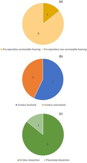
In seven cases, we found no hearing response in the implanted ear; the mean pre-operative PTA in this group was 59 dB HL for bone conduction and 63 dB HL for air conduction. Among these patients, only one had pre-operative serviceable hearing function. The mean schwannoma dimension was 11 mm in this group. Of these seven patients, four had a schwannoma extending to the fundus; the other three lesions did not involve the fundus. The tumour was dissected en bloc in six cases in this group, while only one case required piecemeal removal (Figure 3).
Pie charts for the non-responder group, showing the proportions of: (a) patients with pre-operative serviceable and non-serviceable hearing, (b) vestibular schwannoma cases with and without fundus extension, and (c) en bloc and piecemeal vestibular schwannoma removal procedures.

Responder group
We obtained good hearing results in nine patients; their mean implanted ear PTA after surgery was 51 dB HL, ranging from 34 dB HL to 90 dB HL. The speech detection and recognition intensity levels for five out of these nine patients were determined in an open-set scenario, at the time of data gathering. The mean detection threshold was 46 dB HL while the mean recognition level was 66 dB HL.
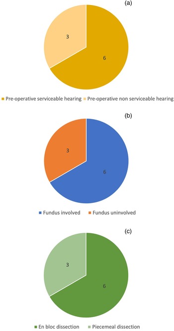
For the responder group in general, the mean PTA before surgery was 41 dB HL for bone conduction and 50 dB HL for air conduction. Six out of nine patients had pre-operative serviceable hearing function, while the other three patients had non-serviceable hearing. The mean lesion dimension was 9.8 mm for the responders. In six cases, the schwannoma extended to the fundus; the other three showed no extension to the fundus. Surgery was carried out en bloc in six cases, while the piecemeal technique was used in three cases (Figure 4).
Pie charts for the responder group, showing the proportions of: (a) patients with pre-operative serviceable and non-serviceable hearing, (b) vestibular schwannoma cases with and without fundus extension, and (c) en bloc and piecemeal vestibular schwannoma removal procedures.

Comparison between responders and non-responders
The morphological criterion used to evaluate the cochlear nerve after vestibular schwannoma dissection was predictive of a good cochlear implantation outcome in more than half of the patients in our study, with a larger number of responders than non-responders.
We found a statistically relevant difference between the two groups in terms of pre-operative serviceable hearing, which was more common in the responder group (p = 0.0362). However, no statistically significant differences were observed in terms of tumour dimension (p = 0.6273), fundus involvement (p = 0.6963) or dissection type (en bloc or piecemeal; p = 0.3827).
Discussion
Hearing rehabilitation achieved via cochlear implantation in patients with vestibular schwannoma has been described in several articles in the literature; these report implantation on the same side as the tumour after radiotherapy or observation, or implantation performed at the same time as surgical removal or after vestibular schwannoma resection.Reference West, Sass and Cayé-Thomasen1,Reference Bartindale, Tadokoro and Kircher2,Reference Sanna, Medina, Macak, Rossi, Sozzi and Prasad4,Reference Roberts, Levin, Sanli, Ferch, Kong and Eisenberg6 Although there are no accepted guidelines regarding cochlear implant positioning in patients with vestibular schwannoma, many clinics worldwide have begun to perform this procedure following expansion of the indications for cochlear implantation in recent years.Reference Bartindale, Tadokoro and Kircher2,Reference Carlson, Sladen, Gurgel, Tombers, Lohse and Driscoll7
In patients indicated for vestibular schwannoma surgical removal, cochlear implantation is usually only performed in cases of bilateral hearing loss or contralateral unstable hearing function, and only in those patients with small tumours. Therefore, the indication for vestibular schwannoma removal and simultaneous or delayed cochlear implantation is rare, and very few case series have been reported in literature. However, because of the aforementioned expansion of the indications for cochlear implantation, some surgeons place a cochlear implant after vestibular schwannoma removal even in patients with good contralateral hearing.Reference Bartindale, Tadokoro and Kircher2,Reference Carlson, Sladen, Gurgel, Tombers, Lohse and Driscoll7
Of the key factors that influence cochlear nerve function after vestibular schwannoma surgery, the dissection procedure is of paramount importance. The procedure must entail the use of sharp detaching tools, avoiding bipolar instruments, to preserve the terminal vascularity of the internal auditory artery. In addition, the dimension of the tumour and its position in relation to the fundus affect the status of the nerve after the procedure.Reference Slattery, Brackmann and Hitselberger8
According to a review by Wick et al., visualisation of cochlear nerve continuity after tumour dissection is the primary means of assessing whether to proceed with cochlear implantation.Reference Wick, Butler, Yeager, Kallogjeri, Durakovic and McJunkin5 Many intra-operative nerve monitoring techniques have been developed in recent years. For approaches that do not preserve hearing function but do preserve the cochlear structure, such as the translabyrinthine approach (the most widely used approach in our case series), electrical auditory brainstem response testing and electrically evoked compound action potential testing represent two means of predicting cochlear implant function.
Nevertheless, the effectiveness of intra-operative nerve monitoring techniques remains highly questionable, as electrically evoked compound action potentials are not always able to detect nerve lesions that are distal to the first neuron,Reference Lassaletta, Polak, Huesers, Díaz-Gómez, Calvino and Varela-Nieto3 while several instances have been reported of patients with electrical auditory brainstem response findings who did not achieve open-set speech recognition.Reference Lundin, Stillesjö and Rask-Andersen9 Moreover, both tests require the opening of a cochlear implant to create the electrically evoked signal, with an increase in costs. In addition, intra-operative cochlear nerve monitoring did not yield significant results in our experience; this lead us to rely on macroscopic observation of the cochlear nerve at the end of schwannoma removal as the main predictor of a good cochlear implant audiological outcome. Therefore, rather than electrophysiological monitoring of the nerve following dissection, an effective morphological rating system is important. This system can be used to evaluate the cochlear nerve's wellbeing and enable positive implant positioning.
Our study is the first scientific work to evaluate the morphological appearance of the cochlear nerve and investigate its impact on hearing function in patients treated via vestibular schwannoma resection and cochlear implantation. Our study primarily examined the outcome of cochlear implantation and vestibular schwannoma resection, while only relying on the morphological appearance of the cochlear nerve. In order to do this, we took advantage of endoscopic magnification of the nerve, and considered three main criteria: (1) the integrity of nerve fibres along the whole course of the cochlear nerve, from its entry zone to the fundus; (2) the nerve colour after a saline solution wash, to establish the presence of possible ecchymotic areas or nerve haematomas that might endanger the nerve itself; and (3) the nerve vascularity from the pontocerebellar angle to the fundus, to assess the terminal vessels nourishing the cochlear nerve. Of our 16 implanted patients who showed a preserved morphology of the cochlear nerve, 9 had a positive hearing outcome, while 7 were ‘non-responders’. This study shows that the morphological appearance of the cochlear nerve can have a bearing on the cochlear implantation decision-making process, as in more than half of our cases it was effective in predicting a good hearing outcome.
However, the limited number of patients in the study must be considered. It is important for future studies to carry out analyses with larger numbers of cases, and to compare these cases with an equal number of patients who are intra-operatively tested only through electrophysiological monitoring, to fully understand which criteria are more useful for predicting hearing outcome and to be able to draw proper guidelines.
We now focus on other possible pre-surgery predictors of the audiological outcome of simultaneous cochlear implant positioning and vestibular schwannoma removal. As secondary endpoints in our study, we analysed the importance of: pre-operative audiological status (serviceable or non-serviceable hearing), tumour size and location, and dissection type (en bloc or piecemeal).
Regarding the pre-operative hearing function of the affected ear, we found six cases of pre-surgery serviceable hearing in the responder group and only one case in the non-responder group. This statistically significant difference indicates that the audiological outcome of cochlear implantation after schwannoma resection is influenced by pre-operative serviceable hearing function. This is in line with the results of West and colleagues’ review of 29 studies on simultaneous cochlear implantation during vestibular schwannoma surgery.Reference West, Sass and Cayé-Thomasen1 The findings showed the effectiveness of simultaneous cochlear implantation, and underlined that better pre-operative hearing function is connected to a better post-operative performance with the cochlear implant. This is because, in cases of a pre-surgery sensorineural hearing loss, the cochlear nerve has probably already been damaged by the vestibular schwannoma itself, with subsequent nerve deterioration. Therefore, the degenerated nerve fibres could not lead to good post-operative function even if the cochlea had been preserved and the surgical dissection of the cochlear nerve had been perfectly carried out. The cochlear nerve damage caused by the presence of the tumour can be explained by the hypoxia caused by vessel compression, and it has recently been suggested that the most noxious factor for the cochlear nerve may be the vestibular schwannoma secreted extracellular vesicles.Reference Zanoletti, Mazzoni, Martini, Abbritti, Albertini and Alexandre10 In addition to the structural damage, there may also be a few functional alterations, such as a decline in neuronal conduction speed or auditory processing.
Focusing now on tumour size, it is clear that simultaneous cochlear implantation and vestibular schwannoma surgery is to be considered only in cases of small tumours (generally smaller than 1.5 cm); in patients with larger tumours, the cochlear nerve and its vascularisation are highly likely to be damaged, and anatomical preservation of the nerve becomes virtually impossible. Despite this, we did not notice a significant difference between the responder and non-responder groups in terms of tumour size, being it very limited in any case.
As far as fundus involvement is concerned, we expected it to negatively impact on the post-operative audiological outcome, as it could make hearing function preservation impossible, because of the greater fragility of the cochlear nerve at the level of its insertion into the modiolus of the cochlea.Reference Slattery, Brackmann and Hitselberger8 However, surprisingly, we found that fundus involvement did not seem to be related to a worse hearing outcome. We considered that our results might be influenced by the small number of cases, but a review involving 45 patients reached the same conclusion.Reference Bartindale, Tadokoro and Kircher2
Regarding the type of intra-operative dissection (i.e. en bloc tumour removal or piecemeal dissection performed because of cochlear nerve adherence), we did not find any statistical difference between the responder and non-responder groups, although this finding could again have been influenced by our limited number of cases.
In conclusion, cochlear implant placement during vestibular schwannoma surgery is still a rarely performed procedure because of its limited indications; hence, only a few studies on this topic are available. This paper reports one of the largest case series in the literature and it highlights how the morphological appearance of the cochlear nerve could be used to predict hearing outcome in more than half of our patients, who showed preserved morphology of the nerve itself. Moreover, our data show that satisfactory cochlear implant hearing results are closely related to pre-surgery serviceable hearing function.
• Simultaneous cochlear implantation and vestibular schwannoma surgery is a relatively new approach
• There are no standardised parameters predictive of good hearing to aid implantation decisions in patients undergoing schwannoma removal
• A morphological criterion based on endoscopic analysis of cochlear nerve fibre integrity, nerve colour and surface vascularity is a reliable marker of implant hearing outcome
• In this study, a positive response to cochlear implantation was significantly related to pre-surgery serviceable hearing
• There were no significant connections between cochlear implantation outcome and tumour size, internal auditory canal fundus involvement or dissection type
In order to identify other pre-operative factors that can predict the effectiveness of this type of hearing rehabilitation, and to prove a connection between cochlear nerve morphology after vestibular schwannoma removal and a good response to cochlear implantation, multicentric prospective studies involving larger numbers of patients are needed.
Competing interests
None declared