Introduction
FAT accounts for up to 10–15% of supraventricular tachycardia (SVT) substrates in patients <30 years. Although focal atrial tachycardia is generally benign, timely diagnosis and appropriate treatment are of utmost importance because focal atrial tachycardia can be incessant and lead to tachycardia-induced cardiomyopathy in a substantial number of patients.Reference Kang, Etheridge and Kantoch1–Reference Manolis, Wang and Estes6
Patients with focal atrial tachycardia are often resistant to pharmacological therapy and tend to respond well to transcatheter ablation – a well-established, safe, and feasible option regarded as the treatment of choice in adult patients with focal atrial tachycardia.Reference Manolis, Wang and Estes6–Reference Lee, Sanders and Kalman8 Nonetheless, there remain limited data on transcatheter ablative therapy in children with focal atrial tachycardia; the studies that do exist include a few case series and one multicentre study.Reference Kang, Etheridge and Kantoch1–Reference Elkiran, Akdeniz, Karacan and Tuzcu5
The radiation burden from the fluoroscopy employed for catheter navigation in ablation procedures has been an important disadvantage, especially in children who are known to be more susceptible to ionising radiation.Reference Balli, Kucuk, Orhan Bulut, Kemal Yucel and Celebi9 Three-dimensional electroanatomic mapping systems have emerged as a feasible alternative for catheter navigation, substantially decreasing the fluoroscopy used in ablation procedures. Nevertheless, experience is limited with transcatheter ablation procedures performed via three-dimensional mapping systems in children with focal atrial tachycardia.Reference Kang, Etheridge and Kantoch1–Reference Elkiran, Akdeniz, Karacan and Tuzcu5
In this study, we aimed to demonstrate the outcome of transcatheter ablation procedures performed through three-dimensional electroanatomic mapping systems using minimal fluoroscopy in a paediatric cohort with focal atrial tachycardia. To the best of our knowledge, this study comprises the largest series of children with focal atrial tachycardia undergoing transcatheter ablation with acute and midterm outcomes at a single centre.
Materials and methods
Patient population
Consecutive patients with focal atrial tachycardia who underwent an electrophysiologic study and a transcatheter ablation procedure in our hospital from September 2014 to February 2020 were included into the study, which was approved by the local ethics committee. Informed consent was obtained from the parents or legal guardians of all patients before the procedures. The diagnosis of focal atrial tachycardia was made according to the electrophysiological criteria previously described.Reference Kang, Etheridge and Kantoch1 Indications for the procedures included patient preference, presence of drug resistant symptoms, frequent and fast tachycardia episodes on Holter electrocardiogram, and systolic dysfunction on echocardiographic evaluations.
Electrophysiologic study
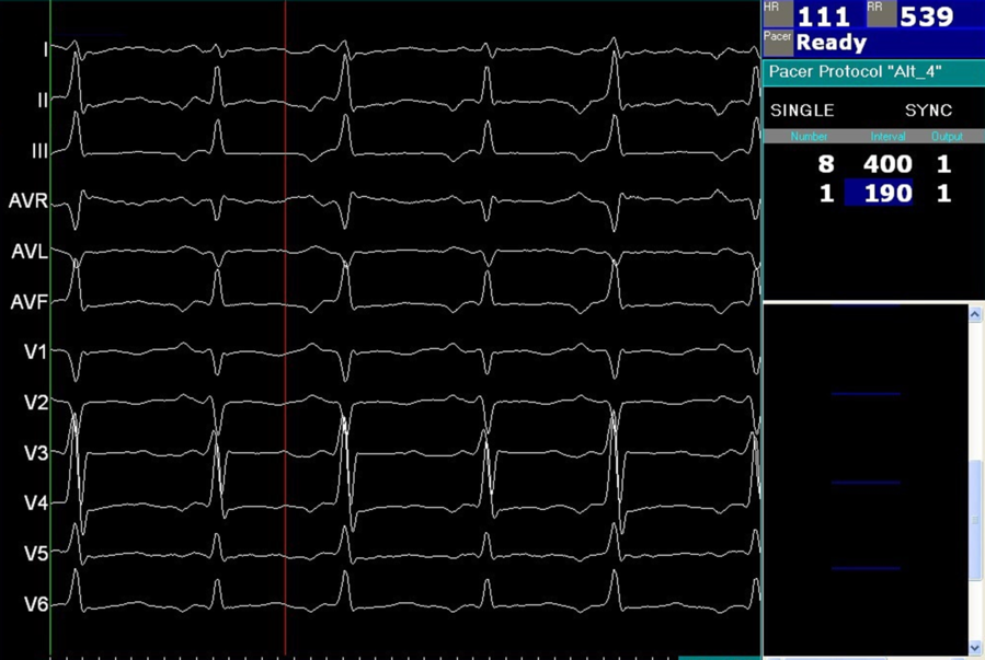
Antiarrhythmic drugs, if any, were ceased for at least five half-lives prior to the procedure except for patients with incessant tachycardia (Fig 1). Local analgesia was obtained through subcutaneous injections of lidocaine in all patients. All procedures were performed under deep sedation regardless of age to avoid map shifts caused by patients’ movements. Endotracheal intubation was not required in any patient. Heparin was used only in patients with left-sided foci. One-hundred U/kg heparin was administered intravenously once the left atrium was accessed through the interatrial septum. Repeated doses, if necessary, were administered to achieve an activated clotting time of >300 seconds. We did not use heparin in patients with right-sided foci. No prophylactic antibiotics were given in any patient.
Twelve channel electrocardiogram showing an incessant focal atrial tachycardia in a 12-year-old girl (negative P waves are noticeable in leads II, III, and AVF).

The procedures were performed with the guidance of an electroanatomic system (EnSite NavXTM system, Abbott, St. Paul, MN, USA) without using fluoroscopy in most patients. The EnSite NavXTM system uses the signal characteristics of cardiac structures to enable the construction of the three-dimensional geometry of cardiac chambers, allowing the display and navigation of catheters from the puncture site to the final destination in the heart. In our study, limited fluoroscopy was employed only to access the left atrium through transseptal puncture in patients with left-sided foci. Transesophageal echocardiography was not required during the transseptal puncture in any patient. Transseptal punctures were performed via fluoroscopy with dye staining of the septum. After the left atrium was accessed, biatrial mapping was completed without fluoroscopy. In addition to the cardiac reconstruction of the atria, respiratory gating technology was used to minimise the effect of patient respiration on the catheters’ localisation in the three-dimensional system.
A 7F sheath was inserted into the right femoral vein, and the left femoral vein was cannulated with two 5 or 6F sheaths. A quadripolar catheter then was advanced into the right atrium to obtain a three-dimensional image and to label the anatomic landmarks, including the superior vena cava, inferior vena cava, His area, and tricuspid valve annulus. We then positioned a decapolar catheter in the coronary sinus and placed two quadripolar catheters at the right ventricle apex and high right atrium. The coronary sinus catheter was used as the system reference. To avoid introducing an extra sheath into children, we did not employ a fourth catheter, instead placing the high right atrium catheter in the His bundle when needed. In patients with a baseline sinus rhythm, we performed standard programmed stimulation and burst pacing manoeuvres to induce focal atrial tachycardia. Isoproterenol infusion (1–5 μg/min) was used to help induce focal atrial tachycardia when needed. Diagnosis of focal atrial tachycardia was confirmed using the following criteria: presence of various degrees of antegrade atrioventricular block during tachycardia; induction or persistence of tachycardia independent of atrioventricular nodal conduction; inability to advance atrial activation by pr-emature ventricular stimuli delivered during tachycardia at a time when His bundle is still refractory; presence of electrogram sequence of atrial-atrial-ventricular following cessation of ventricular burst pacing.Reference Liu, Jacobsen, Pehrson and Chen10
Mapping technique
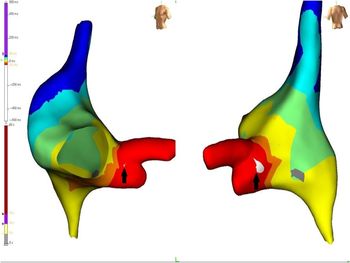
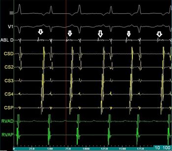
Detailed mapping was performed using the ablation catheter or a flexible quadripolar diagnostic catheter. To locate the potential targets for ablation, we used activation mapping, unipolar signals, and colour-coded electroanatomical mapping (Fig 2). Entrainment mapping also was performed to confirm the underlying mechanism before determining the target for ablation. Activation mapping is based on the prematurity of a local potential compared to the onset of P waves, which are measured point by point until finding the earliest activation point. The ablation target was a local potential ahead of the P wave by at least 20 milliseconds (Fig 3) and a unipolar wave present as a QS wave. If the onset of the P-wave was difficult to discern, we measured the activation time from the onset of the local electrogram to a stable intracardiac reference point, such as the coronary sinus ostium, with a known relationship to the P wave onset. The mechanism was defined as focal if the activation mapping revealed centrifugal spread from a single source and if the spread of local activation times was <70% of the cycle length of the tachycardia.
Right atrial activation map reconstructed during focal atrial tachycardia is shown in the left anterior oblique and posterior projections. The arrows show the earliest site of activation and the target site for ablation.

Low-amplitude, fractioned bipolar signals from the earliest activation site (arrows) are seen on the electrogram screen.

Ablation procedures
Radiofrequency was performed with a multicurve 7F Marinr® radiofrequency catheter (Medtronic Inc., Minneapolis, MN, USA). Radiofrequency was the primary energy source for catheter ablation in most of the patients. Radiofrequency energy was delivered with a target temperature of 55°C and a maximum power output of 30 W for up to 60 seconds. Cryoenergy was preferred in patients with foci close to the AV node, using a steerable 7F Freezor Xtra 6 mm tip catheter (Freezor Xtra, Medtronic, Minneapolis, MN, USA). Cryoablation was performed with a target temperature of −75°C for 240 seconds.
We defined the procedure as successful when the tachycardia was terminated and non-inducible at the end of the procedure, including a 30-minute waiting period. After the ablation procedure, the patients were hospitalised overnight with telemetry monitoring. Symptoms, electrocardiographic and echocardiographic data, 24-hour Holter monitoring, and event recording data were recorded during each follow-up visit in order to assess the success rate of the procedures.
Statistical analysis
SPSS 22.0 (IBM Corporation, Armonk, New York, United States) was used to analyse all data. The Shapiro–Wilk test was performed to determine the conformity of the data to normal distribution, and the Leneve test was conducted to ascertain variance homogeneity. Quantitative data were expressed as mean ± standard deviation. Categorical values were stated as n (number) and % (percentage).
Results
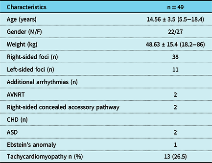
Forty-nine patients with focal atrial tachycardia underwent transcatheter ablation during the study period. The patients’ demographic and clinical characteristics are presented in Table 1. Twenty-two patients were male and 27 were female. The mean weight of the patients was 48.63 ± 15.4 kg (18.2–86), and the mean age was 14.56 ± 3.5 years (5.5–18.4).
Demographic and clinical characteristics of patients

Data presented as mean ± SD.
ASD: atrial septal defect, M/F: male/female, n: number AVNRT: atrioventricular nodal reenterant tachycardia.
The main presenting symptoms included recurrent episodes of palpitation in 33 patients, presyncope or syncope with palpitations in seven, fatigue in five, and haemodynamic collapse in two. Two asymptomatic patients also were subjected to the ablation procedure due to recurrent fast tachycardia episodes detected on Holter electrocardiogram that was performed after an incidental finding of bigeminy supraventricular ectopic beats on electrocardiogram. The presenting arrhythmia was a narrow complex tachycardia in all patients. The tachycardia was defined as incessant in 26 patients. Thirteen patients (26.5%) had left ventricular systolic dysfunction with a mean left ventricular ejection fraction of 38.47 ± 12.4% on echocardiography. Echocardiography also revealed an atrial septal defect in two patients and an Ebstein’s anomaly in one. Two patients (one with the Ebstein’s anomaly) had a right-sided concealed accessory pathway, and another two patients had atrioventricular nodal reentrant tachycardia in addition to focal atrial tachycardia. These additional lesions were successfully ablated as well.
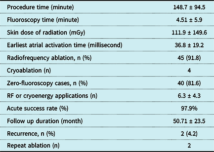
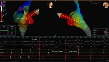
Table 2 provides the patients’ procedural characteristics. The mean procedure time was 148.7 ± 94.5 minutes. No fluoroscopy was needed in ablations performed in the right atrium. Transseptal puncture and thus fluoroscopy were required in nine patients. Left-sided foci were ablated through the patent foramen ovale two patients. The mean age of patients in whom a transseptal puncture was performed was 13.9 ± 4 years. The mean fluoroscopy time in nine patients requiring a transseptal puncture was 4.51 ± 5.9 minutes. The mean skin dose of radiation was 111.9 ± 149.6 mGy. The rhythm at the beginning of the electrophysiologic studies was sinus rhythm in eight patients. In all eight patients, the programmed stimulation, including atrial burst pacing, with or without the use of isoproterenol, induced a sustained focal atrial tachycardia. All ablation procedures were performed during the tachycardia (Fig 4).
Characteristics and outcomes of ablation procedures

Data presented as mean ± SD.
RF: radiofrequency.
Ablation lesions in lateral and posteroanterior views (arrowheads) and the position of the ablation catheter on the three-dimensional map during successful termination of tachycardia are shown. Fine fractional signals from the earliest activation site (arrows) are seen on the electrogram screen.

Thirty-eight patients had right-sided foci, while 11 had focal atrial tachycardia foci in the left atrium. The right atrial sites included the crista terminalis in 22 patients, the coronary sinus ostium in 6, the right atrial appendage in 5, the midseptal region in 2, and the parahisian area in 2. The left atrial sites included the left upper pulmonary vein ostium in four patients, the mitral annulus in three, the left atrial appendage in three, and the right upper pulmonary vein in one. All patients had a single focus except for one who had multiple ectopic foci in the right atrium. Ablation attempts failed to terminate the tachycardia in this patient. The patient responded to chronic sotalol therapy and has been followed for over 40 months with sinus rhythm. Therefore, the acute success rate of the ablation procedures was 97.9%. Radiofrequency energy was used in 45 patients, and cryoablation was performed in 4 patients with foci close to the atrioventricular node. The mean number of radiofrequency or cryoenergy lesions applied was 6.3 ± 4.3. The mean earliest atrial activation time recorded at the successful site of ablation was 36.8 ± 19.2 milliseconds. One patient developed a transient second-degree atrioventricular block, which did not preclude successful ablation. No permanent ablation-related complication occurred.
All patients were routinely evaluated 1 month after discharge and then every 3 months in the first year and every 6 months thereafter. The mean follow-up period was 50.71 ± 23.5 months. Recurrence was noted in two patients (4.2%) 1 and 5 months after the initial procedure during the follow-up period. Both underwent a repeat transcatheter radiofrequency ablation procedure, and recurrent lesions located in the same region as the initial procedure were successfully ablated. The location of recurrent lesions was right-sided (one at the coronary sinus ostium and one at the crista terminalis). Radiofrequency was the type of energy used in patients with recurrence. Left ventricular ejection fraction improved gradually to normalisation over a period of 1 to 6 months in all patients with decreased systolic functions.
Discussion
In this study, we demonstrated the results of three-dimensional electroanatomic mapping-guided transcatheter catheter ablation procedures in children with focal atrial tachycardia. Outcomes of the procedures were encouraging with quite excellent acute success rates and satisfying recurrence rates; the procedures were also performed without any major permanent complications. The procedures were feasible and safe with zero fluoroscopy in patients with right-sided focal atrial tachycardia, and only limited fluoroscopy was required during the transseptal puncture in patients with left-sided foci.
The acute and midterm success rates in our study compared well to those in the study of Toyohara et al (97.9 versus 100%).Reference Toyohara, Fukuhara, Yoshimoto, Ozaki and Nakamura2 Several other studies also reported successful results for transcatheter ablation using three-dimensional electroanatomic mapping in children with focal atrial tachycardia. The acute success rate ranged from 80 to 100% in these studies.Reference Kang, Etheridge and Kantoch1–Reference Elkiran, Akdeniz, Karacan and Tuzcu5 Cummings et al showed that the acute success rate was significantly greater in procedures performed with the guidance of an electroanatomic mapping system compared to those using a two-dimensional mapping technique (77 versus 100%).Reference Cummings, Mahle and Strieper4 They also indicated that more lesions were required to successfully ablate the tachycardia in the two-dimensional mapping group compared to the three-dimensional electroanatomic mapping group (median 9.5 versus 6.5 lesions). The mean number of lesions applied was also relatively low (mean 6.3 ± 4.3 lesions) in our study. In contrast to these studies, Kang et al,Reference Kang, Etheridge and Kantoch1 in their multicentre study analysing 249 children with focal atrial tachycardia from 10 different paediatric centres, reported that the acute success rate with three-dimensional electroanatomic mapping systems did not differ from that of the conventional mapping technique. Nevertheless, the recurrence rate was significantly lower in the electroanatomic mapping group compared to the conventional mapping group (16 versus 35%, p:0.02).Reference Kang, Etheridge and Kantoch1 Similarly, Cummings et al reported greater persistence and/or recurrence rates with the conventional mapping group compared to the electroanatomic mapping group (54 versus 11%, p:0.01).Reference Cummings, Mahle and Strieper4 The recurrence rate ranged from 11.4 to 28.6% in other studies evaluating children who received electroanatomic mapping-guided ablation for focal atrial tachycardia.Reference Toyohara, Fukuhara, Yoshimoto, Ozaki and Nakamura2,Reference Dieks, Müller and Schneider3,Reference Elkiran, Akdeniz, Karacan and Tuzcu5
The recurrence rate in our study was 4.2%. Our study suggests that three-dimensional electroanatomic mapping-guided catheter ablation procedures might be associated with very low recurrence rates in children with focal atrial tachycardia. We thought that the lower recurrence rate in our study compared to previous studies also using three-dimensional mapping systems could be due to the fewer number of focal atrial tachycardia patients with multifoci in our study, as well as the different number of ablation lesions applied. We also believed that performing deep sedation and using respiratory gating technology in all patients might have improved the accuracy of ablation lesions, minimising the effects of patients’ movements and respiration.
High radiation exposure due to the use of fluoroscopy has been a major drawback in the transcatheter treatment of paediatric arrhythmias. Ionising radiation has been known to have deterministic and stochastic effects for both the patient and the operator, including malignancy, skin injury, operator cataract, and genetic defects. Children are particularly susceptible to radiation due to their longer expected lifespan.Reference Elkiran, Akdeniz, Karacan and Tuzcu5,Reference Balli, Kucuk, Orhan Bulut, Kemal Yucel and Celebi9,Reference Kipp, Boynton and Field11 Therefore, we should aim to keep the ionising radiation burden on children as low as possible to reduce long-term cancer risk in this population. Recently, three-dimensional electroanatomic mapping systems have significantly reduced the need to use fluoroscopy for the guidance of transcatheter ablation in paediatric arrhythmias.Reference Balli, Kucuk, Orhan Bulut, Kemal Yucel and Celebi9,Reference Smith and Clark12 However, further systematic studies are needed to demonstrate the efficacy and safety of electroanatomic mapping-guided transcatheter ablation procedures in children with focal atrial tachycardia. Recently, Elkiran et al reported successful results for electroanatomic mapping-guided transcatheter ablation procedures using limited/zero fluoroscopy in children with focal atrial tachycardia.Reference Balli, Kucuk, Orhan Bulut, Kemal Yucel and Celebi9 They suggested that the use of three-dimensional mapping systems enabled them to achieve lower fluoroscopy time during procedures. Fluoroscopy was used for transseptal puncture, passing through the PFO and confirming the catheter position. The mean fluoroscopy time used during transseptal puncture was 7.96 ± 5.44 minutes. Other studies also reported higher fluoroscopy times in children and adults with focal atrial tachycardia who underwent a transcatheter ablation using three-dimensional electroanatomic mapping.Reference Dieks, Müller and Schneider3,Reference Cummings, Mahle and Strieper4,Reference Busch, Forkmann and Kuck13 The mean fluoroscopy time was as high as 40 ± 20 minutes in some studies even with the utilisation of a three-dimensional mapping system.Reference Cummings, Mahle and Strieper4 In our study, fluoroscopy was required only for the transseptal puncture in nine patients with left-sided foci. After the left atrium was accessed, biatrial mapping and ablation were completed without fluoroscopy. We did not employ fluoroscopy to pass through the patent foramen ovale detected in two patients. The mean fluoroscopy time was only 4.51 ± 5.9 in our study. Our study shows that significant reduction of fluoroscopy is feasible in paediatric focal atrial tachycardia patients undergoing transcatheter ablation procedures.
Some significant complications have been associated with catheter ablation of focal atrial tachycardia, including atrioventricular block, sinus node dysfunction, perforation, thrombus formation, pericardial effusion, phrenic nerve damage, thromboembolism, and cardiac arrest.Reference Busch, Forkmann and Kuck13–Reference Kugler, Danford, Houston and Felix15 In our study, we did not encounter any permanent complications. One patient developed a second-degree atrioventricular block during ablation in the parahisian region. The atrioventricular block resolved spontaneously and did not preclude a successful ablation. The energy source used in this patient was cryoenergy. The use of radiofrequency energy in the septum region close to the atrioventricular node or His bundle is associated with increased risk of permanent conduction disturbances. Cryoablation, offering certain advantages as reversible cryomapping and cryoadhesion, has been shown to be safe and efficacious in midseptal and parahisian tachycardias and should be used as a first-line therapy in these patients.Reference Wu, Chiu, Lu, Tseng and Wu16
Conclusion
The outcomes are promising for three-dimensional electroanatomic mapping-guided transcatheter ablation procedures for children with focal atrial tachycardia, as demonstrated by the high acute success, and low recurrence and complication rates. The use of fluoroscopy can be significantly decreased with three-dimensional mapping systems in this group of patients.
Study limitations
Our study presents the experience of a single tertiary paediatric cardiology centre. The lack of a control group and relatively short follow-up duration are the study’s main limitations. However, to the best of our knowledge, this study comprises the largest series of children with focal atrial tachycardia reported from a single centre, and it provides useful data on the feasibility of catheter ablation procedures using near-zero fluoroscopy in children with focal atrial tachycardia.
Acknowledgements
None.
Financial Support
This research received no specific grant from any funding agency, commercial or not-for-profit sectors.
Conflict of Interest
None.