A 25-year-old lady, from a remote Indian village, presented to our hospital with a 7-month history of intermittent fevers, loss of weight, and shortness of breath on exertion. She had been empirically treated for tuberculosis for 3 months at her local primary health centre, without relief of her symptoms. On examination, she was febrile and cachectic, with marked conjunctival pallor. There was a continuous murmur, with a thrill over the pulmonary area. There were no other stigmas of infective endocarditis. A chest X-ray was unremarkable.
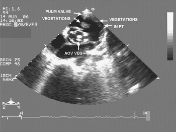
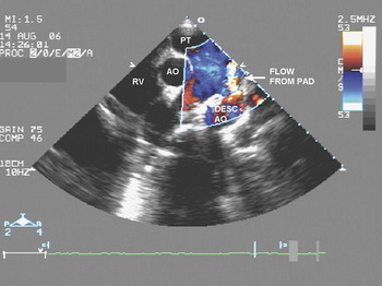
A parasternal short axis view of her echocardiogram at the great arterial level (Fig. 1) showed extensive vegetations affecting all the leaflets of her aortic and pulmonary valves, as well as vegetations attached to the lateral wall of the pulmonary trunk (PULM = pulmonary, AOV VEG = aortic valve vegetations, PT = pulmonary trunk). A persistently patent arterial duct (PAD) was then discovered by colour Doppler. (Fig. 2 – RV = right ventricle; AO = ascending aorta; DESC AO = descending aorta) The tricuspid and mitral valves were unaffected. The patient declined treatment.
Parasternal short axis echocardiogram at the great arterial level showing vegetations on the pulmonary valve, aortic valve and along the wall of the main pulmonary artery.

Colour Doppler confirming presence of a patent arterial duct.

Prior to the advent of cardiothoracic surgery, infective endocarditis was the most common cause of death in adults with persistent patency of the arterial duct.Reference Ozkokeli, Mehmet, Uslu and Akcar1 Our case is a reminder of these times. To the best of our knowledge, only 3 cases of infective endocarditis with all the lesions described above have previously been reported, including one in which the patent arterial duct was diagnosed intraoperatively.Reference Ozkokeli, Mehmet, Uslu and Akcar1 Our echocardiograms uniquely demonstrate all the major vegetations in a single frame.
Acknowledgements
No funding has been acquired by either author.