Introduction
Coronary sinus septal defect, also known as unroofed coronary sinus syndrome, is a rare type of atrial septal defect characterised by an abnormal communication between the left atrium and the coronary sinus. Reference Zieliński, Konka, Hoffman, Różański and Michałowska1 Coronary sinus septal defect accounts for approximately 1% of all atrial septal defect cases and 0.1% of all CHDs. Notably, coronary sinus septal defect is often accompanied by other cardiac anomalies. Reference Kobayashi, Muneuchi, Sugitani, Yamada, Ezaki and Watanabe2 For example, approximately 80–90% of coronary sinus septal defect cases are accompanied by a persistent left superior caval vein. Reference Al-Dairy, Ahmad and Hasan3,Reference Selcuk, Korun and Sasmazel4 Children with coronary sinus septal defect often do not exhibit distinct clinical symptoms or signs. Reference Yarrabolu, Doshi, Douglas, Aly and Balaguru5 As a result, coronary sinus septal defect is often diagnosed in conjunction with associated cardiac anomalies, such as a persistent left superior caval vein. Additionally, it is frequently misdiagnosed due to limited physician experience with its clinical and echocardiographic characteristics. Reference Xie, Yang and Cheng6,Reference Aljizeeri, Dennie, Chan and Beauchesne7 In this study, through a retrospective collection of coronary sinus septal defect cases from the largest cardiovascular centre in South China, we aimed to summarise the clinical and echocardiographic characteristics of coronary sinus septal defect. Additionally, postoperative outcomes were also analysed and reported.
Methods
Study design and participants
This is a retrospective cohort study involving 68 patients aged 18 years or younger who were diagnosed with coronary sinus septal defect based on echocardiography, supplemented by cardiac CT, or intraoperative findings, from 2011 to 2023. Due to the retrospective design, informed consent was waived, and this study was approved by the ethics committee of Guangdong Provincial People’s Hospital (No. KY-Z-2021-643-04). All the procedures were conducted in accordance with the Declaration of Helsinki.
Echocardiographic examination
Echocardiographic examination was performed by certified and experienced paediatric cardiologists from Guangdong Cardiovascular Institute. All the measurements were performed in accordance with the standardised protocol, and all segments were carefully evaluated. All the 68 patients underwent a complete transthoracic echocardiographic examination using a cardiac ultrasound system (Philips IE33, EPIQ 7C, and EPIQ Elite) with a 1.0–9.0 MHz probe. During the examination, the patients were placed in a supine or lateral position, and cardiac structure and function were sequentially evaluated in routine planes including parasternal long-axis and short-axis, apical 4-chamber, subcostal, and suprasternal views. To evaluate the coronary sinus, seven key planes were used, including the parasternal left ventricular long-axis view (Figure 1a), apical 4-chamber view (Figure 1b), post-apical 4-chamber view (Figure 1c), left posterior atrioventricular groove coronal view (Figure 1d), post-subcostal 4-chamber view (Figure 1e), post-subcostal left posterior atrioventricular groove coronal view (Figure 1f), and high aorta short-axis view (Figure 1g).
Echocardiographic planes for CS evaluation. Seven key planes were used for CS evaluation, including the parasternal left ventricular long-axis view ( a ), apical 4-chamber view ( b ), post-apical 4-chamber view ( c ), left posterior atrioventricular groove view ( d ), post-subcostal view ( e ), post-subcostal inferior left atrioventricular groove view ( f ), and high aorta short-axis view ( g ). CS = coronary sinus; LA = left atrium; PLSVC = left superior caval vein; RA = right atrium.

Types of coronary sinus septal defect
Based on the imaging and intraoperative findings, coronary sinus septal defect was divided into four types (Figure 2). Reference Chen, Yu, Wang and Chen8
Illustration of the four types of CSSD. The normal relationship between right atrium, left atrium and coronary sinus ( a ), including left superior caval vein entering to right atrium through coronary sinus ( b ). Complete unroofing of the coronary sinus: type I CSSD with concomitant persistent left SVC ( c ); type II CSSD without left SVC ( d ). Partial unroofing of the coronary sinus: type III CSSD with the defect located in the middle segment of the coronary sinus ( e ); and type IV CSSD with the defect located at the terminal end of the coronary sinus closest to its right atrial orifice ( f ). Both types III and IV could be associated with a persistent left SVC. CS = coronary sinus; IVC = inferior caval vein; LA = left atrium; PLSVC = persistent left superior caval vein; RA = right atrium; SVC = superior caval vein; ☆ = coronary sinus septal defect.

Specifically, Figure 2a shows the normal relationship between right atrium, left atrium, and coronary sinus, and Figure 2b reveals left superior caval vein entering to right atrium through coronary sinus. Complete unroofing of the coronary sinus included type I coronary sinus septal defect with concomitant persistent left superior caval vein (Figure 2c) and type II coronary sinus septal defect without concomitant persistent left superior caval vein (Figure 2d). Partial unroofing of the coronary sinus included type III coronary sinus septal defect with the defect located in the middle segment of the coronary sinus, with or without concomitant persistent left superior caval vein (Figure 2e), and type IV coronary sinus septal defect with the defect located at the terminal end of the coronary sinus closest to its right atrial orifice, with or without concomitant persistent left superior caval vein (Figure 2f).
Statistical analysis
Continuous variables were presented as median (interquartile range), and categorical variables were presented as number (percentage). Descriptive analysis was performed in this study using SPSS 24.0.
Results
Clinical characteristics of coronary sinus septal defect
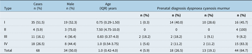
As shown in Table 1, among the 68 patients, 50% were males, the median age was 1.0 (interquartile range 0.42–4.0) years, only 4 cases (5.9%) were diagnosed at prenatal period, and 26.5%, 19.1%, and 64.7% of patients had dyspnoea, cyanosis, and murmur, respectively. The proportion of patients with type I, II, III, and IV coronary sinus septal defect was 51.5%, 5.9%, 16.1%, and 26.5%, respectively.
Clinical characteristics of CSSD

CSSD = coronary sinus septal defect; IQR = interquartile range.
Echocardiographic characteristics of coronary sinus septal defect
As shown in Figure 3, in type I coronary sinus septal defect, the wall of the coronary sinus was unable to be identified in any echocardiographic view (Figure 3a–e). Persistent left superior caval vein was connected to the left atrium between the base of left atrial appendage and the opening of upper left pulmonary vein (Figure 3h). An atrial septal defect was located at the posterior–inferior edge of the atrial septum, corresponding to the original opening of the coronary sinus. (Figure 3c–g). Shunting at the atrial level was observed on colour Doppler (Figure 3f–g). In type II coronary sinus septal defect, the walls of the coronary sinus and persistent left superior caval vein were unable to be identified on any echocardiographic view. Shunting at the atrial level was noted by colour Doppler. In type III coronary sinus septal defect (Figure 4a–d), regardless of presence or absence of persistent left superior caval vein, an enlarged coronary sinus opening into the right atrium was noted. The atrial septum near the coronary sinus opening was intact, but one or more interruptions in the echocardiographic signal were observed in the middle to upper segments of the coronary sinus wall. Shunting from left atrium to coronary sinus was detected on colour Doppler. In type IV coronary sinus septal defect, a defect was present at the end of the coronary sinus, along with an interruption at the posterior–inferior edge of the atrial septum, corresponding to the original opening of the coronary sinus (Figure 5a–e). When a persistent left superior caval vein was present, the coronary sinus was significantly dilated. Colour Doppler revealed shunting between the left atrium, the coronary sinus ostium, and the right atrium (Figure 5d–e). In the absence of a persistent left superior caval vein, the internal diameter of the coronary sinus was normal or slightly reduced.
Echocardiographic view of type I CSSD. The anatomic and haemodynamic features of type I CSSD on echocardiogram CS = coronary sinus; LA = left atrium; LSPV = left superior pulmonary vein; LIPV = left inferior pulmonary vein; PLSVC = left superior caval vein; RA = right atrium; ▲ = atrial septal defect.

Echocardiographic view of type III CSSD. The anatomic and haemodynamic features of type III CSSD on echocardiogram CS, coronary sinus; LA = left atrium; RA = right atrium; ☆ = coronary sinus septal defect.

Echocardiographic view of type IV CSSD. The anatomic and haemodynamic features of type IV CSSD on echocardiogram CS = coronary sinus; LA = left atrium; PLSVC = left superior vena cava; RA = right atrium; ▲ = atrial septal defect; ☆ = coronary sinus septal defect.

Coexisting cardiac anomalies
Coexisting cardiac anomalies of the four types of coronary sinus septal defect are shown in Table 2. Among the cohort, the most common coexisting cardiac anomalies was persistent left superior caval vein (81%), followed by secundum atrial septal defect (54%), ventricular septal defect (35%), atrioventricular septal defect (including complete, transitional, and partial type) (19%), and patent ductus arteriosus (16%). Abnormal cardiac position (15%), pulmonary artery stenosis or atresia (13%), single atrium (9%), atrial isomeric (7%), right ventricular double outlet (6%), abnormal coronary artery origin (6%), and transposition of great vessels (4%).
Coexisting cardiac anomalies

AIVC = absence of inferior caval vein; AI = atrial isomerism; AOCA = anomalous origin of coronary artery; ARSA = aberrant right subclavian artery; ASD = atrial septal defect; CoA = aortic coarctation; CTS = cor triatriatum sinister; ICSSD = isolated coronary sinus septal defect; ILAA = isomerism of left atrial appendage; CM = cardiac malposition; DORV = double outlet right ventricle; AVSD = atrioventricular septal defect; PA = pulmonary atresia; PAPVC = partial anomalous pulmonary vena connection; PDA = patent ductus arteriosus; PLSVC = persistent left superiorcaval vein; PS = pulmonary stenosis; RAA = right aortic arch; SA = single atrium; TAPVC = total anomalous pulmonary vena connection; TOF = Tetralogy of fallot; TGA = transposition of the great arteries; VSD = ventricular septal defect.
Echocardiographic characteristics of cases with missed diagnosis and misdiagnosis
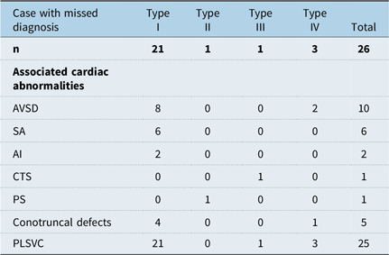
Twenty-six cases were missed diagnosis, and 1 case was misdiagnosed using echocardiographic examination alone, accounting for 39.7% of the overall 68 cases. Among the four types of coronary sinus septal defect, type I was the most frequently missed diagnosis. Associated cardiac abnormalities of each type are shown in Table 3. One case of coronary sinus septal defect was misdiagnosed as inferior caval vein-related sinus venosus atrial septal defect.
Echocardiographic characteristics of cases with missed diagnosis

AVSD = atrioventricular septal defect; SA = single atrium; AI = atrial isomerism; CTS = Cor triatriatum sinister; PS = pulmonary stenosis; PLSVC = persistent left superior vena cava.
Outcomes and types of procedures undertaken
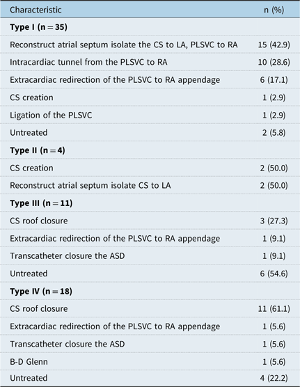
Fifty-four patients (79.4%) underwent surgery, 2 patients (2.9%) underwent transcutaneous intervention, and the rest (12; 17.6%) did not undergo surgery or intervention. The surgical procedures for coronary sinus septal defect included coronary sinus creation and roof closure (draining the coronary sinus to right atrium) and reconstruction of atrial septum. The surgical procedure for persistent left superior caval vein coexist with coronary sinus septal defect included ligation persistent left superior caval vein, intracardiac tunnel and extracardiac redirection drain the persistent left superior caval vein to right atrium, B–D Glenn draining the persistent left superior caval vein to left pulmonary artery. The details of the surgery for each type of coronary sinus septal defect are shown in Table 4. Transcatheter closure was feasible in two patients with Types III and IV (partially unroofed coronary sinus): isolating the coronary sinus to left atrium in one patient (type IV) and the other restoring the integrity of the coronary sinus (type III). At follow-up, two patients with type I coronary sinus septal defect died from multiorgan dysfunction, and one patient underwent reoperation due to narrowing of the extracardiac tunnel. The remaining patients did not experience any major events and recovered well. Of the twelve cases that did not undergo a procedure, the reasons were heterogeneous and as follows: five cases had a coronary sinus septal defect of less than 5 mm with no significant right ventricular overload; three cases involved children under 2 years old who were asymptomatic, and were instead placed under close follow-up; three cases involved complex CHD, and the parents opted not to proceed; and one case presented with severe pulmonary hypertension.
Type of surgery

ASD = atrial septal defect; LA = left atrium; RA = right atrium; PLSVC = persistent left superior caval vein; CS = coronary sinus.
Discussion
To our knowledge, this should be the first large cohort of coronary sinus septal defect in paediatric patients from China. Consistent with prior reports, Reference Tian, Su, Zhu, Zhou, Fang and Hu9,Reference Zieliński, Kowalski, Kuśmierczyk, Hoffman and Michałowska10 type I was the most common type among paediatric patients. The low detection rate during the prenatal period may be attributed to the difficulties in identifying coronary sinus. Paediatric patients often lack typical clinical symptoms and signs, making coexisting cardiac anomalies valuable for improving diagnostic accuracy. Coexisting cardiac abnormalities in type I coronary sinus septal defect are often complex, resulting in severe clinical manifestations. Some patients develop dyspnoea and cyanosis immediately after birth and most require prompt surgical intervention. In contrast, other types of coronary sinus septal defect are typically detected through the presence of a cardiac murmur during routine physical examinations.
Several imaging modalities, including echocardiography, cardiac CT, cardiac magnetic resonance, and cardiac angiography, can be used to diagnose coronary sinus septal defect. Reference Cossor, Addetia, Balkhy, Spencer and Patel11 Echocardiography is preferred as it provides a foundation for further assessment. Prior studies showed that the diagnostic rates with echocardiography ranged from 54 to 73%, Reference Aljizeeri, Dennie, Chan and Beauchesne7,Reference Gajjar, Rao and Desai12 which is consistent with our study’s rate of 62%, suggesting a high rate of missed diagnoses. The typical echocardiographic signs of coronary sinus septal defect include the inability to detect the normal coronary sinus and the right atrium entry in multiple views. Reference Yeager, Chin and Sanders13 However, due to its location in the left posterior atrioventricular groove and has a concealed position, the complete structure of the coronary sinus is often difficult to visualise in routine views, leading to it being overlooked by examiners. Therefore, summarising the anatomic and haemodynamic characteristics of coronary sinus septal defect is clinically relevant for improving diagnosis in paediatric patients.
In the 10 cases (15%) with isolated coronary sinus septal defect, the right atrium was enlarged due to left-to-right shunting at the atrial level. Therefore, in the absence of an atrial septal defect, coronary sinus septal defect is relatively easy to identify. Indeed, no cases of isolated coronary sinus septal defect were missed in our study. An additional 58 cases (85%) were accompanied by other cardiac anomalies, with persistent left superior caval vein being the most common. This finding is consistent with prior studies Reference Goyal, Punnam, Verma and Ruberg14,Reference Rees, Tahir, Ahmad and Subkovas15 and suggests that the presence of persistent left superior caval vein can aid in the detection of coronary sinus septal defect.
Persistent left superior caval vein is easily visualised in the suprasternal notch view, a standard perspective in echocardiographic examinations. It typically drains into the right atrium via an enlarged coronary sinus. However, when it is observed without an enlarged and complete coronary sinus, the presence of coronary sinus septal defect is likely. In such cases, the persistent left superior caval vein may drain through coronary sinus septal defect or directly into the left atrium. Multiple views and the use of colour Doppler can aid in identifying the entry of the persistent left superiorcaval vein into the left atrium. Additionally, in cases with a coexisting persistent left superior caval vein, contrast echocardiography can enhance diagnostic accuracy. A diagnosis can be confirmed if the contrast appears first in the left atrium, then in the coronary sinus, and finally in the right atrium. This method is particularly useful for older children when the imaging qualify of transthoracic echocardiography is suboptimal. Reference Kutty, Biko, Goldberg, Quartermain and Feinstein16 In this study, among the cases with missed diagnoses on echocardiographic examination, 25 cases (96%) had a persistent left superior caval vein. Careful examination of the suprasternal notch view to identify a persistent left superior caval vein could potentially reduce the rate of missed diagnoses.
Coronary sinus septal defect is often accompanied by atrial septal defect. Reference Urmeneta Ulloa, de Célix MDC, Cabrera, Álvarez Vázquez and de Vega17 In this study, 37 cases (54%) were associated with secundum atrial septal defect, 13 cases (19%) with atrioventricular septal defects, and 6 cases (7%) with a single atrium. This phenomenon can be explained from an embryological standpoint. Reference Sugimori, Nakao and Ikegaya18 Specifically, during embryonic development, incomplete or interrupted formation of the atrial septum can lead to various types of atrial septal defects, including secundum atrial septal defect, primum atrial septal defect, coronary sinus septal defect, a combination of these types, or even a single atrium. In our study, coronary sinus septal defect was missed in 77% of cases with a primum atrial septal defect and in 100% of cases with a single atrium. When a persistent left superior caval vein is associated with a primum atrial septal defect, particularly in the presence of a single atrium, there should be a high suspicion for coronary sinus septal defect. However, the pathophysiological characteristics of coronary sinus septal defect are often obscured by coexisting atrioventricular canal malformations, especially in cases with a single atrium. Anatomically, the coronary sinus orifice is located near the primum atrial septal defect, making it easy to confuse and potentially miss the diagnosis during echocardiographic examination. Therefore, when a primum atrial septal defect or single atrium is detected on echocardiography, the coronary sinus septum should be carefully evaluated for the presence of coronary sinus septal defect. If a normal coronary sinus is not visualised, the use of colour and pulsed Doppler to detect blood flow from the persistent left superior caval vein entering the left atrium can aid in confirming the diagnosis. Additionally, the coronary sinus opening is located adjacent to the entrance of the inferior caval vein. An inferior caval vein-related sinus venosus atrial septal defect, located between the inferior caval vein and the entrance of the right lower pulmonary vein, can appear similar to coronary sinus septal defect when viewed from the subxiphoid bi-atrial view on echocardiography. A comprehensive examination should include multiple views to assess the edges of the atrial septum. Sinus venosus atrial septal defects can be observed in standard echocardiographic views, such as the apical four-chamber view, while coronary sinus septal defect cannot. In our study, one case of coronary sinus septal defect was misdiagnosed as an inferior caval vein-related sinus venosus atrial septal defect on echocardiography but was correctly identified preoperatively using cardiac CT.
In cases of atrial isomerism, the persistent left superior caval vein often drains into the left atrium due to the complete absence of the coronary sinus wall. Therefore, when atrial isomerism is present, there is a high suspicion of type I coronary sinus septal defect. However, previous studies suggest that the coronary sinus develops from the left atrial vena fold during embryonic development. Thus, in right atrial isomerism, the coronary sinus is typically absent, and the persistent left superior caval vein drains into the left atrium, which is not classified as coronary sinus septal defect. In this study, 5 cases (7%) had atrial isomerism, all of which involved left atrial isomerism.
In recent years, the preoperative diagnostic rate of coronary sinus septal defect has improved with the use of transoesophageal echocardiography, cardiac CT, and cardiac magnetic resonance. Reference Levy, Thomas, Wethall, Perez, Steurer and Ball19 Transoesophageal echocardiography allows for clear visualisation of posterior heart structures; however, it is invasive and often requires anaesthesia in younger children, which limits its clinical application. Cardiac CT provides clearer images than transoesophageal echocardiography, showing detailed cardiovascular structures with a shorter examination time, making it suitable for children with poor compliance. However, it does carry a risk of radiation exposure. Cardiac MRI, which does not involve radiation, also offers clear images of cardiovascular structures and haemodynamics. Nonetheless, it requires anaesthesia, a longer examination time, and patient cooperation. In this study, cardiac CT was frequently used to confirm the diagnosis, with 10 cases (15%) of coronary sinus septal defect confirmed based on this imaging modality.
Careful intraoperative examination of both intracardiac and extracardiac structures is crucial. Reference Nasr, Markham and Clay20 During surgery, if the persistent left superior caval vein is found to connect directly to the left atrium rather than to the coronary sinus, or if the coronary sinus is dilated or its opening cannot be detected, the presence of coronary sinus septal defect should be suspected. Intraoperatively, the atrial septum should be opened to inspect the left atrium for diagnosis. In addition, during the intraoperative period, the surgeon must maintain a high index of suspicion for unroofed coronary sinus, especially in cases of persistent left superior caval vein. In cases of atrial septal defect, inserting a cardiotomy suction catheter through the coronary sinus and visualising it via the atrial septal defect into the left atrium can help delineate the defect and its margins, thus avoiding any missed cases. In this study, 12 cases (18%) of coronary sinus septal defect were diagnosed during surgery. Unexplained hypoxaemia postoperatively or an increased load on the right ventricle should raise suspicion of coronary sinus septal defect. Two cases in this study were missed during ventricular septal defect repair due to the presence of a persistent left superior caval vein, resulting in significant right-to-left shunting and hypoxaemia. Early reoperation was performed after diagnosing coronary sinus septal defect using echocardiography in the ICU. Additionally, due to a lack of intracardiac exploration during surgery, two other cases were missed during patent ductus arteriosus ligation and percutaneous pulmonary valve balloon dilation. These cases were isolated coronary sinus septal defect, presenting without significant symptoms or cyanosis postoperatively, and were later discovered due to right heart enlargement observed on echocardiographic examination during follow-up.
Age, along with the complexity and severity of associated cardiac anomalies, presents significant challenges for the surgical treatment of coronary sinus septal defect in paediatric patients. Reference van der Bom, Zomer, Zwinderman, Meijboom, Bouma and Mulder21 The surgical approach for coronary sinus septal defect is similar to that used in adults and depends on the type of coronary sinus septal defect, the presence of a persistent left superior caval vein, and the communication between the persistent left superior caval vein and the right superior caval vein. In this study, the rate of severe surgical complications was low, with a surgical mortality rate of 4% and a reoperation rate of 2% due to external conduit stenosis. Specifically, the two deceased cases were type I coronary sinus septal defect accompanied by a ventricular septal defect and a secundum atrial septal defect. Furthermore, the surgery was conducted when the children were only 10 months old, and these factors together might have contributed to the fatalities.
Conclusion
In summary, paediatric patients with coronary sinus septal defect often lack specific clinical manifestations, making preoperative diagnosis challenging. Echocardiography, as the primary diagnostic tool for CHD, is essential for the early detection of coronary sinus septal defect. If a diagnosis is suspected, confirmation can be achieved through cardiac CT and cardiac MRI. Accurate diagnosis and classification of coronary sinus septal defect are crucial for improving disease management. Enhancing our understanding of the anatomic and hemodynamic characteristics of coronary sinus septal defect can further improve the diagnostic accuracy of echocardiography. Precise preoperative and intraoperative diagnosis of coronary sinus septal defect contributes to high surgical success rates and favourable treatment outcomes.
Financial support
This study was supported from the funding of Guangdong Science and Technology Research Project (No. 2021111822056769) for Dr Chengcheng Pang.
Competing interests
None.