Introduction:
Evoked potentials (EPs) are the electric responses of the nervous system (cortical response) to motor or sensory stimulations. Visual, acoustic or auditory (or auditory brain stem) and somatosensory evoked potentials (or VEP, AER/AEP or ABER/ABEP, SSER/SSEP) are reliable diagnostic tests that yield reproducible results today in routine clinical practice.
These tests provide sensitive, quantitative extensions of the clinical neurological examination. They primarily afford numerical data, but sometimes the absence of wave or an abnormal configuration of its potentials also provides useful information Reference Misulis(1,Reference Chiappa, Hill and Chiappa2).
Visual evoked responses (VER/VEP) make it possible to measure the conduction of impulses through the optical nerve pathways.
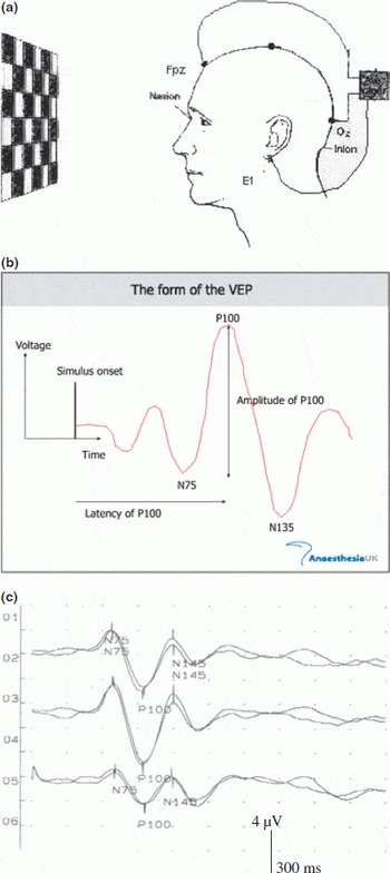
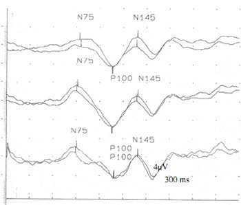
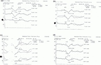
The electrodes convey every cerebral response to an optical stimulus. The results of several repetitions are then added up or computer averaged (Fig. 1a). With a healthy eye, a response to stimulus occurs after 100 to 120 milliseconds (Fig. 1b,c). A characteristic delay of this response can indicate the presence of MS (Fig. 2), and the VEP can provide important diagnostic information regarding the functional integrity of the visual system Reference Odom, Bach, Barber, Brigell, Marmor, Patrizia Tormene and Holder(3)).
a) Visual evoked potential. b) Norma visual evoked potential. c) Normal visual evoked potential.

Prolongation of Ploo latency in patient with multiple Sclerosis.

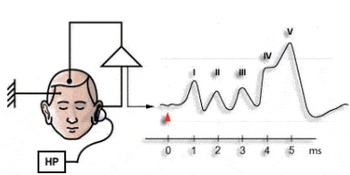
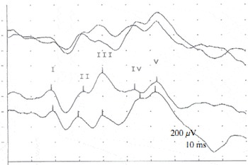
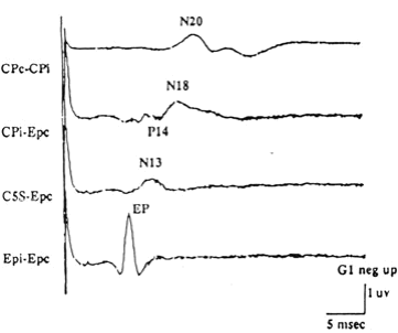
Acoustic or auditory evoked responses potentials (AER/AEP) help a specialist to detect disturbances of the sensory pathway in the auditory or acoustic nerve, as well as disturbances of the cerebellum and of the centripetal pathways. A click stimulus is presented to each ear by earphone and the electrical potential is measured over the corresponding occipital area (Figs 3 & 4). This method of investigation is especially important if no disturbances of cerebral function have been previously clinically identified.
Avditory evoked potential.

Abnormality of avditory evoked potentials in patient with multiple sclerosis.

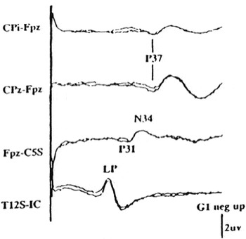
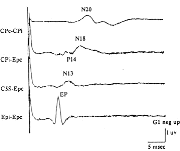
Somatosensory evoked responses (SSER/SSEP) measure sensitivity to touch (tactile sensitivity) on specific parts of the body, usually of the hands and feet, and their conduction of impulses to the brain. The stimulus is applied with light electrical impulses either on the anklebone of the foot above the course of the tibial nerve (a tibial SSER/SSEP) or on the inner side of the wrist joint above the median nerve or ulnar nerve (Figs 5 & 6). The measurement is related to the corresponding area of the brain. Sometimes it is also necessary to measure responses during impulse conduction, over the spinal cord, which is sometimes referred to as fractionated SSER/SSEP.
Normal median nerve sormatosensory evoked potentials.
Normal posterior tibial nerve somatosensory evoked potentials.

Magnetic evoked responses differ from the previously discussed forms of evoked response in that the nerve cells themselves are directly stimulated. Superficial electrodes are used to assess muscular responses on the muscles of the arm or leg, depending on the arm or leg function for which the brain area under stimulus is responsible. The time between the stimulation of the brain cells and the muscle response is measured. Stimuli are also applied over the region of neck vertebrae or of the lumbar vertebrae in order to distinguish between conduction in the brain and conduction in the spinal cord.
Evoked potentials and multiple sclerosis:
These tests can be helpful in making the diagnosis of multiple sclerosis (MS) and other disorders. In MS, signal transmission can slow down and the signal becomes weak. Evoked potentials are used to show abnormalities in the function of nerve pathways that can be caused by MS.
The numerous disorders have been shown to have different abnormalities of visual (VEPs) and auditory evoked potentials (AEPs), and among them multiple sclerosis (MS), Parkinson's disease (PD) and brainstem vascular lesions (BSVLs) (1–4). However, they are most often used in providing additional evidence in support of diagnosis of MS Reference Ellis(5)).
The current diagnostic criteria for MS consider only VEP findings because this particular EP study has been shown to be the most useful (6.7). The VEP is used to identify impaired transmission along the optic nerve pathways, which is a fairly common early finding in MS, even in someone who has never been aware of any visual symptoms Reference Chiappa, Hill and Chiappa(2,Reference McDonald, Compston, Edan, Goodkin, Hartung, Lublin, McFarland, Paty, Polman, Reingold, Sandberg-Wollheim, Sibley, Thompson, van den Noort, Weinshenker and Wolinsky8,Reference Polman, Reingold, Edan, Filippi, Hartung, Kappos, Lublin, Metz, McFarland, O'Connor, Sandberg-Wollheim, Thompson, Weinshenker and Wolinsky9). Age number ofclinical studies attest to the sensitivity of the VEP in revealing demyelinating lesions in the optic nerve (ON) – about 90% of patients who have clear history of ON have abnormal VEPS Reference Chiappa, Hill and Chiappa(2)). In some studies, the proportion is closer to 100%, and VEP was more sensitive than magneting resonance imaging (MRI) Reference Chiappa, Hill and Chiappa(2,Reference Miller, Newton, van der Poel, du Boulay, Halliday, Kendall, Johnson, MacManus, Moseley and McDonald10).
MS is the most common form of central demyelination, characterized pathologically by widespread occurrence in the central nervous system of patches of demyelination followed by gliosis. Clinically, dissemination of lesions in time and space are the characteristics of the condition Reference Misulis(1)). EPs are very useful in establishing evidence of subclinical disease in pathways not known to be affected, so contributing to the clinical criteria of disease disseminated in time and space (11.12). They are also useful in evaluation of clinical course during time and treatment Reference Chiappa, Hill and Chiappa(2)), and can provide not only diagnostic but also prognostic information during evaluation of MS patients Reference Weinstock-Guttman, Baier and Stockton(6,Reference Fischer, Andre-Obadia and Mauguiere7) (Fig. 7). Abnormal VEP parameters in MS patient are typical finding in MS patients, and incidence of VEP abnormalities ranges from a high 96% to a low 47% (Fig. 3) Reference Chiappa, Hill and Chiappa(2,Reference Weinstock-Guttman, Baier and Stockton6,Reference Sinanović11). According to Chiappa and Hill Reference Chiappa, Hill and Chiappa(2)), in 26 clinical series, published in seventies and early eighties, encompassing about 1950 patients with all MS classifications, the average abnormality rate found was 63%.
Follow up of VEPs of multiple sclerosis patient.

There have been a large number of studies of AEPs in patients with MS. Of 1006 patients with varying classifications of MS from some of these studies in eighties, 466 (46%) had abnormal AEPs. Differences in definitions of MS (and diagnostic criteria), patient populations, and techniques account for the variations between studies Reference Chiappa, Hill and Chiappa(2,Reference Sinanović11). Moreover, many types of EPs can help in assisting the diagnosis of multiple sclerosis Reference Sinanović(12,Reference Drislane13,Reference Walsh, Kane and Butler14), and can assist in predicting progression and in a follow up of MS patients Reference Drislane(13,Reference Alshuaib15,Reference Turker, Terzi, Bayrak, Cengiz, Onar and Us18).
In conclusion, VEP studies, being the sole diagnostic tool for EP studies according to the new diagnostic criteria for MS, bear more important for follow up of clinical course of MS (including VEP and AEP) and in the field of differential diagnosis Reference McDonald, Compston, Edan, Goodkin, Hartung, Lublin, McFarland, Paty, Polman, Reingold, Sandberg-Wollheim, Sibley, Thompson, van den Noort, Weinshenker and Wolinsky(8,Reference Sinanović11,Reference Djuricic, Djuricic and Jolic19), although there is debate Reference Misulis(1)) on the diagnostic value of other EP studies in MS.