Introduction
Loiasis and Mansonellasis are endemic in Africa, where they affect between 10 million and more than 100 million individuals (Simonsen et al., Reference Simonsen, Onapa and Asio2011; Zouré et al., Reference Zouré, Wanji, Noma, Amazigo, Diggle, Tekle and Remme2011). Infections with these two helminths constitute an emerging public health problem in Africa. Indeed, the frequent association of loiasis with onchocerciasis or lymphatic filariasis increases the risk of severe life-threatening complications of treatment and slows the implementation of onchocerciasis and lymphatic filariasis control programmes. Moreover, Mansonella perstans and Loa loa have immunomodulatory properties. They interfere with the host's immune response and, therefore, they have an effect on other endemic infectious diseases, such as tuberculosis, malaria and HIV (Metenou et al., Reference Metenou, Babu and Nutman2012; Dolo et al., Reference Dolo, Coulibaly, Dembele, Konate, Coulibaly, Doumbia, Diallo, Soumaoro, Coulibaly, Diakite, Guindo, Fay, Metenou, Nutman and Klion2013; Li & Zhou, Reference Li and Zhou2013; Ndibazza et al., Reference Ndibazza, Webb, Lule, Mpairwe, Akello, Oduru, Kizza, Akurut, Muhangi, Magnussen, Vennervald and Elliott2013). The clinical signs of loiasis are well known, whereas infections with M. perstans, which were previously considered few or not pathogenic, lead to symptoms such as angioedema, pruritus and, in cases of intraocular infections, symptoms mimicking signs specific to loiasis (Bregani et al., Reference Bregani, Tantardini and Rovellini2007; Asio et al., Reference Asio, Simonsen and Onapa2009). In Gabon, these filariases are endemic and frequently found in the same geographical areas. The prevalence of L. loa microfilariae in adults exceeds 20% of the population, resulting in an unacceptable risk of severe adverse events (SAEs) in individuals treated with ivermectin (Boussinesq et al., Reference Boussinesq, Gardon, Gardon-Wendel, Kamgno, Ngoumou and Chippaux1998; Kombila & Richard-Lenoble, Reference Kombila and Richard-Lenoble1998; Akué et al., Reference Akué, Nkoghe, Padilla, Moussavou, Moukana, Mbou, Ollomo and Leroy2011). Cutaneous clinical signs of filariasis are the main reason for consultation or biological testing at the Department of Parasitology–Mycology of the Faculty of Medicine of Libreville, Gabon (Bouyou-Akotet et al., Reference Bouyou-Akotet, Moussavou Boussougou, Ovono-Abessolo, Owono-Medang and Kombila2014).
A method based on a history of subconjunctival adult worm migration, a key clinical symptom of loiasis, was developed for L. loa endemicity and intensity mapping (Wanji et al., Reference Wanji, Akotshi, Mutro, Tepage, Ukety, Diggle and Remme2012). This rapid assessment procedure for loiasis (RAPLOA) was used to generate a global map of the distribution of this filariasis in Africa and to identify hyperendemicity and high post-treatment SAE risk foci (Chippaux et al., Reference Chippaux, Boussinesq, Gardon, Gardon-Wendel and Ernould1996; Gardon et al., Reference Gardon, Gardon-Wendel, Demanga-Ngangue, Kamgno, Chippaux and Boussinesq1997; Takougang et al., Reference Takougang, Meremikwu, Wandji, Yenshu, Aripko, Lamlenn, Eka, Enyong, Meli, Kale and Remme2002; Zouré et al., Reference Zouré, Wanji, Noma, Amazigo, Diggle, Tekle and Remme2011). This risk is considered highly significant for densities of more than 8000 microfilariae (mf)/ml (Twum-Danso, Reference Twum-Danso2003; Mackenzie et al., Reference Mackenzie, Geary, Prichard and Boussinesq2007). However, the association between clinical symptoms and microfilaraemia has been little studied in areas of perennial exposure to infection, in which the subconjunctival migration of adult worms may occur despite the absence of microfilariae in peripheral blood (Pinder et al., Reference Pinder, Leclerc and Everaere1992). In a recent study, it was reported that objective symptoms, such as Calabar swelling and subconjunctival adult worm migration, were more frequent in patients with occult loiasis, a neglected form of L. loa infection (Bouyou-Akotet et al., Reference Bouyou-Akotet, Moussavou Boussougou, Ovono-Abessolo, Owono-Medang and Kombila2014). However, data on the distribution of the objective symptoms among exposed individuals are lacking.
The aim of the present study is to analyse the relationship between the frequency of objective symptoms presented by L. loa microfilaraemic or amicrofilaraemic patients infected, or not, with M. perstans.
Patients and methods
Study site and patients
This study was carried out in Libreville, the capital of Gabon, at the Department of Parasitology–Mycology of the Faculty of Medicine (DPM) at the Université des Sciences de la Santé in Gabon, where outpatient consultations are held and routine screening is carried out for the biological diagnosis of endemic parasite infections. The department has a long track record of filariasis diagnosis and treatment. Since 1978, through consultations and surveys performed by the team throughout the country, the characterization of epidemiological features of endemic filariasis, mostly loiasis, mansonellasis and onchocerciasis, has been routinely performed. Weekly consultations were held with individuals showing symptoms of filariasis and those requiring diagnosis. Socio-demographic data such as age, gender, location, including duration in rural areas, and medical complaints were recorded from January to December 2011 inclusive. All patients underwent clinical examination and laboratory tests. Calabar swelling, subconjunctival or subcutaneous migration of adult worms were classified as objective symptoms, compared with subjective symptoms such as pruritus and itching.
Laboratory procedures
For diagnosis, blood samples were collected between 10.00 and 14.00 hours, before treatment, 4 ml of which were transferred into a tube containing EDTA. A further 3 ml of blood were centrifuged separately to obtain serum samples and then stored at − 20°C for the determination of the specific anti-L. loa antibody titre. Two methods with different sensitivities were used successively to detect microfilariae, and to prevent false-negative results in cases of low microfilaraemia. The first step for the detection and quantification of microfilariae was the microscopic examination of 10 μl of fresh, uncoagulated blood. All microfilariae present on the slide were identified and counted. Parasitaemia is expressed as the number of microfilariae per millilitre (mf/ml) of blood. The detection limit of this method is 100 mf/ml. Then leucoconcentration was carried out according to a standard protocol (Ho Thi Sang & Petithory, Reference Ho Thi Sang and Petithory1963). This technique is more sensitive than direct blood examination. As M. perstans parasite density is often low, leucoconcentration was performed for all patients, regardless of their L. loa microfilaraemia. Leucoconcentration is based on the lysis of red blood cells with saponin to concentrate leucocytes and microfilariae in the pellet. Briefly, 4 ml of the blood collected in the tube containing EDTA were centrifuged. The plasma was removed and 2 ml of 0.9% NaCl and 1 ml of 2% saponin were then added. Reagents were mixed with the pellet and the solution was centrifuged for 10 min. The supernatant was removed, and the same procedure was repeated twice. After the final centrifugation, the pellet containing leucocytes and microfilariae was collected and examined under a light microscope. Parasitic species were identified on the basis of their size and shape (M. perstans is shorter and very much thinner compared with L. loa), the regularity and type of movements between red blood cells during the fresh blood microscopic examination, the presence of a sheath (which does not exist in M. perstans microfilariae), and the caudal end, which is like an end glove for M. perstans (Deluol, Reference Deluol and Varia2004). They were quantified as described above.
Loa loa-specific antibodies were detected in serum samples from patients by indirect immunofluorescence (IIF) with homologous antigens from L. loa microfilariae prepared in the department, as described elsewhere (Walker-Deemin et al., Reference Walker-Deemin, Ferrer, Gauthier, Kombila and Richard-Lenoble2004). The antibody–L. loa specificity threshold was a titre above 1:200.
Data analysis
Data were double-entered in Excel; Stata (StataCorp LP, College Station, Texas, USA) was used for the descriptive and comparative analysis. Data entry was checked before the statistical analysis. Medians with interquartile ranges were calculated for non-normally distributed continuous variables, and means ( ± standard deviation, SD) for normally distributed continuous variables. Parasite densities were assessed only for patients for whom the parasite could be seen under the microscope. For continuous variables, Mann–Whitney U-tests or Kruskal–Wallis tests were used for comparisons. Correlations were analysed with Spearman's rank correlation test. Differences in proportions were assessed with chi-squared or Fisher's exact tests. Multivariate analysis was performed by forward stepwise likelihood ratio logistic regression, with each class of microfilariae considered as the dependent variable, and with variables for which a P value < 0.20 was obtained in the bivariate analysis as independent variables. For all comparisons, a P value < 0.05 was considered significant.
On the basis of parasitological test results (i.e. microfilaraemia), the presence of L. loa-specific antibodies and clinical symptoms, subjects in the study population were assigned to six subgroups: only L. loa microfilariae detected; patients with occult loiasis (clinical symptoms, hypereosinophilia, absence of L. loa microfilariae and presence of L. loa-specific antibodies, titre >1:200); presence of M. perstans microfilariae without loiasis; detection of L. loa microfilariae plus M. perstans microfilariae; dual infection with occult loiasis and the presence of M. perstans microfilaraemia. Hypermicrofilaraemia was defined as a L. loa microfilariae density of more than 8000/ml.
Results
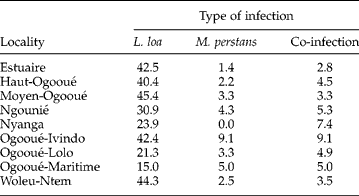
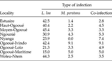
Over a 12-month period, data from 1148 patients were recorded. Up to 66.6% (n= 764) were female. Patients were aged from 4 to 94 years; 64.9% (n= 745) were over the age of 40 years, 8.0% (n= 92) were between 15 and 25 years of age and 2.3% (n= 26) were below the age of 10 years. Age was not recorded for 32 patients. The site of residence was known for 872 (76.0%) patients: 32.7% (n= 285) lived in the province of Estuaire, in which the capital city, Libreville, is located; 18.1% (n= 158) in Woleu-Ntem in the north of the country; 10.8% (n= 94) in Ngounié and 10.2% (n= 89) in Haut-Ogooué, both these regions being located in the south (table 1). Fewer than 3% (n= 26) of patients were visitors from neighbouring countries.
The prevalence (%) of single and concurrent Loa loa and Mansonella perstans infections among 846 patients from nine localities in Gabon.
Detection and prevalence of filarial infections
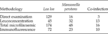
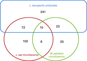
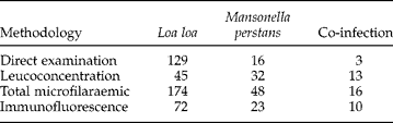
Loa loa infection was more frequently detected by indirect immunofluorescence (n= 346 patients with specific antibodies) compared to microscopy (n= 238 microfilaraemic patients) (table 2). The Ho Thi Sang and Petithory leuconcentration method identified 37.8% additional L. loa infected patients and 70.3% more (n= 45) patients infected with M. perstans; their parasite density was below 100 mf/ml (table 2). The overall rate of blood infection with filariae was 41.7% (n= 479). Loa loa was detected in most cases (n= 454; 39.5%), M. perstans in 5.6% (n= 64) and co-infection in 3.4% (n= 39). The proportion of symptomatic infected patients was highest (>40%) for Ogooué-Ivindo, Moyen-Ogooué, Woleu-Ntem, Estuaire and Haut-Ogooué districts (table 1). Among L. loa-infected patients, occult loiasis predominated (n= 264; 58.2%) and 23 occult loiasis patients had M. perstans microfilaraemia. Overall, 415 (36.1%) patients were infected with L. loa only (174 microfilaremic loiasis and 241 occult loiasis), whereas 25 (2.2%) were infected with M. perstans only. In L. loa microfilaraemic patients, only 43.2% (n= 82/190) had L. loa-specific antibody. Among the 346 patients with detected L. loa-specific antibodies, 105 also had a microfilaraemia, 102 carried only L. loa microfilariae and 10 carried both types of microfilariae (table 2, fig. 1). Hypermicrofilaraemia was found in 18.4% (n= 35/190) of patients with L. loa microfilaraemia (3.0% of the total study population) and nine patients (0.7% of the total study population) had more than 30,000 mf/ml, with a maximum density of 94,400 mf/ml.
Diagnostic methods used for the detection of single or concurrent L. loa and M. perstans infections in 479 patients.
The number (n) of single and concurrent Loa loa and/or Mansonella perstans infected individuals.

Filarial infection, clinical symptoms and intensity of Loa loa microfilaraemia
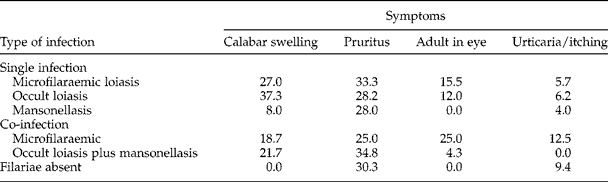
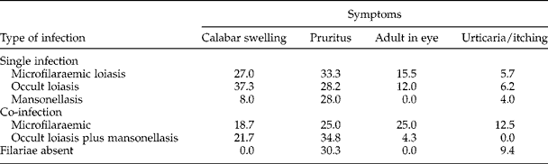
The main clinical sign was general pruritus (n= 348; 30.3%), the prevalence of which was similar in infected (n= 145/479; 30.3%) and uninfected individuals (n= 203/669; 30.3%) (table 3). Urticarial papules or itching were not significantly associated with the presence or absence of filarial infection. Calabar swelling was present in 147 (30.7%) patients, with only two patients having a Mansonella infection. This symptom was significantly more frequent in patients with occult loiasis (OL) (36.0%; n= 95/264) than in those with L. loa microfilaraemia (26.3%; n= 50/190) (P= 0.03). Overall, 61 (5.3%) patients had adult worm or history of migration across the eye; 56 had L. loa infection alone and the frequency of subconjunctival migration was similar for L. loa microfilaraemic (16.3%; n= 31) and OL (11.4%; n= 30) patients (P= 0.13) (table 3). No objective symptoms prevailed in cases of co-infection (table 3).
The frequency (%) of clinical symptoms caused by single or concurrent L. loa and M. perstans infections in Gabon.
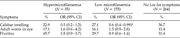
Logistic regression analysis showed that Calabar swelling (3.4 (2.0–5.9); P< 0.01) and adult worm migration across the eye (4.1 (2.1–8.0); P< 0.01) were strong predictors of loiasis. However, the prevalence of Calabar swelling and adult worm migration across the eye were not predictive of hypermicrofilaraemia, even in patients with a very high microfilaraemia (>30,000 mf/ml) (table 4).
The relationship between the frequency (%) of clinical symptoms and microfilaraemia in patients with loiasis; N, number of patients examined; OR, odds ratio; CI, 95% confidence interval.
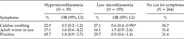
*, P= 0.03.
Adult worm migration across the eye was reported in 2 of the 9 patients (22%) with more than 30,000 mf/ml, 4 of 26 (15.4%) patients with microfilaraemia between 8 and 30,000 mf/ml and 25 of 155 (16.1%) patients with low microfilaraemia (P= 0.9). For the 90 infected individuals with L. loa microfilaraemia values below 100 mf/ml, frequencies of objective symptoms were similar to those of the total population: 29% had general pruritus, 29% Calabar swelling and 18% had adult worm migration in the eye. No specific objective symptoms were noted in 5 (14.3%) out of 35 hypermicrofilaraemic patients.
Discussion
The relationship between loiasis-specific clinical symptoms and the amicrofilaraemic form of this parasitic infection, occult loiasis, has rarely been investigated in exposed individuals. The main aim of this study was to use weekly consultations performed at the DPM to investigate this relationship.
In areas in which the prevalence of Onchocerca volvulus is low and limited to well-defined endemic foci, and lymphatic filariasis is absent, the key issue is the burden represented by L. loa and M. perstans infections and the impact of these infections on disability-adjusted life years. Both parasitic infections affect populations in resource-limited areas, thereby increasing the poverty by decreasing local economic productivity (Agbolade & Akinboye, Reference Agbolade and Akinboye2001; Hotez, Reference Hotez2011). Detailed information is required for the characterization of endemicity, because these filarial worm species are common, associated with high levels of morbidity and influence other co-endemic infections (Muhangi et al., Reference Muhangi, Woodburn, Omara, Omoding, Kizito, Mpairwe, Nabulime, Ameke, Morison and Elliott2007; Metenou et al., Reference Metenou, Babu and Nutman2012; Li & Zhou, Reference Li and Zhou2013; Ndibazza et al., Reference Ndibazza, Webb, Lule, Mpairwe, Akello, Oduru, Kizza, Akurut, Muhangi, Magnussen, Vennervald and Elliott2013). This information is also important due to the increasing control of soil-transmitted helminths by mass or targeted chemotherapy with ivermectin and albendazole, which may cause serious adverse reactions after treatment of L. loa-infected patients with high microfilaraemia. Therefore, loiasis merits greater attention than it currently receives.
The overall prevalence of L. loa was 39.5%, similar to the prevalence obtained with the RAPLOA estimation model (Zouré et al., Reference Zouré, Wanji, Noma, Amazigo, Diggle, Tekle and Remme2011). These data also confirm the high burden of loiasis in the north of the country (Woleu-Ntem), on the border with Cameroon, and throughout semi-urban and rural areas along the national road, which begins in the province of Estuaire, where Libreville is located, and extends into the south-east (Haut-Ogooué) (Akué et al., Reference Akué, Nkoghe, Padilla, Moussavou, Moukana, Mbou, Ollomo and Leroy2011). This highway facilitates access of inhabitants from the main cities, who carry out agricultural activities in villages located along this road, to these areas, where they are exposed to infective Chrysops bites. The observed prevalence (20–51.5%) differs from those reported in earlier surveys performed in the country, which varied from 12% to 33% according to the geographic area (Akué et al., Reference Akué, Nkoghe, Padilla, Moussavou, Moukana, Mbou, Ollomo and Leroy2011). There are two principal explanations for such high prevalences. First, a combination of three methods was used for the detection of filarial worms. The direct examination of 10 μl of fresh whole blood, a method commonly used in endemic countries, led to the diagnosis of loiasis in only 30% of truly infected patients; and in only one-quarter of those infected with M. perstans. Mansonella perstans infection was identified mostly (45 of 64 individuals) after Ho Thi Sang and Petithory leucoconcentration of 4 ml of blood. This more sensitive technique allowed the detection of an additional 20% of infected individuals. Indirect immunofluorescence was positive in 346 of 454 patients with loiasis, and occult loiasis was the main clinical form in patients with symptoms (57%). If each microscopy technique was used alone, the prevalence obtained would not reflect the real level of parasitism, given the high frequency of infected patients with low or no microfilaraemia. Screening to determine both prevalence and incidence of filariasis could be improved by the standardization of diagnostic methods, followed by the validation of an accurate and specific cost-effective method. Serological methods (indirect immunofluorescence or enzyme-linked immunosorbent assay (ELISA)), and DNA amplification by conventional polymerase chain reaction (PCR) or with loop-mediated isothermal amplification (LAMP) technology, have provided promising results. These tools should therefore also be considered if their sensitivity is found to be equivalent to that of the combination of the three methods used here (Toure et al., Reference Toure, Bain, Nerrienet, Millet, Wahl, Toure, Doumbo, Nicolas, Georges, McReynolds and Egwang1997; Fink et al., Reference Fink, Kamgno and Nutman2011; Fernández-Soto et al., Reference Fernández-Soto, Mvoulouga, Akue, Abán, Santiago, Sánchez and Muro2014). The second explanation is that the study population consisted of symptomatic individuals who had a higher probability of being infected than asymptomatic individuals. The survey was not community-based and, therefore, did not take into account the non-negligible proportion of asymptomatic infected individuals. The effect of parasite density on the occurrence of specific symptoms may therefore have been masked. These explanations could also justify the overall low prevalence of M. perstans-infected patients. This infection is asymptomatic most of the time and is discovered fortuitously in blood samples. Individuals are contaminated during agricultural activities throughout the day in plantations where they are more frequently exposed to Chrysops bites. Culicoides, vectors of M. perstans, bite early in the morning and during twilight.
The specific symptoms frequently presented by infected individuals are the subconjunctival and subcutaneous migration of macrofilariae and Calabar swellings in hands, wrists and lower forearm (Carme et al., Reference Carme, Mamboueni, Copin and Noireau1989; Boussinesq et al., Reference Boussinesq, Gardon, Gardon-Wendel, Kamgno, Ngoumou and Chippaux1998). No relationship was found between these signs and microfilaraemia intensity in symptomatic patients, or in previously described individuals living in forested areas of Gabon (Van Hoegaerden et al., Reference Van Hoegaerden, Chabaud, Akue and Ivanoff1987).
Occult loiasis, the main form of L. loa infection in the study population, was described to be predominant in immigrants (compared to continuously exposed individuals who were supposed to be either asymptomatic or with adult worm migration). Objective symptoms have frequently been associated with microfilarial density. But present data confirm that they are also found in patients with little or no microfilaraemia. Whether or not symptoms occur in an individual depends mainly upon the degree of host immune response to infection, since most disease manifestations of loiasis are considered to be hypersensitivity reactions to adult worms (Pinder et al., Reference Pinder, Leclerc and Everaere1992, Klion et al., Reference Klion, Massougbodji, Sadeler, Ottesen and Nutman1991). Moreover, Calabar swelling, which is the predominant objective symptom, is significantly more frequent in patients without microfilaraemia. Indeed, during OL, infective larvae L3 are able to progress their development to give adult worms (L4). Then, when L4 females release microfilariae in peripheral blood, these microfilariae are destroyed by the immune system and patients become occults: they do not have microscopic detectable peripheral microfilaraemia although they carry adult worms.
The absence of a relationship between the presence of high microfilaraemia and adult worms has already been reported (Akué et al., Reference Akué, Nkoghe, Padilla, Moussavou, Moukana, Mbou, Ollomo and Leroy2011). Indeed, adult filariae may be present at any site, causing a broad spectrum of clinical signs (Pion et al., Reference Pion, Demanou, Oudin and Boussinesq2005). For example, subconjunctival females containing microfilariae are frequently removed from patients who had no detectable microfilariae in the blood a few years previously (Carme et al., Reference Carme, Mamboueni, Copin and Noireau1989). Other clinically significant signs, such as neuropathies, are more frequent in people exposed to the parasite during adulthood, as reported for other signs of loiasis (Gobbi et al., Reference Gobbi, Boussinesq, Mascarello, Angheben, Gobbo, Rossanese, Corachán and Bisoffi2011). In a study comparing the population living in endemic areas and visitors, only 10% of temporary residents were microfilaraemic, whereas 95% of these individuals had oedema (Klion et al., Reference Klion, Massougbodji, Sadeler, Ottesen and Nutman1991). Moreover, several individuals, who were amicrofilaraemic and had not recently had antifilarial treatment, were found to have had confirmed ocular passage of an adult L. loa in other settings (Nutman et al., Reference Nutman, Miller, Mulligan and Ottesen1986; Dupont et al., Reference Dupont, Zue-N'dong and Pinder1998). It is also important to note that some patients with high microfilaraemia did not present any objective symptoms used to implement, or not, mass chemotherapy.
General pruritus and oedema were detected in 10 out of 25 patients with single M. perstans infection. This species is now of clinical importance, as demonstrated in patients seen in our department 5 years ago (Bouyou-Akotet et al., Reference Bouyou-Akotet, Moussavou Boussougou, Ovono-Abessolo, Owono-Medang and Kombila2014). This study focused on L. loa-specific signs, such as joint pain, headache and itching, that are not included in the panel of signs frequently associated with mansonellasis (Asio et al., Reference Asio, Simonsen and Onapa2009; Simonsen et al., Reference Simonsen, Onapa and Asio2011). Their real frequency is therefore not assessed, although they should be investigated because they are troublesome and unpleasant, inducing some degree of disability. Indeed, itching is the main sign presented by infected symptomatic patients attending the weekly consultations at the DPM, as reported for a Nigerian population (Anosike et al., Reference Anosike, Dozie, Onwuliri, Nwoke and Onwuliri2005; Bouyou-Akotet et al., Reference Bouyou-Akotet, Moussavou Boussougou, Ovono-Abessolo, Owono-Medang and Kombila2014). Moreover, another reason for considering and studying this filariasis is its impact on other prevalent infectious diseases in Gabon: malaria, tuberculosis and other helminthiases (Dolo et al., Reference Dolo, Coulibaly, Dembele, Konate, Coulibaly, Doumbia, Diallo, Soumaoro, Coulibaly, Diakite, Guindo, Fay, Metenou, Nutman and Klion2013; Li & Zhou, Reference Li and Zhou2013; Ndibazza et al., Reference Ndibazza, Webb, Lule, Mpairwe, Akello, Oduru, Kizza, Akurut, Muhangi, Magnussen, Vennervald and Elliott2013).
Loiasis is the main filariasis responsible for clinical symptoms observed in an exposed population living in Gabon, although 2% of the population had single M. perstans infection. Infected patients live in rural as well as urban cities of Gabon. The presence of adult eye worm migration as a strong predictor of high microfilarial density would obscure the real burden of L. loa hypermicrofilaraemia. The mapping of loiasis in endemic areas should also take into account the group of patients with occult loiasis for the characterization of L. loa endemicity and control strategies.
Acknowledgements
We are grateful to the patients and the laboratory staff of the Department of Parasitology, particularly to Mr Migueba Valentin, for collecting and preparing the samples.
Financial support
Funds of the Department of Parasitology.
Conflict of interest
None.
Ethical standards
All tests performed were part of the routine diagnosis of filarial infection in the DPM. Oral informed consent for the use of data was obtained from each participant after the study had been explained.