Introduction
The species is the fundamental unit in biology and biodiversity conservation, and consequently the accurate assessment of species-level diversity is essential to systematic research, conservation risk-assessment and the use of species as biodindicators of environmental conditions. Lichen taxonomy has traditionally used a morphospecies concept, that is individuals grouped on the basis of a shared set of phenotypic characters. However, this definition may be inadequate when morphological characters are subject to phenotypic plasticity and/or mask the presence of different species with the same morphological appearance. Species falling in the last category are now generally referred to as “cryptic species” (Hawksworth & Rossman Reference Hawksworth and Rossman1997; Bickford et al. Reference Bickford, Lohman, Sodhi, Ng, Meier, Winker, Ingram and Das2007).
The term ‘semi-cryptic species’ has also been proposed to refer to species which cannot be clearly diagnosed by their morphology, but which are separated by other characters, such as by their ecology and/or distribution (Vondrák et al. Reference Vondrák, Říha, Arup and Søchting2009). ‘Cryptic species' is also often used for species with subtle morphological differences, but that application of the term does not reflect its current use in species-concept literature. In cases of cryptic speciation, where there are no visible distinguishing phenotypic differences, molecular studies are necessary to estimate properly the real biodiversity of organisms of all kinds (Herbert et al. 2004; Saez & Lozano Reference Sáez and Lozano2005). Taylor et al. (Reference Taylor, Jacobson, Kroken, Kasuga, Geiser, Hibbett and Matthew2000) contrasted the operational phylogenetic species concept to the theoretical species concept, offering a phylogenetic approach to recognize fungal species based on a concordance of multiple gene genealogies. To delimit species on the basis of this criterion, new operational concepts have been incorporated in an attempt to clarify and recognize the real diversity in fungi.
It is remarkable how molecular studies are revolutionizing our understanding of species circumscriptions in lichen-forming fungi, where molecular data have repeatedly demonstrated the existence of cryptic species in lichens (Crespo & Pérez-Ortega Reference Crespo and Pérez-Ortega2009; Crespo & Lumbsch Reference Crespo and Lumbsch2010). However, it is estimated that much of the undescribed diversity in fungi could remain masked behind a species complex that includes two or more independent evolutionary lineages (Hawksworth Reference Hawksworth2001), especially in those with wide geographical distribution.
Several studies using molecular markers have led to the discovery of cryptic species in lichen-forming fungi. For example, Kroken & Taylor (Reference Kroken and Taylor2001) recognized at least six phylogenetic species within Letharia rather than the two previously accepted species. Högnabba & Wedin (Reference Högnabba and Wedin2003) noted that there are at least two well-supported monophyletic groups that could be interpreted as phylogenetic species within the Sphaerophorus globosus complex, and Myllys et al. (Reference Myllys, Stenroos, Thell and Ahti2003) found three different clades in Cladonia arbuscula. Divakar et al. (Reference Divakar, Amo De paz, del Prado, Esslinger and Crespo2007) reported two monophyletic clades in the Physconia distorta complex, one of which was considered to be a cryptic species. Furthermore, the measure of genetic distances is providing an additional valuable tool for species identification when morphological characters are scarce, as in the case of cryptic species (e.g. Hebert et al. Reference Hebert, Penton, Burns, Janzen and Hallwachs2004; Zemlack et al. Reference Zemlak, Ward, Connell, Holmes and Hebert2009; Del-Prado et al. Reference Del-Prado, Cubas, Lumbsch, Divakar, Blanco, Amo de Paz, Molina and Crespo2010).
Parmelia sulcata is a common lichen recolonizing temperate urban areas following reductions in sulphur dioxide levels (Hawksworth & McManus Reference Hawksworth and McManus1989; Crespo et al. Reference Crespo, Divakar, Argüello, Gasca and Hawksworth2004).This species, first described by Taylor (Reference Taylor and Mackay1836) from Ireland, is characterized by squarrose rhizines and a grey pseudocyphellate, sorediate upper surface (Hale Reference Hale1987; Divakar et al. Reference Divakar, Upreti and Elix2001); it contains atranorin, chloroatranorin, salazinic acid, and consalazinic acid (Galloway & Elix Reference Galloway and Elix1983). The species grows in a wide range of ecological environments, most frequently on bark or wood, but can also be found on siliceous rocks, especially if they are mossy. Reproduction is predominantly asexual from soredia, although apothecia are not exceptionally rare in rural areas. It is widely distributed, and perhaps the most common parmelioid lichen in Europe. The first intra-specific molecular studies on P. sulcata showed an unexpectedly high genetic variability (Crespo et al. Reference Crespo, Bridge and Hawksworth1997), and Crespo et al. (Reference Crespo, Bridge, Hawksworth, Grube and Cubero1999) reported a differential distribution of haplotypes in relationship to recolonizing populations following ameliorations of sulphur dioxide levels in north-west and west London. Divakar et al. (Reference Divakar, Molina, Lumbsch and Crespo2005a) confirmed this variability, and described the new species, P. barrenoae, from central Spain based on a combination of molecular and morphological characters.
Molecular data have supported the establishment of other phylogenetic species within Parmelia. Within P. saxatilis, Mediterranean populations were described by Crespo et al. (Reference Crespo, Molina, Blanco, Schroeter, Sancho and Hawksworth2002) as a separate species, P. serrana, based on strong molecular evidence and also on some weak morphological, and a few ecological, features (Molina et al. Reference Molina, Crespo, Blanco, Lumbsch and Hawksworth2004); a group I intron in position 1516 in the nSSU of P. saxatilis never appeared in P. serrana. Feuerer & Thell (Reference Feuerer and Thell2002) described P. ernstiae as distinct from P. saxatilis s. lat. on the basis of molecular markers and phenotypic characters, and later Molina et al. (Reference Molina, Crespo, Blanco, Lumbsch and Hawksworth2004) confirmed the monophyly of that species and showed phylogenetic relationships between P. ernstiae and P. saxatilis s. str.
In all cases where genetic complexity is discovered in established morphospecies as a result of molecular studies, there is a problem in fixing the application of the specific name. The problem is to determine which halpotype the name-bearing type of the species name belongs to, especially when no DNA can be recovered from ancient reference specimens. For example, in the case of P. saxatilis, an interpretative type (i.e. an epitype) was selected to fix the application of the name (Crespo et al. Reference Crespo, Molina, Blanco, Schroeter, Sancho and Hawksworth2002). In this contribution, we similarly typifiy P. sulcata.
In order to examine the genetic diversity, phylogeny and species concepts in P. sulcata, we used extensive taxon sampling from four continents (America, Africa, Europe and Asia), including the lectotype area in Ireland. We selected as molecular markers nuITS, nuIGS rDNA, and β-tubulin (partial gene). Begerow et al. (Reference Begerow, John and Oberwinkler2004) reported on the value of β-tubulin for inferring the phylogeny of basidiomycetes, though paralogous genes have been described in this molecular marker (e.g. Jackson et al. Reference Jackson, Vaughan and Gull2006; Msiska & Morton Reference Msiska and Morton2009), even in parmelioid lichens where Thell et al. (Reference Thell, Feuerer, Kärnefelt, Myllys and Stenroos2004) reported the occurrence of multiple copies. Despite the problem of paralogous markers, O'Donnell et al. (Reference O'Donnell, Cigelnik and Nirenberg1998) reported that there was 3·5 times more phylogenetic information in β-tubulin genes than in the mitochondrial SSU rRNA genes, and so proposed it as a useful marker for studying close relationships in Fusarium. Myllys et al. (Reference Myllys, Lohtander and Tehler2001) and Molina et al. (Reference Molina, Crespo, Blanco, Lumbsch and Hawksworth2004) have demonstrated the value of this gene as a molecular marker over ITS nuDNA in lichen-forming fungi. The conserved β-tubulin gene has proved a good phylogenetic molecular marker in several lichenological studies (Myllys et al. Reference Myllys, Lohtander and Tehler2001; Articus et al. Reference Articus, Mattsson, Tibell, Grube and Wedin2002; Crespo et al. Reference Crespo, Molina, Blanco, Schroeter, Sancho and Hawksworth2002; Thell et al. Reference Thell, Stenroos, Feuerer, Kärnefelt, Myllys and Hyvönen2002; Molina et al. Reference Molina, Crespo, Blanco, Lumbsch and Hawksworth2004). In our study we also use, for the first time in Parmelia, nuIGS rDNA as a molecular marker since it has been found to be of value for species distinction in other groups of lichens (Wirtz et al. Reference Wirtz, Printzen and Lumbsch2008).
Materials and Methods
Taxon sampling
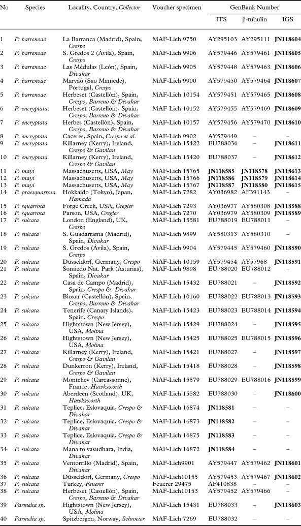
Sequence data of the nuITS, nuIGS rDNA and β-tubulin partial gene were analyzed in 37 Parmelia s. str. samples. The specimens were collected from four different continents (viz. Africa, Asia, America and Europe). Details of the material, area of collection, location of voucher specimens, and GenBank accession numbers are presented in Table 1. Three sequences of P. mayi were used as outgroup following our previous study (Molina et al. Reference Molina, Del-Prado, Divakar, Sánchez-Mata and Crespo2011).
Specimens of Parmelia used in this study, with location, reference collection detail and GenBank accession numbers. Newly obtained sequences are shown in bold

Molecular methods
Small samples prepared from freshly collected and frozen specimens were ground with sterile plastic pestles. Total genomic DNA was extracted using the DNeasy Plant Mini Kit (Qiagen) according to the manufacturer's instructions, but with modifications as described in Crespo et al. (Reference Crespo, Blanco and Hawksworth2001). Dilutions of 1:10 of the total DNA were used for PCR amplifications of the gene in the nuITS rDNA region. Fungal nuITS rDNA was amplified using the primers ITS1F (Gardes & Bruns Reference Gardes and Bruns1993), ITS4 (White et al. Reference White, Bruns, Lee, Taylor, Innis, Gelfand, Sninsky and White1990) and ITS1-LM (Myllys et al. Reference Myllys, Lohtander, Källersjö and Tehler1999), and ITS2-KL (Lohtander et al. Reference Lohtander, Myllys, Sundin, Källersjö and Tehler1998). The partial β-tubulin sequence was obtained using Bt3-Lm and Bt10-LM primers (Myllys et al. Reference Myllys, Lohtander and Tehler2001), and nuIGS rDNA was amplified using IGS1-2a (Carbone & Kohn Reference Carbone and Kohn1999) and NSSU31T (Frank Kauff). When the first PCR products were negative, we used 4 µl as the template in a new amplification reaction using NCS1R (Carbone & Kohn Reference Carbone and Kohn1999) as internal primer.
Amplifications were performed in 50 µl volumes containing a reaction mixture of 5 µl 10X DNA polymerase buffer (Biotools) (containing MgCl2 2 mM, 10 mM Tris-HCl, pH 8·0, 50 mM KCl, 1 mM EDTA, 0·1% Triton X-100), 1 µl of dinucleotide triphosphate (dNTPs), containing 10 mM of each base, 2·5 µl of each primer (10 µM), 1·25 µl of DNA polymerase (1U µl−1) and 27·5 µl dH2O. Finally, 40 µl of this mixture was added to 10 µl of DNA from each specimen.
The amplifications were carried out in an automatic thermocycler (Techne Progene) and performed using the following programs. For the ITS we used an initial denaturation at 94°C for 5 min, and 30 cycles of: 94°C for 1 min, 54–58°C for 1 min, 72°C for 1·5 min; and a final extension at 72°C for 5 min. For the β-tubulin gene we used an initial denaturizing at 94°C for 5 min, and 30 cycles of: 94°C for 1 min, 55–58°C for 1 min, 72°C for 1·5 min; and a final extension at 72°C for 5 min. Finally, for the IGS we used an initial denaturizing at 94°C for 5 min, and 35 cycles of: 94°C for 1 min, 52°C for 1 min, 72°C for 1·5 min; and a final extension at 72°C for 5 min.
The PCR products were then cleaned either using the Bioclean Columns kit (Biotools) according to the manufacturer's instructions, or adding 5 µl of ExoSAP-IT® (Exonuclease 1- Shrimp Alkaline Phosphatase) to the PCR products, followed by a heat treatment of 15 min at 37°C and 15 min at 80°C. The cleaned PCR products were sequenced using the same amplification primers. The ABI Prism™ Dye Terminator Cycle Sequencing Ready reaction kit (Applied Biosystems) was used and the following settings were applied: denaturation for 3 min at 94°C, 25 cycles at 96°C for 10 s, 50°C for 5 s, and 60°C for 4 min. Sequencing reactions were electrophoresed on a 3730 DNA analyzer (Applied Biosystems). Partial nuSSU rDNA, including an intron at the end of the 3′ (SSU), were removed before the alignment. Sequence fragments obtained were assembled with SeqMan 4.03 (DNAStar) and manually adjusted.
Sequence alignments
Previously published sequences (Molina et al. Reference Molina, Crespo, Blanco, Lumbsch and Hawksworth2004; Divakar et al. Reference Divakar, Molina, Lumbsch and Crespo2005a) were obtained from GenBank and included in the present study, together with the newly produced ones. Sequences were aligned using the Q-INS-i algorithm (Katoh & Toh Reference Katoh and Toh2008a) of the multiple sequence alignment software MAFFT version 6.611 (Katoh et al. Reference Katoh, Misawa, Kuma and Miyata2002; Katoh & Toh Reference Katoh and Toh2008b), following Wedin et al. (Reference Wedin, Wiklund, Jørgensen and Ekman2009), but aligning sequences in a single step. Major insertions and ambiguous regions were identified and eliminated by Gblocks version 0.91b (Castresana Reference Castresana2000) with the following parameters: minimum number of sequences for a conserved position = 17, minimum number of sequences for a flank position = 17, maximum number of contiguous non-conserved positions = 10, minimum length of a block = 5, and allowed gap positions = “with half”, following Talavera & Castresana (Reference Talavera and Castresana2007).
Phylogenetic analyses
We checked the congruence among the topologies recovered with each individual marker analyzing the three datasets separately. Conflict was considered as significant if a significantly supported clade (bootstrap support ≥ 70%; Hillis & Bull Reference Hillis and Bull1993) for one marker was contradicted with significant support by another marker. No incongruence was found and the three markers were combined for the analyses.
A maximum parsimony (MP) analysis was performed using PAUP 4.0b10 (Swofford Reference Swofford2003). A heuristic search of 1000 random taxon addition replicates was conducted treating gaps as a ‘fifth character’ with TBR branch-swapping and the MulTrees option in effect; 1000 trees were allowed to be saved in each replicate. Parsimony-uninformative characters were excluded from the analyses. Nonparametric bootstrap (Felsenstein Reference Felsenstein1985) was used to assess robustness of clades, running 1000 pseudoreplicates with the same settings as in the heuristic search, but with 100 random addition replicates. Bootstrap support above 70% was considered as significant support (Hillis & Bull Reference Hillis and Bull1993).
The datasets were also analyzed using a Bayesian approach (Huelsenbeck et al. Reference Huelsenbeck, Rannala and Masly2000). MrBayes 3.1.2 (Huelsenbeck & Ronquist Reference Huelsenbeck and Ronquist2001) was employed to sample trees using a Markov Chain Monte Carlo (MCMC) method. The evolutionary models for Bayesian analyses were selected for each dataset using the Akaike Information Criterion (AIC) as implemented in jModeltest (Guindon & Gascuel Reference Guindon and Gascuel2003; Posada Reference Posada2008). The AIC was used to select only among the 24 models implemented in MrBayes, by setting the jModeltest analyses to: three substitution schemes, allowing base frequencies to be fixed or not (F), allowing the possibility of a proportion of invariable sites (I), allowing a gamma distributed rate heterogeneity between sites (G) modelled by four discrete categories and using full ML optimization. The analyses used three partitions with the following models: SYM+G for the ITS, HKY for the β-tubulin, and GTR+G for the IGS. The partitioned Bayesian analysis of the combined datasets was conducted by applying the previously determined models to each dataset with all parameter values, except branch lengths and tree topologies, unlinked. The number of discrete gamma categories was kept as default four. Bayesian prior distributions included treating all tree topologies as equally likely, a uniform (0·50) distribution for the gamma shape parameter, a uniform (0, 1) distribution for the proportion of invariable sites, a flat (1, 1, 1, 1, 1, 1) Dirichlet for the rate matrix, and a flat (1,1,1,1) Dirichlet for the state frequencies (except when the model dictated state frequencies to be equal). For the combined dataset, two parallel runs were executed, each with four chains, three of which were incrementally heated with a temperature of 0·15. The analysis was diagnosed for convergence every 100 000 generations, and was set to halt automatically when the average of the standard deviation between runs descended to below 0·01. Every 100th tree was saved into a file. Using the last 50% of the posterior tree sample, a majority rule consensus tree was assembled and the posterior probabilities were calculated with the sumt option. Posterior probabilities ≥ 95% were considered as strong support. Phylogenetic trees were drawn using the program TREEVIEW (Page Reference Page1996).
Hypothesis testing
Since the phylogenetic analysis is incongruent with the current concept of Parmelia sulcata in suggesting that clade B1 (i.e. P. encryptata) is a different species, we tested whether our data are sufficient to reject the following hypotheses: 1) monophyly of P. sulcata s. str. (clade A) + clade B, 2) monophyly of P. sulcata s. str. (clade A) + clade B1, 3) monophyly of P. sulcata s. str. (clade A) + clade B2, and 4) monophyly of P. sulcata s. str. (clade A) + P. squarrosa (clade C). Such a topology might be present in suboptimal trees not sampled or not present in the 50% majority-rule consensus tree of the MCMC sampling, which may not be significantly worse than the obtained topology. For hypothesis testing two different methods were employed: 1) Shimodaira–Hasegawa (Reference Shimodaira and Hasegawa1999) SH test; and 2) expected likelihood weight (ELW) test following Strimmer & Rambaut (Reference Strimmer and Rambaut2002). The SH and ELW tests were performed using Tree-PUZZLE 5.2 (Schmidt et al. Reference Schmidt, Strimmer, Vingron and von Haeseler2002) with the combined dataset on a sample of 200 unique trees, the best trees agreeing with the null hypotheses, and the unconstrained ML tree. These trees were inferred in Tree-PUZZLE employing the GTR+I+G nucleotide substitution model.
Calculation of genetic distances
Pairwise maximum likelihood distances (given as number of nucleotide substitutions per site), among the ITS rDNA sequences included in the analysis, were calculated with TREE-PUZZLE 5.2 (Schmidt et al. Reference Schmidt, Strimmer, Vingron and von Haeseler2002) using the GTR model of nucleotide substitution, assuming a discrete gamma distribution with six rate categories (Lumbsch Reference Lumbsch2002; Del-Prado et al. Reference Del-Prado, Cubas, Lumbsch, Divakar, Blanco, Amo de Paz, Molina and Crespo2010).
Chemistry
Extrolites (i.e. ‘secondary metabolites’) were identified by thin layer chromatography (Elix & Ernst-Russell Reference Elix and Ernst-Russell1993) in the Parmelia sulcata groups A, C and D using solvent system C (Orange et al. Reference Orange, James and White2001).
Results
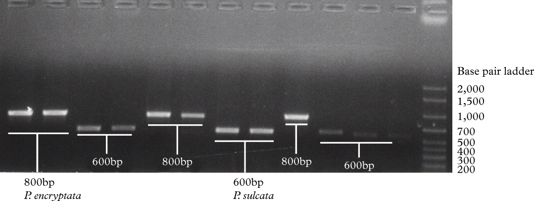
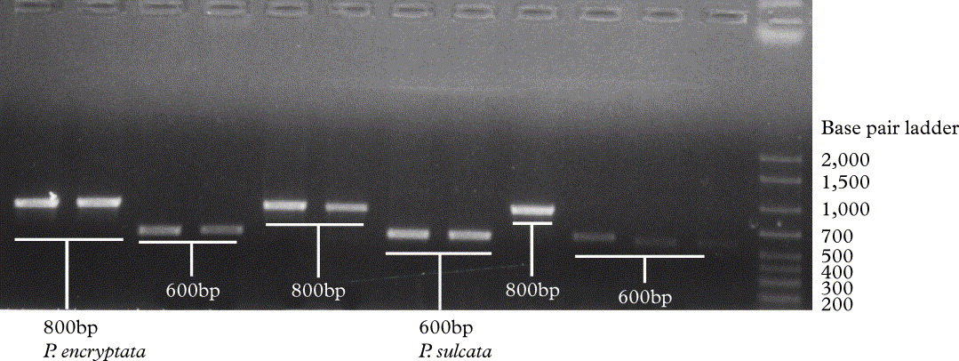
Seven ITS, 28 IGS rDNA, and 3 β-tubulin sequences were generated. In addition, 33 ITS, and 22 β-tubulin sequences were downloaded from GenBank and aligned with the former ones, resulting in an alignment of 40 OTUs (Table 1). Of the 40 OTUs, 15 lack β-tubulin and 12 lack IGS (Table 1). The size of the ITS PCR product obtained ranged between 600–800 bp (Fig. 1). The differences in size were due to the presence or absence of an insertion of about 200 bp which was identified as a Group I intron (DePriest & Been Reference DePriest and Been1992). The intron was located in the 3′ end of the small ribosomal DNA subunit (SSU), and was removed from the analysis.
Parmelia encryptata (MAF-Lich 10152, 10157, 9902, 15420, 15422) and P. sulcata (MAF-LICH 15581, 9899, 10159, 15432, 10160, 15418, 15429), gel electrophoresis showing size of different PCR products.
The matrix contained 1973 aligned characters (ITS: 1–514; β-tubulin: 515–1290; IGS:1291–1973), from which 139 unambiguously aligned parsimony informative sites were used in the parsimony analysis. The analysis resulted in 486 501 most parsimonious trees of 201 steps, with a CI = 0·796 and an RI = 0·926.
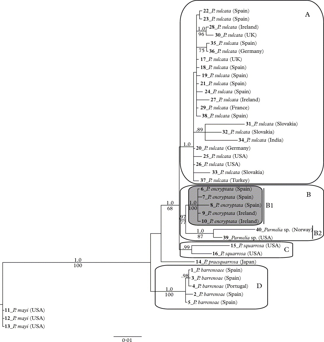
The Bayesian analyses of the three combined datasets was halted after 600 000generations, the last half of each tree sample being stationary. A majority rule consensus tree was constructed from the 6000 trees of the stationary tree sample. Since there were no conflicts between the overall topologies of the MP and B/MCMC analyses, only the 50% majority-rule consensus tree is shown with MP bootstrap values added (Fig. 2).
50%-majority-rule Bayesian consensus tree based on 6000 trees from the combined analysis of ITS, IGS and β-tubulin. Bayesian posterior probabilities equal or above 0·95 are indicated above branches. Bootstrap values ≥ 70% obtained in the parsimony analyses are indicated below the branches. Boxes represent monophyletic clades A, B, C and D. Numbers in bold to the left of the terminals correspond to specimen number in Table 1.
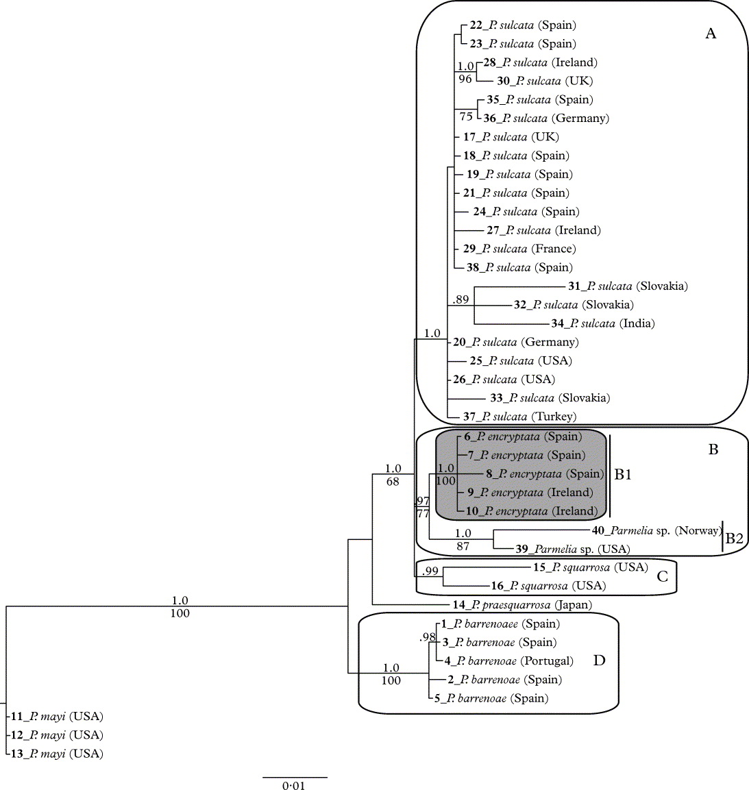
The 50% majority-rule consensus tree of the combined analysis (Fig. 2) showed five monophyletic clades (BPP ≥ 1·00). One clade included the P. sulcata s. str. group specimen from near the lectotype locality in Ireland (clade A; Fig. 2). The other main clade (clade B; Fig.2) included specimens initially identified as P. sulcata based on their morphological characters, but which did not form a monophyletic group together with the rest of the P. sulcata samples, and were instead located in an inclusive monophyletic clade joining P. sulcata s. str., P squarrosa, and P. sulcata specimens in clade B (1 and 2). Therefore, we introduce the new species name P. encryptata to accommodate this previously unrecognized taxon (clade B 1).
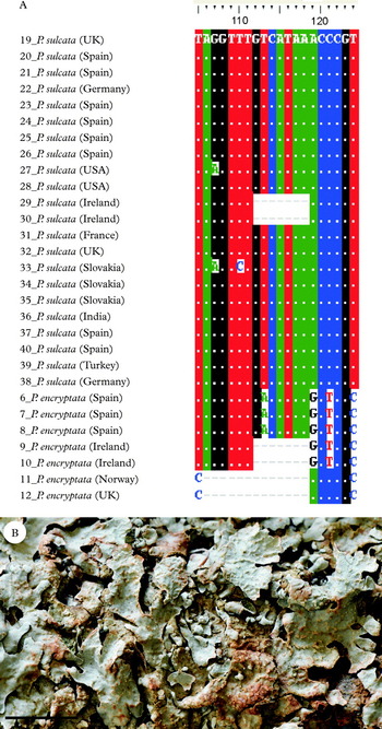
A, Parmelia sulcata and P. encryptata, ITS rDNA sequence alignment showing autapomorphic nucleotides at positions 113, 119, 122 and 124 and a short deletion between 106–119 positions. Numbers indicated to the left of the taxa correspond to specimen numbers in Fig. 2 (phylogenetic tree) and Table 1; B, Parmelia encryptata, habit (MAF-Lich 10157—holotype) Scale = 2 mm.

Clade B includes two sister clades (B1 and B2). Group B1 comprises several specimens from Spain and Ireland, all of which have an extra length in the ITS region, caused by the insertion of single tracts of about 200 base pairs. The strict consensus tree of the parsimony analysis shows one additional clade with bootstrap support (81%) grouping the three specimens from Spain within clade B1 (data not shown). Group B2 nests two specimens, one from New Jersey (USA) and the other from Spitzbergen (Norway). Both sequences have a deletion of 13 nucleotides in position 105 in the ITS1 of the ITS rDNA (Fig. 3A).
Clade C consists of two specimens of P. squarrosa. Parmelia praesquarrosa appears as the sister of the previous group, though this relationship needs to be confirmed. Group D includes all the specimens of P. barrenoae, and is positioned at the base of the three.
The results of the four tests for probabilities of alternative hypotheses are shown in Table 2. Monophyly of P. sulcata s. str. (clade A) and P. encryptata (clade B1), and P. sulcata s. str. (clade A) and clade B2, are both significantly rejected by the ELW test, while the SH test failed to reject this topology. All tests failed to reject a topology that has P. sulcata s. str. (clade A) and clade B in a monophyletic clade. SH and ELW also failed to reject P. sulcata s. str. (clade A) and P. squarrosa (clade C) in a monophyletic cluster.
Probabilities of two phylogenetic hypotheses being correct

* = significant, NS = non significant.
Pairwise distances between the haplotypes of Parmelia sulcata s. str. (clade A) ranged from 0·002 to 0·015 nucleotide substitutions per site (s/s), with a mean value of 0·008 ±0·004 s/s.
Pairwise distances between the haplotypes of clade B ranged from 0·002 to 0·029 s/s, with a mean value of 0·017 ± 0·010 s/s. Pairwise distance between two haplotypes comprising clade B1 is 0·002 s/s, and 0·009 in clade B2. Distances between the haplotypes included in clade B1 and clade B2 ranged from 0·017 to 0·029 s/s, with a mean of 0·023 ± 0·005 s/s.
Pairwise distances between the haplotypes of P. sulcata s. str. (clade A) and clade B ranged from 0·015 to 0·040 s/s, with a mean value of 0·026 ± 0·006 s/s. Distances between the haplotypes of clades A and B1 ranged from 0·022 to 0·040 s/s with a mean value of 0·028 ± 0·005, and between clades A and B2 ranged from 0·015 to 0·029 s/s, with a mean of 0·021 ± 0·005.
The results of the chemical analyses show all specimens of P. sulcata and P. encryptata contain atranorin, chloratranorin, salazinic acid and consalazinic acid. No automorphic phenotypic feature was detected characterizing separately clades A and B.
Taxonomy
Parmelia sulcata Tayl
in Mackay, Fl. Hib. 2: 145 (1836); type: Ireland, Co. Kerry, Lough Bray, 1812, T. Taylor [hb. Taylor Sheet no. 1128] (FH 00302186—lectotype designated by Hale & Kurokawa Reference Hale and Kurokawa1962: 5); Dunkerron, 16 June 2006, A. Crespo & R. Gavilán (MAF-Lich 15421—epitypus hic designatus).
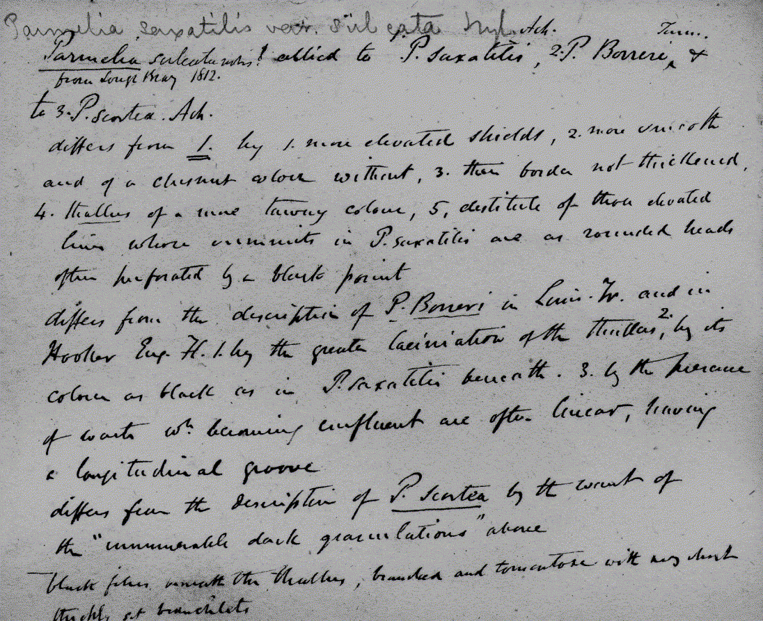
The typification of Taylor's name proved not to be straightforward and to require some careful investigation. Taylor (Reference Taylor and Mackay1836), in his account of lichens growing in Ireland, states that the species is “On siliceous rocks, also on trees; not uncommon” and then adds that it is “far more common on rocks in the County of Kerry than on trees”. No precise locality names are provided and while there is mention that, according to information he received from William Borrer, the species did occur in Tunbridge (Kent) though it is not clear if he had then seen those specimens himself. Hale & Kurokawa (Reference Hale and Kurokawa1962) designated as lectotype a specimen from Lough Bray in Co. Kerry in FH but did not discuss the matter further. That collection is one of two in Taylor's herbarium in FH under this name and was collected in 1812 (sheet no. 1128); the other was from “Gortymullen Hill”, which is also in Co. Kerry and close to Dunkerron, and collected in 1835 (sheet no. 1127). The choice of the Lough Bray specimen as lectotype is unequivocally supported by a note that accompanies it, which is in Taylor's handwriting (M. Schmull, in litt.), and seems to have been his working notes while he wondered at its naming; he left a blank after the genus name, discussed its differences from three other species, and afterwards squeezed into the blank space “sulcata nobis! from Lough Bray 1812” (Fig. 4).
Parmelia sulcata type FH 00302186 text by Taylor.

We see no reason to deviate from that choice as the Lough Bray specimen agrees morphologically and microchemically with the concept of Parmelia sulcata s. lat. However, with no explanation, Hale (Reference Hale1987: 47) states that the lectotype of this species is from “Dunkerron, Kerry” in FH, and that there are isolectotypes in BM and US. Today, there are no specimens labelled as being from Dunkerron in FH, either in the Taylor or the Tuckerman herbaria (M. Schmull, in litt.). There is a specimen labelled as from Dunkerron in BM, which was annotated by Hale in 1963, but it has no collection date and the handwriting on the label is evidently a later version of Taylor's hand (M. Schmull, in litt.); there is thus no evidence that the BM specimen was studied by Taylor before the species was described, and therefore that is not a candidate for lectotype selection. We did not manage to obtain any information on the specimen stated to be in US, and have to conclude that Hale's (Reference Hale1987) statement as to the lectotype being from Dunkerron was a “lapsus”.
The Lough Bray lectotype has apothecia, and we measured the ascospores as 16–19 ×8–10·5 µm. This ascospore size range is much larger than the 11–14 × 6–8 µm given by Hale (Reference Hale1987). Ranges given in other published works are: 12–18 × 8–13 µm (Lynge Reference Lynge1921), 13–15 × 5·5–6 µm (Hillmann Reference Hillmann1936; Kopaczevskaja et al. Reference Kopaczevskaja, Makarevich, Oxner and Rassadina1971), 13–15 × 5–6 µm (Ozenda & Clauzade Reference Ozenda and Clauzade1970), 11–14 × 6–8 µm (Nash et al. Reference Nash III, Ryan, Gries and Bungartz2002), 11–14(–15) × 6–8 µm (Smith et al. Reference Smith, Aptroot, Coppins, Fletcher, Gilbert, James and Wolseley2009: 654). Ascospores size ranges 13–16 × 8–9·5 µm in the specimens we studied in the P. sulcata s. str. lineage (Fig. 2). It therefore seems likely that there are two ascospore-size categories in the complex, and that Lynge may well have had material of both. The most common taxon would appear to have spores that rarely exceed 15 µm in length. No material of P. encryptata with apothecia was found, so that its ascospore size range is currently unclear. If it should prove to have ascospores regularly over 16 µm in length, the issue of whether to reconsider the epitypification will need to be addressed.
We failed to obtain any sequences from the Lough Bray lectotype, and therefore selected as an epitype for the name a specimen growing close to the lectotype locality in Co. Kerry in order to fix the application of the name.
Parmelia encryptata A. Crespo, Divakar & M. C. Molina sp. nov
MycoBank No.: MB 561715
Similis Parmeliis sulcatis sed differt in intron group I, et sequencis ACATAAGCTCGC in [gene ITS 1] at positio 113–124 in alignment.
Typus: Spain, Castellón, Herbès, Sierra de la Creu, alt. 1000 m, on Quercus, 14 October 2003, A. Crespo, P. K. Divakar & E. Barreno (MAF-Lich 10157— holotypus).
(Fig. 3B)
Remarks. This is a cryptic species segregated from Parmelia sulcata and it is morphologically similar to P. sulcata s. str. but genetically different. In the molecular phylogenetic tree (Fig. 2), all samples of Parmelia encryptata clustered in a strongly supported monophyletic group forming an independent lineage. Additionally, it has group I intron at 1516 position and the species can be distinguished easily by gel electrophoresis in having 800bp PCR product (Fig. 1) and sequencing is not required to distinguish the two clades. Furthermore, the new taxon has automorphic nucleotide differences in ITS 1 at positions 113, 119, 122 and 124 in the alignment (Fig. 3A). In addition, pairwise genetic distances between P. sulcata s. str. and P. encryptata were found sufficient to support their separation as distinct species (ranged from 0·022 to 0·040 s/s). The new taxon is sympatric with P. sulcata s. str., and so far we have not seen material with apothecia.
Discussion
Our results show that Parmelia sulcata, as it has been circumscribed, does not form a monophyletic group. It is possible to distinguish at least two different haplotypes separated in well-supported monophyletic lineages (Fig. 2). Group A contains the majority of specimens studied, including ones from Africa, Asia, Europe and North America. The specimen we collected close to the lectotype locality (Dunkerron, Co. Kerry, Ireland) is placed within this clade and, therefore, we consider that the name P. sulcata s. str. should be applied to this lineage; this haplotype has a wide distribution. Crespo et al. (Reference Crespo, Bridge, Hawksworth, Grube and Cubero1999) previously demonstrated, in a population study on P. sulcata, that this haplotype was the most common among three different sized PCR products in an area of lichen recolonization, following a reduction in mean sulphur dioxide levels in the London area. Other specimens identified as P. sulcata on the basis of chemical and morphological characters, formed a distinct independent monophyletic group (clade B; Fig. 2).
The molecular data presented demonstrate that there are at least two distinct lineages in this ‘species’ as generally circumscribed. Most interesting is that the two phylogenetic groups do not have distinctive geographical distributions; in the same areas in Ireland, Spain and the USA, both haplotypes are sympatric, and they can be found on the same phorophytes. However, in other cases geographical differences have been found to corroborate phylogenetic lineages (e.g. Argüello et al. Reference Argüello, del Prado, Cubas and Crespo2007; Divakar et al. Reference Divakar, Figueras, Hladun and Crespo2010).
Moreover, within clade B, two sister groups can be recognized. One (clade B2), including specimens from the USA and Norway, has a deletion of 13 nucleotides in ITS1. The other (clade B1), comprising samples from Spain and Ireland, is characterized by the presence of an autocatalytic group I intron in 1516 position nuSSU of approximately 200 bp.
Therefore, as currently circumscribed, P. sulcata does not satisfy the phylogenetic species concept as a monophyletic lineage (Donoghue Reference Donoghue1985; de Queiroz Reference de Queiroz2005a, b), since the clade clusters with P. squarrosa. This is especially so since P. squarrosa is a reproductively isolated lineage and also genetic distances clearly separate this from other Parmelia species (Del-Prado et al. Reference Del-Prado, Cubas, Lumbsch, Divakar, Blanco, Amo de Paz, Molina and Crespo2010). Several authors underline the need to identify the presence of the same clades in different single-locus genealogies, which can be taken into account as evidence that the clades represent reproductively isolated lineages (Dettman et al. Reference Dettman, Jacobson and Taylor2003; Pringle et al. Reference Pringle, Baker, Platt, Wares, Latge and Taylor2005). Our results show a concordance genealogy (Avise Reference Avise2000; Taylor et al. Reference Taylor, Jacobson, Kroken, Kasuga, Geiser, Hibbett and Matthew2000), where the phylogenetic tree topologies obtained from three independent molecular markers (nuITS, IGS and partial β-tubulin gene) is congruent.
Molecular analyses on other organisms have provided evidence for autocatalytic group I intron vertical inheritance over millions of years with few losses (Simon et al. Reference Simon, Fewer, Friedl and Bhattacharya2003; Besendahl et al. Reference Besendahl, Qiu, Lee, Palmer and Bhattacharya2004), and, on many occasions, they have been used for phylogenetic reconstructions (Grube et al. Reference Grube, Gutmann, Arup, de los Ríos, Mattson and Wedin1999; Thell et al. Reference Thell, Stenroos and Myllys2000; Crespo et al. Reference Crespo, Molina, Blanco, Schroeter, Sancho and Hawksworth2002; Linke et al. Reference Linke, Hemmerich and Lumbsch2003; Molina et al. Reference Molina, Crespo, Blanco, Lumbsch and Hawksworth2004). However, remarkable examples of horizontal transfer of genes (group I intron) between closely related species have also been reported in other organisms (Nishida et al. Reference Nishida, Tajiri and Sugiyama1998; Oksanen et al. Reference Oksanen, Lohtander, Sivonen and Rikkinen2004; Simon et al. Reference Simon, Moline, Helms, Friedl and Bhattacharya2005). In Parmeliaceae, group I introns have received particular attention. Thell & Miao (Reference Thell and Miao1999) concluded that intron absence/presence could not be used as a phylogenetic species marker in the cetrarioid genera of Parmeliaceae. More recently, other approaches studying 1506 and 1516 intron positions at 3′ end of nuclear SSU ribosomal DNA concluded that 1506 introns could have been horizontally inherited, whereas 1516 introns were vertically transmitted (Gutiérrez et al. Reference Gutiérrez, Blanco, Divakar, Lumbsch and Crespo2007). In fact, 1516 introns had been used as an additional marker to segregate phylogenetic species in Parmelia (Molina et al. Reference Molina, Crespo, Blanco, Lumbsch and Hawksworth2004). Therefore, the 1516 group I intron could be considered as phylogenetically informative for the recognition of monophyletic groups.
Pairwise genetic distances also support clade A and B1 as distinct lineages. Del-Prado et al. (Reference Del-Prado, Cubas, Lumbsch, Divakar, Blanco, Amo de Paz, Molina and Crespo2010) established inter- and intra-specific pairwise distance thresholds, between 0·015–0·017 nucleotide substitutions per site (s/s), in parmelioid lichens using ITS sequences. However, the results we obtained revealed a clear difference between P. encryptata and P. sulcata s. str (clade A). The values obtained are within the interspecific ranges which would support P. encryptata (clade B1) as a species different from others so far described. Furthermore, while the distance also separates clade B1 and B2 as distinct lineages, the minimum distance values of 0·015s/s between clade A (P. sulcata s. str.) and clade B2 are at the margin of the threshold and thus the taxon sampling of this clade should be increased prior to making any decision because the other values are in the interspecific range.
Additionally, the alternative hypothesis test (ELW) significantly rejects P. sulcata s. str. (clade A) and clade B1 as constituting a monophyletic cluster. These results provide evidence that clade B1 is a reproductively isolated lineage and so merits species-level recognition.
We therefore concluded that clade B1 should be segregated as the new cryptic species, P. encryptata. Since clade B2 and P. sulcata s. str. lack group I intron and the minimum value of pairwise genetic distances between these two clades are at the edge of interspecific threshold, more comprehensive taxon sampling with additional molecular markers should be undertaken before making a formal taxonomic decision on this question, and thus we leave clade B2 unnamed here.
Clade D includes all specimens of P. barrenoae (Fig. 2), recognized as an independent lineage within the P. sulcata complex (Divakar et al. Reference Divakar, Molina, Lumbsch and Crespo2005a) characterized by the simple, rather than squarrose, rhizines. That species was described from the Iberian Peninsula, but we can now extend the known distribution to Morocco where it shares habitats with P. sulcata s. str. (Fig. 2, Table 1).
The taxonomic decisions on clades A, B1 and B2 will cause a problem for those wishing to identify P. sulcata in the absence of molecular information, and in order to make clear if P. sulcata or P. encryptata is intended in any study, we suggest that the form P. sulcata aggr. is used, as proposed by Hawksworth (Crespo & Lumbsch Reference Crespo and Lumbsch2010). This is especially important in relation to ecophysiological and bioindication studies as the two species may behave differently and ecologists need to be aware that they may be dealing with different biological entities. In the study of material of this complex, Crespo et al. (Reference Crespo, Bridge, Hawksworth, Grube and Cubero1999) found that, although in other areas of England the intron-containing taxon (i.e. P. encryptata) was widespread, in London the recolonizing thalli following amelioration of sulphur dioxide levels, only the intron-lacking taxon (i.e. P. sulcata s. str.) was expanding through the area.
Recent molecular studies have demonstrated that vegetatively reproducing lineages can also present high genetic diversity (Kroken & Taylor Reference Kroken and Taylor2001; Divakar et al. Reference Divakar, Molina, Lumbsch and Crespo2005a, b; Ferher et al. Reference Fehrer, Slavikova-Bayerova and Orange2008; Wirtz et al. Reference Wirtz, Printzen and Lumbsch2008; Del-Prado et al. Reference Del-Prado, Divakar and Crespo2011; Molina et al. Reference Molina, Del-Prado, Divakar, Sánchez-Mata and Crespo2011). Traditionally, it was thought that these lineages were clones and evolutionarily dead-ends (see Tehler Reference Tehler1982). However, even in the absence of sexual reproduction, lichens are able to exchange photobionts (Piercey-Normore Reference Piercey-Normore2006; Nelsen & Gargas Reference Nelsen and Gargas2008) which could provide opportunities for gene transfer. This could be one of the plausible explanations of presenting high genetic diversity in vegetatively reproducing lineages. It has been shown that the presence of cryptic species in lichenized fungi does not correspond to any particular reproductive mode. Especially in Parmeliaceae, cryptic lineages have been reported in apotheciate taxa such as Parmelina quercina, Melanelixia glabra (Argüello et al. Reference Argüello, del Prado, Cubas and Crespo2007; Divakar et al. Reference Divakar, Figueras, Hladun and Crespo2010), the isidiate Parmela saxatilis, Parmelina tiliacea (Molina et al. Reference Molina, Crespo, Blanco, Lumbsch and Hawksworth2004, Reference Molina, Del-Prado, Divakar, Sánchez-Mata and Crespo2011; Nunez-Zapata et al. Reference Nunez-Zapata, Divakar, Del Prado, Cubas, Hawksworth and Crespo2011), and the sorediate Parmelia sulcata, and Parmotrema reticulatum (Divakar et al. Reference Divakar, Molina, Lumbsch and Crespo2005a, b).
This study provides a compelling example of genetic diversity and species-level polyphyly in sorediate morphs and adds to a growing literature suggesting that traditional methods for species delimitation in lichenized fungi may underestimate the actual species diversity. Thorough inclusions of multigenic data are required to draw species boundaries in the P. sulcata species complex with confidence. We suggest that P. sulcata s. lat. is a morpho-model in an early stage of diversification, where P. encryptata and P. sulcata s. str. are examples of cryptic speciation at the molecular level.
We are particularly indebted to Mark R. D. Seaward (University of Bradford) and Michaela Schmull (Harvard University) for assistance in elucidating Taylor's handwriting styles, and also to Michaela for detailed information on Taylor material in FH and the loan of the lectotype. We acknowledge the Ministerio de Ciencia e Innovación (CGL2010-21646) for providing main funds for this research, to grant CGL2008-01600 from the same ministry for facilitating the involvement of DLH, Ramón y Cajal grant (RYC02007-01576) to PKD, and the Universidad Complutense de Madrid for all complementary facilities. Sequencing was carried out at the Unidad de Genómica (Parque Científico de Madrid, UCM) and we thank especially M. Isabel Garcia Saez.