Case report
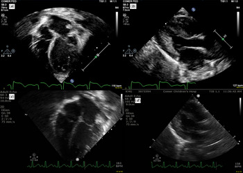
We report the case of a 10-month-old Caucasian girl with developmental delay and epileptic encephalopathy requiring multiple anti-epileptic therapies (phenobarbital, vigabatrin, pyridoxine, levetiracetam, and ketogenic diet). She was initially evaluated at 2 months of age for a systolic heart murmur. An echocardiogram was obtained and showed moderate left atrial and left ventricular dilation (Fig 1, Panels A and B). There were no intra- or extracardiac shunts identified and biventricular function was normal. The main and branch pulmonary arteries measured normal in size. Given the unexplained left atrial and ventricular dilation, she subsequently underwent a cardiac CT angiogram which confirmed the echocardiographic findings (moderate to severe left atrial and moderate left ventricular dilation). No atrial or ventricular septal defects or aortopulmonary window were visualised. The coronary artery origins appeared normal. A complex network of small systemic-to-pulmonary artery collateral vessels was visualised bilaterally in the paraesophageal and perihilar regions. A few of these collateral vessels originated from the descending aorta but it was difficult to definitively ascertain complete source and drainage of the entire vascular networks (Fig 2). The left heart enlargement was therefore attributed to systemic-to-pulmonary artery collateral mediated left-to-right shunting. Of note, the patient had undergone an echocardiogram at 2 weeks of age at another centre which showed only a patent foramen ovale and no evidence of left heart enlargement.
Moderate left atrial and left ventricular dilation (M-mode: left ventricular internal dimension [diastolic] = 2.8 cm, Z score = 3.8) is noted in apical 4-chamber (Panel A) and parasternal long-axis (Panel B) views at 2 months of age. The same views show complete regression of left atrial and left ventricular (M-mode: left ventricular internal dimension [diastolic] = 2.9 cm, Z score = 2.0) enlargement at 7 months of age (Panels C and D).

A meshwork of small systemic-to-pulmonary artery collaterals is noted to arise from ascending and descending aorta and arch arteries on this CT angiographic image with 3D reconstruction.

A brain MRI scan with spectroscopy, obtained at 2 months of age, showed delayed myelination for age with a thin corpus callosum. Non-specific small foci of susceptibility in the cerebellar hemispheres reflecting chronic microhaemorrhages or mineralisation were noted. MR spectroscopic findings were within normal limits for her age. A video electroencephalographic study obtained during awake and sleep state at 3 months of age showed abnormal background activity with striking discontinuity, abundant independent multi-focal epileptiform discharges, and numerous electroclinical seizures. In addition, the study revealed multiple diffuse epileptic seizures which were clinically characterised as asynchronous tonic seizures and isolated epileptic spasms. The findings were consistent with a severe early infantile encephalopathy, such as Ohtahara syndrome. Within 1 month, the electroencephalographic background became more continuous, but the patient developed migrating focal seizures occurring multiple times daily. Ultimately, with a combination of anti-epileptic medications and ketogenic diet, the seizures were reduced to 0–5 isolated, brief focal seizures arising from multiple regions.
An epilepsy panel (Invitae, San Francisco, CA) was obtained and revealed a pathogenic variant in KCNT1 (c1420C > T; p. Arg474Cys, R474C) which is associated with intractable seizures and severe cognitive and motor impairment. A likely pathogenic variant was identified in the PNKP (polynucleotide kinase 3 prime-phosphatase) (MIM 605610) (partial deletion exon 14). The PNKP is associated with autosomal recessive early infantile epileptic encephalopathy. However, PNKP-related conditions are known to be only caused by the co-occurrence of two pathogenic variants. Variants of unknown significance were identified in PIGN (phosphatidylinositol glycan anchor biosynthesis) (MIM 606097) (c2486C > A; p. Ala829Asp) and PRRT2 (proline-rich transmembrane protein 2) (MIM 614386) (gain, entire coding sequence, copy number = 3) genes.
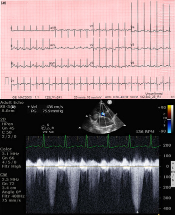
Upon follow-up, she was noted to have intermittent prolongation of corrected QT interval (QTc: 450–509 ms) (Fig 3, Panel A) between 5 and 6 months of age. The patient did well until 9 months of age without any tachypnea, cough, or haemoptysis. Serial echocardiograms during this period showed progressive normalisation of left atrial and ventricular sizes (Fig 1, Panels C and D). The most recent echocardiogram, however, has shown severe pulmonary hypertension (Fig 3, Panel B). Her seizures are well controlled on the current therapeutic regimen (<5 seizures per day) but she does have marked developmental delay. Recent electrocardiograms (over the past 2 months) have shown a normal corrected QT interval. She has been started on pulmonary vasodilator therapy and is being closely monitored.
Borderline prolonged corrected QT interval is noted on this electrocardiogram (Panel A). Continuous wave Doppler interrogation of the tricuspid regurgitation jet reveals severe elevation in right ventricular pressure (Panel B).

Discussion
KCNT1 (also known as SLACK, SLO2.2, or KCa4.1) gene, which is strongly conserved over evolution, encodes a sodium-activated potassium (KNa) channel which is widely expressed in the nervous system where it contributes to the slow hyperpolarisation that follows repetitive firing of neurons. KCNT1, therefore, regulates the rate of burst firing and enhances the accuracy with which action potentials lock to incoming stimuli. In addition, KNa channels also play a role in protecting cells from injury under ischemic conditions. Reference Barcia, Fleming and Deligniere1,Reference Bhattacharjee, Gan and Kaczmarek2
KCNT1-encoded KNa channel is also expressed in cardiac myocytes where its precise role has not been defined. Reference Bhattacharjee, Gan and Kaczmarek2,Reference de Los Angeles Tejada, Stolpe, Meinild and Klaerke3 However, antiarrhythmics, such as quinidine, clofilium tosylate, and bepridil, have been found to inhibit KNa channel suggesting that this channel may play a role in regulation of cardiac electrical activity and arrhythmogenesis. Reference de Los Angeles Tejada, Stolpe, Meinild and Klaerke3
KCNT1 (c1420C > T; p. Arg474Cys, R474C) variant, which affects the carboxy-terminus regulator of potassium (RCK) domain of the protein, Reference Lim, Ricos, Dibbens and Heron4 has previously been reported as a de novo change in six individuals with severe infantile-onset epilepsy syndromes. Reference Ohba, Kato and Takahashi5–Reference Kuchenbuch, Barcia and Chemaly7 This variant has also been reported in a 2-day-old Japanese girl with epilepsy, heart failure, and systemic-to-pulmonary artery collaterals who died of massive haemoptysis despite several catheter-based interventions on collaterals. Reference Kawasaki, Kuki and Ehara8 This substitution occurs at a position which is conserved across the species and was not observed in approximately 6500 individuals of European and African American ancestry in the NHLBI Exome Sequencing Project, indicating it is not a common benign variant in these populations. Moreover, in silico analysis predicts this variant is probably damaging to the protein structure/function. Finally, alternative missense variants at the same residue (c.1421 G > A; p.R474H) or other missense variants in the nearby residues (c.1420 C > T; p.R474C) (R477T) have been reported in association with KCNT1-related disorders, Reference Barcia, Fleming and Deligniere1,Reference Kuchenbuch, Barcia and Chemaly7,Reference Stenson, Mort, Ball, Shaw, Phillips and Cooper9 supporting the functional importance of this region of the protein.
KCNT1 mutations have been associated with a broad range of neurological phenotypes such as malignant migrating partial seizures in infancy, autosomal-dominant nocturnal frontal lobe epilepsy, leucoencephalopathy, and early-onset epileptic encephalopathy including Ohtahara and West syndromes. Reference Barcia, Fleming and Deligniere1,Reference Lim, Ricos, Dibbens and Heron4 Most of these epilepsy syndromes have onset early in infancy and are frequently characterised by refractoriness to anti-epileptic medications and poor outcome. Reference Kuchenbuch, Barcia and Chemaly7,Reference McTague, Nair and Malhotra10
Less is known about the cardiac effects of KCNT1 mutations. In 2017, Kawasaki et al reported three Japanese infants with de novo KCNT1 mutations who had bilateral profuse systemic-to-pulmonary artery collaterals which originated from aortic arch, descending aorta, and subclavian arteries. Reference Kawasaki, Kuki and Ehara8 Interestingly, one of these three patients carried the same missense mutation as our patient and another had a missense mutation at an adjacent residue (c.1420 C > T; p.R474C). Two of the three reported infants had heart failure symptoms and all three had left heart enlargement and several episodes of haemoptysis which recurred despite multiple catheter-based interventions on collaterals. More importantly, haemoptysis was reported to be the cause of death in two of these patients. Since then, a few other patients have been found to have systemic-to-pulmonary artery collaterals in association with KCNT1-associated epilepsy (verbal communication from a colleague at the Children’s Hospital of Philadelphia), but the exact prevalence and the natural history of systemic-to-pulmonary “collateralopathy” in these patients remains unknown. These findings are intriguing as systemic-to-pulmonary artery collaterals are typically seen at birth in complex CHDs such as tetralogy of Fallot with pulmonary atresia and can be acquired in those with single-ventricle physiology. In tetralogy of Fallot with pulmonary atresia, these collateral vessels augment pulmonary blood flow and may be the sole source of pulmonary blood flow to certain segments of the lungs but are considered an unreliable source as they can undergo dynamic changes, including stenosis and complete involution. Primary cardiac disease due to systemic-to-pulmonary artery collaterals, as seen in patients with KCNT1 mutations, has not been reported before.
Even though the data are sparse, the clinical course and the echocardiographic findings in our patient suggest that spontaneous regression of systemic-to-pulmonary artery collateral-mediated left heart dilation in carriers of KCNT1 mutations may be an indicator of elevated pulmonary vascular resistance and pressures. Whether all carriers of KCNT1 mutations should be screened for collateral-associated heart disease and the optimal modality for such screening (echocardiography versus CT angiography or cardiac MRI) remains unknown. Once the collaterals are identified, the risk of life-threatening complications such as haemoptysis or pulmonary hypertension, and optimal timing for intervention are important questions that beg an answer.
Other vascular abnormalities such as an arteriovenous fistula between the hepatic artery and the left portal branch have also been reported in these patients. Reference Kuchenbuch, Barcia and Chemaly7 These clinical findings suggest that KCNT1-encoded KNa channel may play a yet unknown role in cardiovascular angiogenesis.
In addition to structural abnormalities such as “collateralopathy”, KCNT1 mutations have also been associated with electrical abnormalities. A high incidence of sudden unexpected death has been reported in these patients, suggesting an underlying propensity to malignant arrhythmias. Reference Kuchenbuch, Barcia and Chemaly7 Specific arrhythmogenic phenotypes have been reported in some patients. A patient with Brugada syndrome leading to arrhythmogenic syncope was found to have KCNT1 carboxy terminus c.3317 G > A; pArg1106Gln mutation. An additional patient with the same mutation and multi-drug refractory seizures has also been reported. Reference Lim, Ricos, Dibbens and Heron4,Reference Juang, Lu and Lai11,Reference Barcia, Chemaly and Kuchenbuch12 This patient was diagnosed at 12 years of age and has had a normal 24-hour Holter evaluation and has done well from a cardiac standpoint. An unspecified cardiac arrhythmia has been reported in a young patient with familial KCNT1 c.2782 C > T; p. Arg928Cys mutation. Reference Møller, Heron and Larsen13 Our patient had intermittent prolongation in corrected QT interval despite not being on any QTc prolonging medications and normal serum potassium, calcium, and magnesium concentrations. This finding has previously not been reported in KCNT1 mutations carriers.
Quinidine, a class IA antiarrhythmic drug, has been shown to reverse KCNT1-encoded KNa channel over activity in vitro. Reference Milligan, Li and Gazina14 These encouraging in vitro findings have led to evaluation of quinidine in patients with KCNT1-associated epilepsy syndromes with mixed results. While some patients have shown improvement, evidence for a convincing neurological response is lacking in the majority. Serious adverse effects such ventricular arrhythmias have, however, been reported with quinidine use. Of note, all of these trials are limited by a small sample size which makes drawing any conclusions difficult. Reference Fitzgerald, Fiannacca and Smith15–Reference Numis, Nair and Datta17 The effects of quinidine on cardiovascular features of KCNT1 mutations have not been systematically evaluated.
In summary, KCNT1 mutations, which encode a gain-of-function KNa channel, are associated with a unique systemic-to-pulmonary artery collateral-mediated heart disease or “collateralopathy” which is characterised by a net left-to-right shunt leading to left heart enlargement. This “collateralopathy” can clinically present as heart failure, life-threatening haemoptysis, or pulmonary hypertension. Though only a handful of patients have been reported, the natural history of KCNT1 mutation-associated “collateralopathy” appears variable with most of the reported patients experiencing an unrelenting course with recurrent episodes of haemoptysis despite multiple catheter-based interventions. As seen in our patient, spontaneous regression of systemic-to-pulmonary collateral-mediated left heart dilation is certainly possible in KCNT1 mutation carriers; however, these patients should be carefully monitored for pulmonary hypertension. It is possible that the reported patients represent the “tip of the iceberg” and milder subclinical forms of this condition with left heart enlargement on imaging but no clinical symptoms also exist. KCNT1 mutations are also associated with a higher risk of sudden death suggesting pre-disposition to malignant arrhythmias. The precise substrate leading to this increased risk has not been defined but may include cardiac phenotypes, such as Brugada syndrome and possibly prolonged corrected QT interval. Further studies are needed to systematically evaluate the cardiovascular effects of KCNT1 mutations.
Acknowledgements
None.
Financial Support
This research received no specific grant from any funding agency, commercial or not-for-profit sectors.
Conflict of Interest
None.
Ethical Statement
Informed consent was obtained from all individual participants included in the report. This report does not include any human or animal experimentation.