Introduction
Nasopharyngeal tuberculosis is a rare entity, even in endemic tuberculosis areas.Reference Percodani, Braun, Arrue, Yardeni, Murris-Espin and Serrano1–Reference Srirompotong, Yimtae and Jintakanon3 Primary nasopharyngeal tuberculosis is described as isolated infection of the nasopharynx in the absence of pulmonary or systemic disease. It is an even more unusual condition, with just a few case reports in the English language literature.
We describe a case of primary nasopharyngeal tuberculosis in a patient from an area in which tuberculosis was not endemic, who presented with the symptoms resembling exacerbation of previously diagnosed chronic rhinosinusitis.
Case report
A 48-year-old man presented to our department with symptoms resembling exacerbation of previously diagnosed chronic rhinosinusitis. The patient was a heavy smoker and came from a low socioeconomic background. He had suffered from the symptoms of chronic rhinosinusitis for more than five years, and had been treated conservatively. He had been referred to our out-patient division after six weeks of antibiotic treatment in another institution, with no improvement. His major complaints were extreme postnasal drip and an unpleasant nasal odour. General symptoms were absent.
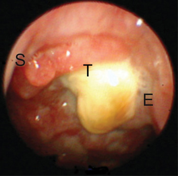
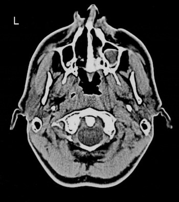
Endoscopic examination of the nose and nasopharynx revealed irregular thickening of the left lateral and posterior wall of the nasopharynx, partially covered with crusts and necrotic tissue (Figure 1). This irregular thickening was confirmed on computed tomography (CT) (Figure 2).
Endoscopic view of the left side of the nasopharynx. S = choanal edge of septum; T = thickening of the nasopharyngeal wall; E = eustachian tube orifice

Axial computed tomography scan showing irregular soft tissue thickening of the left (L) lateral and posterior wall of the nasopharynx, without bone involvement. Also note the obvious radiological signs of chronic rhinosinusitis in the right maxillary sinus.

To rule out nasopharyngeal carcinoma, multiple biopsies were taken under local anaesthesia. However, histopathological findings were consistent with nonspecific chronic inflammation.
Finally, the patient was admitted to hospital for nasopharyngeal curettage under general anaesthesia.
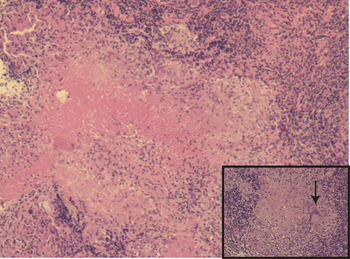
Histopathological study of curetted tissue showed typical giant cell epithelioid granulomas with caseous necrosis (Figure 3). Direct examination after Ziehl–Neelsen staining was positive for tuberculosis. The patient had no cervical lymphadenopathy and all pulmonary tests were negative; therefore, a diagnosis of primary nasopharyngeal tuberculosis was made.
Photomicrograph showing typical tuberculous granuloma with an area of central caseous necrosis surrounded by epithelioid cells and occasional multinucleated Langerhans-type giant cells (arrow). (H&E; ×20 (inset ×40))

Five weeks later, the diagnosis was definitively confirmed by positive culture of biopsy material. Culture of the patient's sputum remained negative.
After six months of antituberculous triple therapy, endoscopic examination revealed a completely normal nasopharynx. There was no evidence of recurrence over two-year follow up.
Discussion
Nasopharyngeal tuberculosis is a very uncommon disease. In a large study on 843 cases of tuberculosis, only 1.8 per cent of patients had upper respiratory tract disease, with just one case of nasopharyngeal involvement.Reference Rohwedder4 A literature search identified only a few publications on nasopharyngeal tuberculosis, mainly case reports originating from endemic regions.
Nasopharyngeal tuberculosis has a slight female predominance, with a peak of incidence between 50 and 60 years of age.Reference Percodani, Braun, Arrue, Yardeni, Murris-Espin and Serrano1, Reference Bath, O'Flynn and Gibbin5 Smoking and low socioeconomic status have been recognised as potential risk factors.Reference Percodani, Braun, Arrue, Yardeni, Murris-Espin and Serrano1 In our patient, both these factors were present.
Two potential pathways of infection have been described. In primary nasopharyngeal tuberculosis, infection occurs directly via nasal ventilation. In secondary disease, infection spreads from the primary site (most often pulmonary) through the airway via mucus flow, or via haematogenous or lymphogenous spread.Reference Percodani, Braun, Arrue, Yardeni, Murris-Espin and Serrano1, Reference al Serhani, al Rikabi and Assiry6
The most common clinical presentation of nasopharyngeal tuberculosis is cervical lymphadenopathy.Reference Tse, Ma, Chan, Ho, King and Fung2, Reference Srirompotong, Yimtae and Jintakanon3 The upper cervical lymph nodes are predominantly affected, making the clinical picture almost indistinguishable from that of nasopharyngeal carcinoma. Otological symptoms (e.g. hearing loss, tinnitus and otalgia) are the second most common type of complaint, while, surprisingly, rhinological symptoms (e.g. nasal obstruction, mucopurulent or haemorrhagic nasal discharge and postnasal drip) contribute less than 20 per cent of presenting symptoms. Rare cases have presented with diplopia, snoring and obstructive sleep apnoea.Reference Sithinamsuwan, Sakulsaengprapha and Chinvarun7–Reference Yosunkaya, Ozturk, Maden and Cetin9 Our patient presented with symptoms resembling an exacerbation of chronic rhinosinusitis. Not surprisingly, his symptoms were initially mistaken for an exacerbation of this condition, delaying accurate diagnosis and treatment.
• Primary nasopharyngeal tuberculosis is isolated infection of the nasopharynx in the absence of pulmonary or systemic disease
• Primary nasopharyngeal tuberculosis is extremely rare, with only a few English language reports
• A case is presented of primary nasopharyngeal tuberculosis in a patient previously diagnosed with chronic rhinosinusitis
• The diagnostic difficulties in this case are discussed
• To avoid inappropriate diagnosis and treatment, nasopharyngoscopy is recommended for all patients with exacerbation of sinonasal symptoms
The diagnosis of nasopharyngeal tuberculosis is based on the histopathological and microbiological findings from biopsy material.Reference King, Ahuja, Tse, van Hasselt and Chan10 Histopathological examination typically reveals granulomatous inflammation with epithelioid giant cells and caseous necrosis. Ziehl–Neelsen staining may directly detect acid-fast bacilli. After four to six weeks of appropriate microbiological culture, the drug sensitivities of the infecting strain may become apparent. Unfortunately, this ideal scenario is rarely seen in practice, and multiple, repeated biopsies are often needed to confirm the diagnosis and initiate appropriate therapy. However, it is in exactly such difficult cases that we see the remarkable role of new diagnostic technologies, such as polymerase chain reaction analysis to detect bacterial DNA.Reference Noussair, Bert, Leflon-Guibout, Gayet and Nicolas-Chanoine11
The treatment of choice for nasopharyngeal tuberculosis is antituberculous triple therapy combining isoniazid, rifampicin and ethambutol, or quadritherapy with additional pyrazinamide, for at least six months.Reference Tse, Ma, Chan, Ho, King and Fung2, Reference Srirompotong, Yimtae and Jintakanon3
When treated properly, nasopharyngeal tuberculosis carries an excellent prognosis, and complete resolution of disease is the rule.
To our best knowledge, the presented case represents the first published report of primary nasopharyngeal tuberculosis in a patient previously diagnosed with chronic rhinosinusitis. These two disease entities have many overlapping symptoms, and there is obviously huge potential for misdiagnosing such a patient and introducing inappropriate treatment. Therefore, we suggest that all patients suffering from exacerbation of sinonasal symptoms should undergo flexible or rigid nasopharyngoscopy. If a nasopharyngeal mass is detected, it should be considered to be nasopharyngeal carcinoma until proven otherwise. Histopathological examination of multiple (and sometimes repeated) biopsies is needed, both to exclude malignancy and to confirm the diagnosis of nasopharyngeal tuberculosis.
Acknowledgment
The authors thank Prof Sven Seiwerth MD PhD for his contribution in obtaining the histopathological diagnosis for this patient.