I. INTRODUCTION
For high-temperature waste heat conversion applications, low-dimensional layered oxides as well as perovskite-related phases have been reported to have relatively high efficiency (Misture and Edwards, Reference Misture and Edwards2012). The efficiency and performance of thermoelectric (TE) energy conversion or cooling is related to the dimensionless figure of merit (ZT) of the TE materials, given by ZT = S 2 T/ρκ, where T is the absolute temperature, S is the Seebeck coefficient or TE power, ρ is the electrical resistivity, and k is the thermal conductivity (Nolas et al., Reference Nolas, Sharp and Goldsmid2001). Examples of these oxides include NaCoO x (Terasaki et al., Reference Terasaki, Sasago and Uchinokura1997; Misture and Edwards, Reference Misture and Edwards2012), Ca2Co3O6 (Mikami et al., Reference Mikami, Funashashi, Yoshimura, Mori and Sasaki2003; Mikami and Funahashi, Reference Mikami and Funahashi2005), Ca3Co4O9 (Masset et al., Reference Masset, Michel, Maignan, Hervieu, Toulemonde, Studer, Raveau and Hejtmanck2000; Minami et al., Reference Minami, Itaka, Kawaji, Wang, Koinuma and Lippmaa2002; Grebille et al., Reference Grebille, Lambert, Bourée and Petricek2004; Hu et al., Reference Hu, Si, Sutter and Li2005), and Bi2Sr2Co2O y (Wang et al., Reference Wang, Venimadhav, Guo, Chen, Li, Soukiassian, Schlom, Pan, Wong-Ng, Vaudin, Cahill and Xi2009).
The search for oxide compounds with superior TE properties continues. Pervoskite materials such as La1−x A x CoO3 (A = Ca and Sr) (Sehlin et al., Reference Sehlin, Anderson and Sparlin1995; Wang et al., Reference Wang, Sui, Ren, Wang, Wang, Su and Fan2010) and RCoO3 (Moon et al., Reference Moon, Masuda, Seo and Koumoto2001) have demonstrated large Seebeck coefficient and low resistivity. Double-perovskites with lanthanides ordered on one subset of the octahedrally coordinated B sites have recently been of crystal chemical interest (Harada et al., Reference Harada, Wakeshima and Hinatsu1999; Kanaiwa et al., Reference Kanaiwa, Wakashima and Hinatsu2002; Wiebe et al., Reference Wiebe, Greedan, Luke and Gardner2002). Sr2 RSbO6 is a series of double-pervoskite materials that may have potential for TE applications. Using neutron diffraction studies, Karunadasa et al. (Reference Karunadasa, Huang, Ueland, Schiffer and Cava2003) observed in the structure of Sr2 RSbO6 (R = Dy, Ho, and Gd), the magnetic lanthanide sublattice displays a classical fcc arrangement, and edge-shared network of lanthanide octahedra are known to result in geometric magnetic frustration. However, they did not observe magnetic ordering in these compounds down to temperature of 2 K.
Since X-ray diffraction (XRD) is a non-destructive technique for phase identification, XRD patterns are especially important for phase characterization. The main goal of this investigation is to determine the crystal structure, crystal chemistry, and experimental X-ray patterns for Sr2 RSbO6 (R = Pr, Nd, Sm, Eu, Gd, Dy, Ho, Y, Er, Tm, Yb, and Lu) at room temperature, and to make them widely available through submission to the Powder Diffraction File (PDF, 2013).
II. EXPERIMENTAL DETAILS
A. Sample preparation
All samples were prepared by heating a stoichiometric mixture of SrCO3, R 2O3 (R = Pr, Nd, Sm, Eu, Gd, Dy, Ho, Y, Er, Tm, Yb, and Lu), and Sb2O3 in air. R 2O3 were first heat-treated at 550 °C overnight prior to use to ensure the absence of carbonates and hydroxides. Samples were weighed out, well-mixed and calcined overnight at 850 °C, followed by heat treatments at 950 °C for 50 h, 1200 °C for 40 h, and finally at 1400 °C for another 20 h, with intermediate grindings. During each heat treatment, the samples were furnace cooled. The phase purity of the samples was established by powder XRD.
B. X-ray Rietveld refinements and powder reference patterns
The Sr2 RSbO6 powders were mounted in zero-background holders on a Bruker D8 Advance diffractometer equipped with a VANTEC-1 position-sensitive detector. [The purpose of identifying the equipment in this article is to specify the experimental procedure. Such identification does not imply recommendation or endorsement by the National Institute of Standards and Technology.] Patterns were measured using the Bragg–Brentano mode (CuKα radiation, 40 V, 40 , 0.3° divergence slit) from 5° to 145° 2θ in 0.007 297° steps.
The Rietveld refinement technique (Rietveld, Reference Rietveld1969) with the software suite GSAS (Larson and von Dreele, Reference Larson and von Dreele2004) was used to determine the structure of Sr2 RSbO6. The Thompason–Cox–Hastings pseudo-Voigt profile function was used; GSAS #2. For the major phase, both the Lorentzian size and strain-broadening coefficients were refined. For impurity phases, only LX was refined. A common specimen displacement coefficient was refined in each case. A six-term shifted Chebyshev polynomial was used to model the background. A scale factor and the lattice parameters were refined for each phase. All U iso were fixed at 0.01 Å2. The Sb–O distances were refined subject to a restraint of 2.00(2) Å and the R–O distance restraints were varied depending on the size of the R. A restraint of 90(3)° was applied to the cis O–Sb–O angles. Refinement of the cation occupancies resulted in values insignificantly different from unity; so all cation occupancies were fixed at 1.00.
Reference patterns were obtained with a Rietveld pattern decomposition technique. Using this technique, the reported peak positions were derived from the extracted integrated intensities and positions calculated from the lattice parameters. When peaks are not resolved at the instrument resolution function, the intensities are summed, and an intensity-weighted d-spacing is reported. They are also corrected for systematic errors both in d-spacing and intensity. In summary, these patterns represent ideal specimen patterns.
C. Bond valence sum (V b ) calculations
The bond valence sum values, V b , for Sr, R, and Sb were calculated using the Brown–Altermatt empirical expression (Brown and Altermatt, Reference Brown and Altermatt1985; Brese and O'Keeffe, Reference Brese and O'Keeffe1991). The V b of an atom i is defined as the sum of the bond valences v ij of all the bonds from atoms i to atoms j. The most commonly adopted empirical expression for the bond valence v ij as a function of the interatomic distance d ij is v ij = exp[(R 0 –d ij )/B]. The parameter, B, is commonly taken to be a “universal” constant equal to 0.37 Å. The values for the reference distance, R 0: Sr–O 2.118, Sb–O 1.942, Pr–O 2.135, Nd–O 2.117, Sm–O 2.088, Eu–O 2.076, Gd–O 2.065, Dy–O 2.036, Ho–O 2.023, Y–O 2.014, Er–O 2.010, Tm–O 2.000, Yb–O 1.985, and Lu–O 1.971 (Brown and Altermatt, Reference Brown and Altermatt1985; Brese and O'Keeffe, Reference Brese and O'Keeffe1991). When two different cations occupying the same site, the resulting V b value is computed as the weighted sum of the contributions from both cations.
III. RESULTS AND DISCUSSION
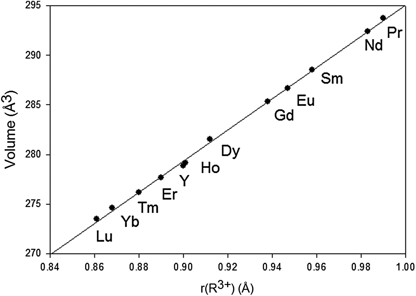
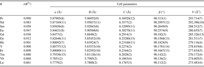
Table I lists the lattice parameters for Sr2 RSbO6. Contrary to the report by Wittmann et al. (Reference Wittmann, Rauser and Kemmler-Sack1981) that while the compounds with R = Nd, Sm, Eu, and Dy are monoclinic, and those with smaller R (R = Y, Yb, and Lu) are orthorhombic, we found that the structures of the entire Sr2 RSbO6 series are monoclinic, with space group P21/n. These materials have lower symmetry as compared to the Ba analogs. The Ba variants can be best described by the cubic doubled perovskite cell, space group Fm-3m. In the Sr variants, from R = Lu to Pr, the lattice parameters “a” ranges from 5.7779(2) to 5.879 05(8) Å, “b” ranges from 5.7888(2) to 5.969 52(9) Å, “c” ranges from 8.1767(3) to 8.369 20(12) Å, “β” ranges from 90.112(2) to 90.313(1) Å, and “V” ranges from 273.483(4) to 293.714(7) Å3. Figure 1 gives the plot of the unit-cell volumes, V, of Sr2 RSbO6 vs. Shannon ionic radius of r(R 3+) (Shannon, Reference Shannon1976). V decreases across the lanthanide series from Pr to Lu, or with the decreasing size of the ionic radius, which is known as the lanthanide contraction of R at the octahedral site.
Plot of unit-cell volume of Sr2 RSbO6 (R = Pr, Nd, Sm, Eu, Gd, Dy, Ho, Y, Er, Tm, Yb, and Lu vs. r(R 3+) (where “r” is the Shannon ionic radii (Shannon, Reference Shannon1976).

Crystallographic data for Sr2 RSbO6 (monoclinic P21/n); r(R 3+) is the Shannon ionic radius using VI coordination.

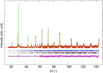
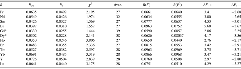
Table II gives the refinement residuals for Sr2 RSbO6 (R = Pr, Nd, Sm, Eu, Gd, Dy, Ho, Y, Er, Tm, Yb, and Lu). Figure 2 provides a sample Rietveld plot for Sr2DySbO6 . The observed (crosses), calculated (solid line), and difference XRD patterns (bottom) for Sr2DySbO6 are illustrated. The difference pattern is plotted at the same scale as the other patterns. Above 60° 2θ, the vertical scale has been magnified ten times. The row of tick marks indicates the calculated peak positions. The refinement residuals mainly reflect variations in the counting times, and the presence of traces of impurities.
(Color online) Observed (crosses), calculated (solid line), and difference XRD pattern (bottom) for Sr2DySbO6 by Rietveld analysis technique (1969). The difference pattern is plotted at the same scale as the other patterns up to 60° 2θ. At higher 2θ angles, the scale has been magnified ten times. The row of tick marks refers to the calculated peak positions.

Sr2 RSbO6 refinement residuals.

The number of variables for Gd- and Dy-compounds is higher because of the presence of a small amount of impurities in these two phases (two polymorphs of Gd2O3, SrGd2O4, and Sb2O4 for Sr2GdSbO6; and Dy2O3, SrDy2O4, and Sr1.45Sb0.55O3-x for Sr2DySbO6.
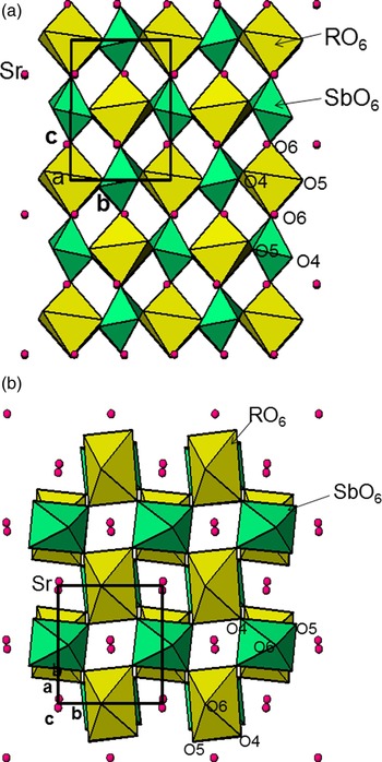
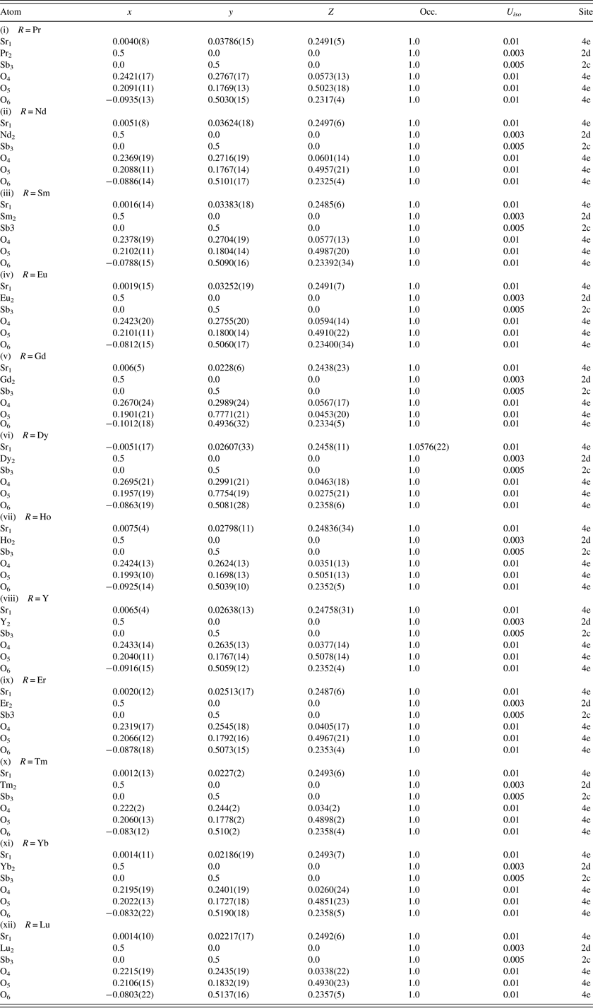
The atomic coordinates, displacement parameters for the structures of Sr2 RSbO6 are given in Table III. Refinement of the cation occupancies resulted in values insignificantly different from unity; therefore all cation occupancies were fixed at 1.00. The perovskite ABO3structure consists of a three-dimensional (3D) lattice of corner-sharing BO6octahedra enclosing a cavity in which the larger atom, A, is 12-coordinated to oxygen. Comparing with the ABO3 pervoskite structure, the double-perovskites, A 2 BB′O6, have chemical supercells in which two kinds of ions are found in the B sites in alternating octahedra. In this case, B = R and B′ = Sb. In other words, the trivalent lanthanides and pentavalent antimony are found to be fully ordered in the double-perovskite arrangement of alternating corner-sharing octahedra. Figures 3(a) and 3(b) give the structure of Sr2 RSbO6 viewing down along the a-axis and along the c-axis, respectively. The corner-sharing alternate SbO6 and RO6 units form zigzag chains along the c-axis.
(Color online) Crystal structure of Sr2DySbO6 at room temperature showing the unit-cell outline and the B-site octahedra, (a) view down along the a-axis and (b) view down along the c-axis.

Atomic coordinates and isotropic displacement factors for Sr2 RSbO6 (R = Pr, Nd, Sm, Eu, Gd, Dy, Ho, Y, Er, Tm, Yb, and Lu). Values inside brackets are standard deviations. Space group monoclinic P21/n.

The Sb–O distances are all similar but decrease slightly toward the end of the lanthanide series as the size of R 3+ become relatively small. Table IV gives the bond distances of Sr–O, R–O, and Sb–O, and bond valence sum values (V b ) of the cation sites. The results of bond valence calculations confirm that from R = Pr to R = Lu, Sb atoms in the octahedral sites are all of 5+ valence. The V b values of Sb5+ sites with r(R 3+) < r(Er3+) are all around the values of 5.0, but those values with R > R Er 3+ increase gradually from 5.1 to 5.3, indicating these sites experience compressive stress, or overbonding (in an undersized cage environment). However, the V b values for the Sr site in the Sr2 RSbO6 series are substantially <2.0 (1.71–1.93), indicating that these sites are under tensile stress. These tensile stress decreases as the size of r(R 3+) decreases. Note that since all R sites are >3.0 (ideal V b value of 3.0), ranging from 3.3 (R = Lu) to 3.8 (R = Pr), representing a large compressive stress or overbonding for the R sites. Although the results of the current calculations provide approximate values for the stress state of the site (Karpinski et al., Reference Karpinski, Kazakov, Angst, Mironov, Mali and Roos2001), they give a useful relative trend for comparison.
Bond distances and bond valence sums (V b ) for Sr2 RSbO6. Values inside brackets are standard deviations.

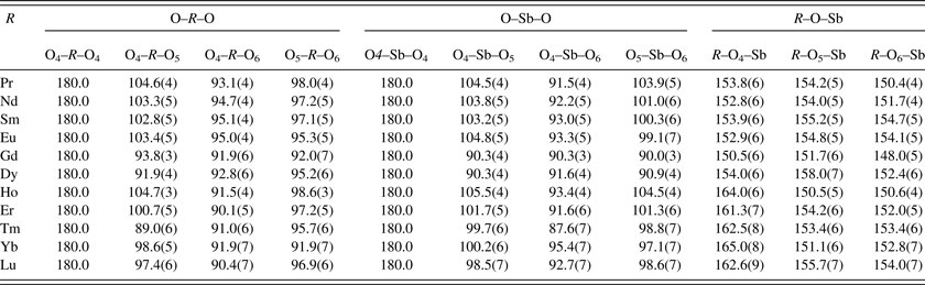
In monoclinic Sr2 RSbO6, there are three R–O–Sb angles (R–O4–Sb, R–O5–Sb, and R–O6–Sb) that are between different SbO6 and RO6 octahedral units (Table V). While the R–O6–Sb angle is a measure for the deviation from ideal value of 180° along the c-axis, the R–O4–Sb and R–O5–Sb bendings are related to the rotation of the octahedra with respect to the ab-plane (Figures 3(a) and 3(b)). These R–O–Sb angles all deviate rather significantly from 180° (approximately between 150° and 165°), the R–O4–Sb angle increases about 10° (closer to 180°) as r(R 3+) decreases from Pr to Lu, indicating lesser degree of rotation. One notes that the R and Sb sites in the SbO6 and RO6 octahedra also have distorted environment, as evidenced from different R–O and Sb–O distances (Table IV), and different O–R–O and O–Sb–O bond angles (Table V). For example, while the O4–R–O4 and O4–Sb–O4 angles are 180°, the remaining O–R–O and O–Sb–O angles all deviate from 90°.
Selected bond angles (°) for Sr2 RSbO6. Values inside brackets are standard deviations.

A. Reference XRD pattern
The reference powder patterns for Sr2 RSbO6 have been submitted and published for inclusion in the PDF: (R = Pr, 56–1402; R = Nd, 56–1403; R = Sm, 56–1404: R = Eu, 56–1430; R = Gd, 56–1410; R = Dy, 56–1485; R = Ho, 56–1399; R = Y, 56–1409; R = Er, 56–1398; R = Tm, 56–1400; R = Yb, 56–1401; and R = Lu, 56–1408). In these patterns, the symbols “M” and “ + ” refer to peaks containing contributions from two and more than two reflections, respectively. The symbol * indicates that the particular peak has the strongest intensity of the entire pattern and has been designated a value of “999”. The intensity values reported are integrated intensities rather than peak heights.
IV. SUMMARY
Crystal structure, crystal chemistry, and X-ray powder reference patterns of the Sr2 RSbO6 (R= Pr, Nd, Sm, Eu, Gd, Dy, Ho, Y, Er, Tm, Yb, and Lu) series of compounds have been determined. The structure consists of corner-sharing alternate SbO6 and RO6 units in the form the zigzag chains along the c-axis. Sr in general has a 12-coordination environment. Bond valence sum calculations indicated that all Sb adopt 5+ valence state in these compounds. The large compressive stress has been found at the R 3+ site throughout the series (V b ranges from 3.3 to 3.8). Similarly, large compressive stress has also been found in the Sb5+ sites in compounds consisting of relatively small size of R 3+ (R = Er, Tm, Yb, and Lu). The reference powder patterns for Sr2 RSbO6 have been submitted for inclusion in the PDF.
Acknowledgement
Partial financial support from the ICDD Grant-in-Aid Program is acknowledged.