Introduction
Solitary sinonasal fibrous tumours are extremely rare, mesenchymal neoplasms which were first described in 1931 as primary pleural tumours.Reference Klemperer and Rabin1 Also referred to as benign mesotheliomas or submesothelial fibromas, they have been described particularly in serous membranes, although they also originate in other regions such as the mediastinal space, lungs,Reference Witkin and Rosai2 vulva,Reference Nielsen, O'Connell, Dickersin and Rosenberg3 orbit,Reference O'Donovan, Bilbao, Fazi and Antonyshyn4 thyroid,Reference Parwani, Galindo, Steinberg, Zeiger, Westra and Ali5 nasopharyngeal region,Reference Witkin and Rosai6 larynx and infratemporal fossa.Reference Rayappa, McArthur, Gangopadhyay and Antonius7 The nasal cavity and paranasal sinuses are an extremely rare location for this type of neoplasm. We have found only 27 cases published in the literature, most of which have been reported during the past 15 years.
In histological terms, solitary fibrous tumours are solid tumours made up of fusiform cells and a magma of diffuse sclerotic tissue. Immunohistochemical techniques aid differentiation from other soft tissue tumours.Reference Corina, Volante, Carconi and Contucci8
Although radiotherapy and chemotherapy have been used to manage tumours of this sort, surgery is still the treatment of choice. Although several different modalities have been used, sinonasal endoscopic surgery appears to be the preferred approach in the great majority of cases.
We present here a case of a large solitary fibroma of the paranasal sinus, which we treated by sinonasal endoscopic surgery. We describe its clinical and histopathological features, we discuss the endoscopic technique utilised to deal with large sinonasal masses penetrating the pterygomaxillary and infratemporal fossae, and we offer a brief survey of the literature.
Case report
A 38-year-old woman presented with an approximately one-year history of nasal obstruction, without any other typical clinical manifestations of sinonasal tumours (such as rhinorrhoea, epistaxis or headache). The patient presented in a generally good state of health, without any personal antecedent conditions of interest. The neurological examination was normal.
Nasal endoscopy revealed an irregularly shaped, friable, reddish mass which occupied the whole of the right nasal fossa and was apparently attached to the lateral wall of the fossa.
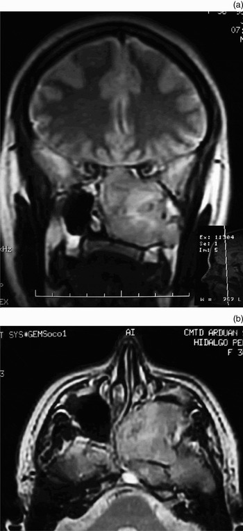
A magnetic resonance imaging (MRI) scan of the paranasal sinuses revealed a large, 7 × 8.5 × 6 cm mass which occupied the anterior and posterior ethmoids and the maxillary and sphenoid sinuses, displacing the septum and penetrating the pterygomaxillary fossa, having destroyed the lateral wall of the right nasal fossa (Figure 1). There were no signs of bone damage at the base of the cranium, nor of intracranial effects. No cervical lymph nodes were detected.
(a) Coronal and (b) axial flair magnetic resonance T2-weighted images showing tumour in the paranasal sinuses and pterygomaxillary and infratemporal fossae.

The tumour was resected in a number of fragments by means of sinonasal endoscopic surgery. The lesion was of a firm consistency, with abundant vascularisation and edges in intimate contact with adjacent bony structures.
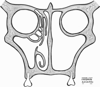
Surgery was undertaken as follows. An endoscopic medial maxillectomy was performed with the aid of 0°, 30° and 70°, 4 mm forward oblique telescopes, with the patient in the supine decubitus position with 25–30° cephalic flexure. We commenced the procedure with a total inferior turbinectomy and ample medial meatotomy, in order to create adequate space and to identify the relevant anatomy. We then made a mucoperiosteal incision in the nasal wall, using an electric scalpel (Stryker Colorado Needle; Stryker, Craniomaxillofacial, Portage, Michigan, US) at a distance of 1 cm from the tumour. After exposing the maxillary bone using an elevator and periosteal stripper, we commenced bone removal and the extirpation of the tumour with the aid of chisels, burrs and a paranasal sinus shaver handpiece. We then progressively demolished the part of the nasal wall in contact with the tumour and began to remove large sections of the tumour, following previously marked limits, until we had achieved complete anterior and posterior ethmoidectomy and sphenoidectomy, including the middle turbinate. The frontal sinus also required evaluation in order to ensure that complete extirpation of the lesion had been achieved. (If the tumour involved the frontal sinus, a Draf II approach would have been needed.) The limits of the resection were as follows: inferior, the floor of the nasal fossa; posterior, the posterior wall of the maxillary sinus; superior, the orbit; and anterior, the anterior wall of the maxillary sinus (Figure 2). In such cases, systematic catheterisation of the lacrimal system is not generally required, except when post-surgical obstructive pathology presents, in which case dacryocystorhinostomy may have to be performed.
Diagram of extirpation of the lateral nasal wall, showing limits of the endoscopic medial maxillectomy.

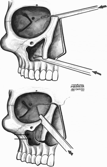
In endoscopic approaches of this sort, when radical tumour extirpation is required, it is necessary to eliminate the lateral nasal wall by means of large osteotomies: inferior, superior and a union of the two (Figure 3). The extension of the inferior osteotomy and the demolition of the posterior wall of the maxillary sinus allow access to the pterygomaxillary region and infratemporal fossa.
Diagram of the osteotomies employed: superior, inferior and a union of the two. Extirpation of the posterior wall of the maxillary sinus (arrows) was required in order to reach the pterygomaxillary and infratemporal region.

Following extirpation of the tumour and cleaning of the surgical cavity, we proceeded to thorough haemostasis. The cavity was filled with Surgicel® (Haemostatic bandage) (Johnson Medical Ltd, Gargrave, North Yorkshire, UK) and impregnated with antibiotic cream, and the nasal fossa was lightly covered with porous packing material.
Post-operative progress was good, with no immediate or delayed complications. During follow up, we performed regular nasal endoscopies, as well as computed tomography (CT) scans one and six months post-operatively. The patient's clinical condition improved markedly, with disappearance of the nasal obstruction, although there were occasional episodes of mild headache.
At the time of writing, a year and a half following surgery, no suspicious recurrence had been observed via nasal endoscopy. Post-operative CTs revealed diffuse enlargement of the nasal cavity mucosa, post-surgical changes and retention of post-cicatrisation secretions (Figure 4).
(a) CT coronal and (b) axial images in bone window. Six months post-operatively, showing post-surgical changes and no radiological evidence of tumour recurrence.

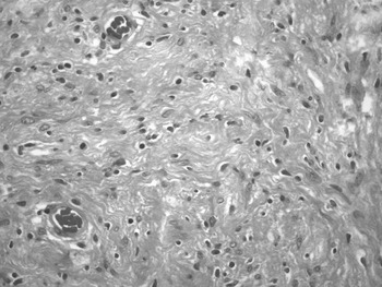
Pathological examination (of the bloc resection specimen) revealed a large, solid, reddish-brown, unencapsulated, 5 × 5 × 6 cm mass. Optical microscopy revealed a variable pattern, with zones of both hypo- and hypercellularisation, composed of spindle cells and areas with abundant collagen deposits (Figure 5). Immunohistochemical studies were positive for vimentin and CD34. Analysis for the proteins bcl-2, CD-99 and S-100 was negative. These findings strongly support our diagnosis of solitary fibrous tumour.Reference Witkin and Rosai6, Reference Fletcher9
Histological analysis. The tumour was variable in cellularity, consisting of a mixture of haphazard, storiform or short fascicular arrangement of soft spindle cells and less cell-dense collagenous bands. The spindle cells had plump, fusiform or elongated vesicular nuclei without pleomorphism, and a small to moderate amount of pale, eosinophilic cytoplasm. A haemangiopericytoma-like growth pattern was focally present (H&E; ×160).

Discussion
Solitary fibrous tumours or benign mesotheliomas are well known growths which arise in the mesenchymal tissue of the serous membranes. Most originate in the pleura. They can also be found in a wide range of other locations,Reference Witkin and Rosai2–Reference Rayappa, McArthur, Gangopadhyay and Antonius7 although much more rarely. Such a tumour in the nasal cavity site is extremely rare.
During the past few years, a number of cases of solitary fibrous tumours originating in the nasal cavity have been described. In the course of our work to date, we have found a total of 27 previously published cases of solitary fibromas of different sizes, originating in the paranasal fossae and sinuses. Within the nasal cavity the location of these tumours varies, in that they originate in both the anteriorReference Alobid, Alos, Blanch, Benitez, Bernal-Sprekelsen and Mullol10 and posteriorReference Pasquini, Cantaroni, Salfi, Tamburini, Marchi and Sciarretta11 ethmoid region and the sphenoid sinus.Reference Abe, Murakami, Inove, Ohde, Yamaguchi and Watanabe12
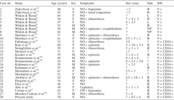
The clinical findings of the 28 published cases (17 women, 11 men) of solitary fibrous tumour are summarised in Table I. This Table shows that the most common clinical symptom and the principal reason for consultation was nasal obstruction continuing for a number of weeks, although various other signs (such as epistaxis, exophthalmos or rhinorrhoea) have also been described. The mean age of the patients was 48.9 years (range 30 to 71 years). The tumours were usually described as reddish masses with a fibrous texture, highly vascularised, circular or oval in shape, encapsulated and well delimited. They ranged in size from 1.7 to 8.5 cm in their largest dimension; the present case is the largest that has come to our notice.
Published cases of solitary fibrous tumour: clinical, epidemiological and immunohistochemical characteristics

No = number; IHC = immunohistochemistry; F = female; M = male; NO = nasal obstruction, R = right; L = left; NP = nasopharynx; ? = not defined; V+ = positive for vimentin; CD34+ = positive for CD34
Most of the published studies agreed on the need to maintain stringent diagnostic criteria in order to avoid confusing these tumours with other, more aggressive neoplasias.Reference Alobid, Alos, Blanch, Benitez, Bernal-Sprekelsen and Mullol10, Reference Abe, Murakami, Inove, Ohde, Yamaguchi and Watanabe12
In the published cases, imaging results were usually not specific enough to enable diagnosis, but were important as a means of evaluating tumour size and planning surgery. Computed tomography and MRI scans showed occupation of the sinuses and nasal fossae by isodense masses, with a greater or lesser degree of remodelling or effects on bone, according to the size of the tumour.
In histological terms, these reported tumours were formed by fusiform cells surrounded by bands of collagen fibres. In the great majority of cases, the cells reacted positively to vimentin and CD34 and negatively to S-100. It should be noted that CD34 is not strictly specific for fibromas, since it is expressed by a wide range of fusiform cell neoplasms, such as dermatofibrosarcomas.Reference Abe, Murakami, Inove, Ohde, Yamaguchi and Watanabe12
• This paper describes a rare large, solitary fibrous tumour of the maxillary sinus, anterior and posterior ethmoid cells and pterygomaxillary and infratemporal fossae, presenting only with nasal obstruction
• Tumours of this type tend to be benign and slow growing but locally invasive. Treatment is surgical, as they are resistant to chemoradiation therapy
• In the presented case, surgical treatment consisted of endoscopic medial maxillectomy with extension to the pterygomaxillary and infratemporal fossae
According to the literature, some 10 per cent of extrathoracic solitary fibrous tumours, including nasal tumours, present signs of malignancy, such as manifest local invasion, hyperplasia, atypia, necrosis, elevated rates of recurrence and remote metastasis.Reference Abe, Murakami, Inove, Ohde, Yamaguchi and Watanabe12, Reference Vallat-Decouvelaere, Dry and Fletcher25
The definitive therapy for such tumours is complete surgical extirpation. If successful, the probability of recurrence is very low. A wide range of surgical procedures has been utilised to deal with this type of tumour in the paranasal fossae and sinuses: lateral rhinotomy, medial maxillectomy, midface degloving and ethmoidectomy. At the present time, we consider endoscopic techniques to be the approach of choice for tumours of this sort. Moreover, the size of the lesion does not contraindicate in principle the use of nasal endoscopic surgery, as exemplified by the present case. However, other external factors need to be considered when choosing between this and other available techniques, such as: anatomical features, the patient's general condition, the available instrumentation and the surgeon's experience.
In some cases, alternative or supplementary treatments to surgery have been employed; these have included radiotherapy and embolisation of the tumour's arterial supply.
Conclusions
We report here a case of solitary fibroma, possibly the largest to date that has been treated by an endoscopic approach. Endoscopic techniques are currently the modality of choice for the treatment of such tumours of the sinonasal cavity and pterygomaxillary and infratemporal regions. The size of the lesion does not contraindicate in principle the use of endoscopic sinonasal surgery as a curative treatment, as exemplified by the present case. It is necessary to take other external factors into account when choosing the treatment technique, such as: anatomical features, the patient's general condition, the available instrumentation and the surgeon's experience.
In the treatment of large, tumourous masses, endoscopic medial maxillectomy with extension to the pterygomaxillary and infratemporal regions offers advantages over traditional surgical techniques (e.g. sublabial, midfacial degloving or lateronasal rhinotomy). The endoscopic approach may be the safest and most effective treatment, as it results in fewer post-surgical complications and better functional and aesthetic results. It can be combined with other external approaches (e.g. degloving) when treating lesions that are incapable of resection by means of endoscopic techniques alone. This approach offers better control of the surgical field.