




No CrossRef data available.
Article contents
The role of acidic organelles in the development of schistosomula of Schistosoma mansoni and their response to signalling molecules
Published online by Cambridge University Press: 01 November 2004
Abstract
The cercariae of Schistosoma mansoni become transformed into schistosomula during host skin penetration. We have found that large acidophilic compartments are detected in schistosomula but not in cercariae or in any other stages of the parasite by use of the fluorescent dye LysoTracker, a dye specific for mammalian lysosomes. Some of these large acidic compartments incorporated monodansylcadaverine, a specific dye for autophagosomes. We have used potent inhibitors (wortmannin and 3-methyladenine) and a potent inducer (starvation) of autophagy to show that the pathway to the formation of the acidic compartments requires specific molecular signals from the environment and from the genome. Certain doses of ultraviolet light inhibited significantly the formation of the acidic compartments, which may indicate disruption of the lysosome/autophagosome pathway. We have also defined two proteins that are commonly associated with lysosomes and autophagosomes in mammalian cells, the microtubule-associated membrane protein (MAP-LC3) and lysosome-associated membrane protein (LAMP-1), in extracts of schistosomula. We suggest that the autophagy pathway could be developed in transformed schistosomula.
Information
- Type
- Research Article
- Information
- Copyright
- 2005 Cambridge University Press
INTRODUCTION
Schistosomes have evolved a variety of mechanisms to invade man and survive in the blood circulation. Schistosomula modulate their surface membranes and cytoplasmic inclusions to establish an environment suitable for their survival (Hockley & McLaren, 1973; Wilson & Barnes, 1974a; Skelly & Shoemaker, 2001). In previous studies we have shown that large acidophilic compartments are detected in the schistosomula, but not in the cercariae of S. mansoni (Al-Adhami et al. 2001; Carneiro-Santos et al. 2001). We presented evidence that the acidic compartments in transformed schistosomula exhibit some properties of lysosomes. We suggested that these compartments might represent autophagosomes that develop in transformed schistosomula as a protective or remodelling physiological mechanism accompanying the process of cercarial to schistosomular transformation (Al-Adhami et al. 2003).
Autophagy is a mechanism utilized by cells to re-cycle redundant cell elements during differentiation, starvation and apoptosis (De Duve & Wattiaux, 1966; Dorn, Dunn & Progulske-Fox, 2002). The morphology and regulation of autophagy have been studied in yeast and mammalian cells (Kim & Klionsky, 2000), but little is known about this mechanism in multicellular organisms. Threadgold & Arme (1974) demonstrated the process of autophagy in adult worms of Fasciola hepatica. Autophagy occurs in the gastrodermis of adult S. mansoni after starvation (Bogitsh, 1975) or drug treatment (Clarkson & Erasmus, 1984). In C. elegans, the autophagy pathway is essential for dauer development and life-span extension of the worm (Melendez et al. 2003). In this work we have developed a morphological and biochemical assay to investigate the hypotheses that the acidic compartments have some properties of autophagosomes and that classical regulators of the autophagy pathway including drugs and starvation can affect their formation. We have also demonstrated alterations in the acidic compartments of schistosomula from irradiated cercariae. The possible roles of these compartments during parasite growth and development are discussed.
MATERIALS AND METHODS
Parasite
A Puerto Rican strain of S. mansoni was maintained in Biomphalaria glabrata snails and TO mice. Miracida were prepared by hatching eggs from the guts of 8-week infected mice. Cercariae were collected by shedding snails and schistosomula were obtained by mechanical transformation of cercariae using the syringe method of Colley & Wikel (1974). Lung forms of the parasite were obtained from mice 7 days after infection (Clegg, 1965) and adult worms were recovered by perfusion of 8-week infected mice (Smithers & Terry, 1965).
Labelling of the parasite with monodansylcadaverine and LysoTracker Red
Working solutions of monodansylcadaverine (MDC) (3·3 mg/ml in dimethyl sulphoxide) and LysoTracker Red (LTR) (10 μM in water) were prepared and kept on ice. Approximately 50–100 miracidia of S. mansoni were suspended in 1 ml of copper-free water. Miracida were incubated simultaneously with 5 μl of MDC working solution (50 μM) and 50 μl of LTR working solution (0·5 μM) for 10 min at 37 °C. After incubation, the miracidia were kept on ice, washed 3 times with water and then immediately analysed by fluorescence microscopy. Miracidia were determined to be viable from their swimming activity and the movement of the cilia.
Schistosomula, lung forms and adult worms were washed and kept in fresh, sterile Glasgow minimum essential medium (GMEM, Sigma). These stages were labelled with MDC and LTR as described above but the labelling process was undertaken in GMEM instead of copper-free water for 30 min at 37 °C. The labelled parasites were washed 3 times with GMEM and analysed by quantitative fluorescence microscopy.
All labelled stages were viewed under a Leitz Orthoplan microscope using the following filter set (590 nm emission/450 nm excitation) for the LTR and (525 nm emission/380 nm excitation) for the MDC. Photography was performed using a Leitz Wild MPS48/52 photoautomat camera fitted to a Laborlux S microscope (Redman & Kusel, 1996). The parasites were paralysed with carbachol (10 mM) to obtain the photographic images. In some experiments the schistosomula were quantified by using the OpenLab (Improvision, UK), which is a fluorescence microscope, provided with software for scientific imaging applications and calibrated image measurements (Al-Adhami et al. 2003).
Drug treatments of the schistosomula
To achieve drug treatment, cercariae of S. mansoni were transformed into schistosomula in GMEM containing 20 μM wortmannin (Sigma), 20 mM 3-methyladenine (Sigma) or 50 μM N-ethylmaleimide (Sigma). Schistosomula were incubated with the drugs for 2 h at 37 °C in a CO2 incubator. After treatment, schistosomula were labelled with MDC and LTR for 30 min at 37 °C. Schistosomula were washed 3 times with GMEM and examined by fluorescence microscopy.
Another group of cercariae were transformed in the absence of the drugs and schistosomula were immediately exposed to the above-mentioned drugs separately and then incubated in the presence of the drugs for 2 h and also up to 24 h at 37 °C. Schistosomula were washed and labelled with MDC and LTR as described before.
Starvation experiments
To obtain starvation conditions, schistosomula were washed and cultured in Earl's balanced salt solution (EBSS, Sigma) supplemented with 10% foetal calf serum (FCS, Sigma) plus 100 I.U./ml penicillin and 100 μg/ml streptomycin (Gibco). Another group of schistosomula was incubated in EBSS containing the antibiotics supplement, but lacking the FCS. Both groups were considered as the starved schistosomula. Control (fed) schistosomula were cultured in GMEM containing 10% FCS. Both fed and starved groups were incubated for 24 h at 37 °C in a CO2 incubator. Following the incubation period, schistosomula were examined under the light microscope and the damaged worms were removed with a fine pipette, whereas viable parasites were maintained for labelling. Both starved and control groups were washed 3 times with EBSS or GMEM, and then simultaneously labelled with MDC (50 μM) and LTR (0·5 μM) for 30 min at 37 °C. Starved and fed schistosomula were washed and analysed by fluorescence microscopy. Also, schistosomula starved for 24 h were re-fed in full nutrient medium (GMEM+10%FCS) and sampled after a further 24 h incubation.
Adult worms were cultured and labelled as described before with the schistosomula. All starved and control groups were assayed in triplicate.
Electron microscopy
Schistosomula were processed for electron microscopy as described in our previous work (Al-Adhami et al. 2003). Schistosomula were prepared by mechanical transformation of cercariae in GMEM and incubated for 2 h at 37 °C. After incubation, schistosomula were washed 3 times in GMEM and fixed in 2·5% glutaraldehyde cacodylate (0·1 M, pH 7·2) containing 0·2 M sucrose for 1 h at 4 °C. Schistosomula were washed with acetate buffer, post-fixed for 1 h in osmium tetroxide (0·2 M) at pH 7·2, followed by washing in distilled water, dehydration in ethanol and embedding in Araldite. Sections were cut, stained with 2% methanolic uranyl acetate for 5 min followed by lead citrate for 5 min, and examined with a transmission microscope at 80 kV (Zeiss 902TEM, Germany).
Labelling of the irradiated schistosomula with monodansylcadaverine and LysoTracker Red
To study the effect of irradiation on the development of acidic compartments in schistosomula, cercariae were exposed to UV light as a source of irradiation. The procedure was adapted from the method described by Gui et al. (1993). Short-wave UV irradiation from a UVGL-58 Mineralight lamp was utilized in this experiment. The intensity of the generated UV light was measured using a digital UVX radiometer and, on average, produced a light of power 350 μW.cm−2 and wavelength 254 nm. A platform was set up 92 mm below the lamp, and a position was marked where UV intensity was at maximum with 350 μW.cm−2. Four Petri dishes (diameter 36 mm) were coated with 1% gelatine solution for 5 min then rinsed in distilled water. One ml of aquarium water containing concentrated cercariae (approximately 1000/ml) was added to each Petri dish. Irradiation was carried out for the required times which were 90 sec, 3 min and 5 min supplying 475–1750 μW.min.cm−2. The fourth Petri dish was kept without irradiation and considered as control. After irradiation, cercariae were immediately transferred to sterile test tubes and kept on ice. Five ml of GMEM was added to each tube and cercariae were transformed using the syringe method. Schistosomula were divided into 2 groups. In the first group, fresh schistosomula were incubated for 2 h at 37 °C and then labelled with MDC (50 μM) and LTR (0·5 μM). In the second group schistosomula were transferred to separate wells of a 24-well plate. Each well contained 2 ml of fresh GMEM and 10% foetal calf serum. Schistosomula were incubated for 24 h at 37 °C. After incubation schistosomula were rinsed 3 times in fresh GMEM, labelled with MDC and LTR as described above. Fresh (2-h-old) and cultured (24-h-old) schistosomula were quantified by fluorescence microscopy.
Western blotting
Expression of the lysosomal proteins LAMP-1 and MAP-LC3 was investigated in homogenates of cercariae and schistosomula of S. mansoni. Cercariae or schistosomula were homogenized in Tris-buffer containing 0·2 M sucrose. The homogenates were analysed for protein content using the bicinchoninic acid (BCA) assay (Smith et al. 1985). Twenty-five μg of protein from each homogenate was subjected to SDS-PAGE (10% gel from Bio-Rad). After electrophoresis, proteins were transferred to nitrocellulose membrane (Amersham) which was subsequently blocked with 20 mM Tris buffer, pH 7·2, 15 mM NaCl, 0·2% Tween 20 and 5% milk (blocking solution) for 1 h at room temperature. The blot was incubated overnight at 4 °C with mouse anti-LAMP-1 (dilution 1[ratio ]100, Santa Cruz, USA), goat anti- MAP-LC3 (dilution 1[ratio ]50, Santa Cruz, USA) or rabbit anti-schistosome glycocalyx (dilution 1[ratio ]100, prepared in our laboratory at Glasgow University). Membranes were washed 3 times for 15 min in a copious volume of wash solution (20 mM Tris buffer, pH 7·2, 15 mM NaCl, 1% milk). Subsequently, incubation with the appropriate horseradish peroxidase-coupled antibodies (HRP-Abs) was performed for 1 h at room temp, followed by washing 3 times for 15 min in the wash solution and twice for 15 min in 20 mM Tris buffer, pH 7·2, 150 mM NaCl (high salt wash solution). Blots were finally analysed by the colorimetric method using the 4-chloro-1-naphthol substrate or by the chemiluminescent substrate using the Lumi-glo kit (Cell Signalling Technology, UK).
Statistical analysis
Statistical analysis was performed using the analysis of variance test (ANOVA) with P<0·05 as the criterion of significance. The MINITAB programme version 11 was utilized for this purpose. Images shown represent at least 3 experiments.
RESULTS
Acidic compartments are labelled with monodansylcadaverine
In previous work, we have shown that the acidic compartments have the properties of lysosomes as they were labelled with the LysoTracker Red and we suggested that some of them might be autophagosomes (Al-Adhami et al. 2003). To test this hypothesis we used monodansylcadaverine (MDC) to label the schistosomula in conjunction with the LysoTracker Red. It has been reported that MDC accumulates as a selective fluorescent marker for autophagic vacuoles in mammalian cells (Biederbick, Kern & Elsasser, 1995). To assess the uptake of MDC by the parasite, 2-h-old schistosomula were labelled with MDC and sampled at different time intervals. Schistosomula were incubated with the dye for 1 min, 5 min, 10 min, 15 min, 20 min, 30 min and 1 h. The results showed significant uptake of the dye as the incubation time increased (Fig. 1A). The acidic compartments were visualized 5 min after incubation with MDC. In the 10 min samples, the number of labelled compartments increased and significant uptake of the dye was noticed in the 30 min and 1 h incubated samples. Therefore, the 30 min incubation time was adopted for labelling the parasite with MDC.

Fig. 1. (A) Two-h-old schistosomula were labelled with monodansylcadaverine (MDC) and quantified at 5 min, 10 min, 15 min, 20 min, 30 min and 1 h after incubation. The data represent mean fluorescence/schistosomulum. Error bars are standard errors (n=30). (B–F) Different stages of Schistosoma mansoni simultaneously labelled with the LysoTracker Red (LTR) and MDC. The LTR-labelled compartments are displayed in red (left panel) and the MDC in green (middle panel). Arrows indicate some of the compartments showing overlapping in the merged images (right panel). The labelled stages are the miracidium (B), schistosomulum (C), lung stage (D), fresh adult worm (E) and cultured adult worm (F).
In order to investigate the formation of acidic compartments with respect to the development of the parasite, the following stages of S. mansoni were labelled with LTR and MDC: (i) miracidia, (ii) schistosomula, (iii) lung stage and (iv) adult worms. Results are shown in Fig. 1B,C and D. The acidic compartments were detected in the schistosomula, few were seen in the lung forms, but they were absent in both the miracidia and adult worms. In previous work, we showed that the acidic compartments do not develop in the cercariae until after transformation into schistosomula (Al-Adhami et al. 2003). Fresh adult worms showed no acidic compartments but culturing these worms in amino acid-rich medium containing foetal calf serum has led to the development of acidic compartments (Fig. 1E and F).
Regulators of the autophagy pathway affect the labelling of schistosomula with monodansylcadaverine and LysoTracker Red
It is known that wortmannin and 3-methyladenine (3-MA) suppress the formation of the autophagosomes in yeast and mammalian cells by inhibiting class III PI-3 kinases (Kim & Klionsky, 2000). To determine if the formation of the acidic compartments was regulated by the PI-3 kinases, we transformed cercariae into schistosomula in the presence of 20 μM wortmannin or 20 mM 3-MA and labelled them with the LTR and MDC after 2 h incubation at 37 °C. Results are shown in Fig. 2. We performed quantitative fluorescence analysis of the wortmannin-treated and the 3-MA-treated schistosomula. Results showed significant decrease in the mean area (μ2)/compartment (5·7±0·3; 8·5±0·5), the quantity of fluorescence (pixels)/compartment (1478·9±113·4; 1670·5±120·5) and the mean number of compartments/schistosomulum (6·4±1; 12·5±0·9) of the wortmannin-treated and 3-MA- treated schistosomula as compared to their control groups (area 14·2±1·8, 13·9±2; fluorescence 1774·2±83·6, 2170·9±89; number 26·5±1·1, 29·5±1, P<0·05) respectively (Fig. 2A,B and C). By fluorescence photography, we also demonstrated that the incorporation of the LTR and MDC was significantly inhibited in the schistosomula treated with wortmannin (Fig. 2E) or 3-MA (Fig. 2F) when compared to the control group (Fig. 2D).

Fig. 2. Effect of drugs on the acidic compartments of 2-h-old schistosomula. (A–C) Effect of wortmannin (W), 3- methyladenine (3-MA) and N-ethylmaleimide (NEM) on the area (μm2)/compartment, the quantity of fluorescence (pixels)/compartment and the total number of compartments/schistosomulum. Schistosomula were quantified at 590 nm. Measurements were made with the OpenLab. Histograms represent mean±S.E.M. (n=30). (D–G) Schistosomula labelled with the LysoTracker Red (left panel) and monodansylcadaverine (middle panel) in the absence of drugs (D, control) or in the presence of wortmannin (E), 3-methyladenine (F) or N-ethylmaleimide (G). Some of the compartments overlapped in the merged images (right panel).
The N-ethylmaleimide (NEM)-sensitive protein (NSF) is an essential component of the transport machinery in mammalian cells (Woodman, 1997). Hydrolysis of ATP by NSF results in the disassembly of cis-SNARE complexes allowing the formation of trans-SNARE complex and subsequent membrane fusion. NEM inhibits vesicular transport by abrogating the ATPase activity of NSF. It has been shown that NSF is required for the early (Munafo & Colombo, 2001) and late (Kim & Klionsky, 2000) sequestration in the autophagy pathway in mammalian cells. To investigate the possibility that an NEM-sensitive protein is required during the formation of the acidic compartments we transformed the schistosomula in GMEM containing 50 μM NEM. Results showed that the area (3·2±0·6), the quantity of fluorescence (1050±96) and the number of compartments (10·5±0·6) were significantly decreased in the treated schistosomula when compared to the same measurements of the control group (12·3±1·3; 1850·4±100·2; 28·7±0·8, P=0) respectively (Fig. 2A,B and C). Figure 2G shows that treatment of schistosomula with NEM blocked the formation of the acidic compartments.
In the last experiment we assessed the schistosomula treated with various drugs to ensure they are healthy and alive. The possibility of drug-induced toxicity was excluded by the following observations. First, the morphology of treated schistosomula was monitored visually under the microscope and no damage was observed regarding the tegument and tissue integrity. Second, when drug-treated schistosomula were washed, cultured for 24 h and then labelled with LTR and MDC, the uptake of the dyes was similar to that obtained with the non-treated control group. This result also indicated that the effect of drugs is reversible. Third, schistosomula that were transformed, incubated for 2 h and then treated with the various drugs showed no significant differences concerning the development of acidic compartments when compared to the control non-treated group. This result was recorded by quantitative analysis in addition to photography (data not shown).
Starvation affects the labelling of schistosomula with monodansylcadaverine and LysoTracker Red
In eukaryotic cells nutrient limiting conditions i.e. starvation, decrease protein synthesis and increase rates of protein degradation by the autophagy pathway (Klionsky & Emr, 2000). Therefore, we studied changes in the acidic compartments of schistosomula subjected to amino acid deprivation as a physiological stimulus of autophagy. In previous work, we have shown that schistosomula cultured in GMEM supplemented with 10% FCS exhibited significant increase in the area, fluorescence and number of acidic compartments as compared to the fresh 2-h-old schistosomula (Al-Adhami et al. 2003). In this work, we found that schistosomula cultured in EBSS which is free of amino acids in the presence or absence of 10% FCS (starved) showed significant development of the LTR and MDC-labelled compartments as compared to schistosomula cultured in GMEM which contains amino acids and 10% FCS (Fed) (Fig. 3A and B). This result indicated that starvation induced the formation of acidic compartments in the schistosomula after 24 h incubation in amino acid-deprived medium in the presence or absence of FCS. To examine the reversibility of the starvation effect, starved schistosomula were cultured in GMEM containing 10% FCS for a further 24 h at 37 °C (re-fed). The pattern of labelling of acidic compartments of the re-fed schistosomula was similar to that of the fed schistosomula. The re-feeding experiment showed that schistosomula were physiologically normal and the observed morphological changes in acidic compartments during starvation were not due to pathological status such as damage of the surface membrane.

Fig. 3. Effect of starvation on the acidic compartments in 24-h-old schistosomula and adult worms. (A–B) Schistosomula were incubated in the amino acid-rich medium GMEM (A) or in the starvation medium EBSS (B) for 24 h at 37 °C. Following this incubation, schistosomula were labelled with the LysoTracker Red (LTR) (left panel) and monodansylcadaverine (MDC) (middle panel) and then washed and analysed by fluorescence microscopy. Some of the acidic compartments were overlapped in the merged images (right panel). (C–D) Adult worms were incubated in GMEM (C) or in EBSS (D) for 24 h at 37 °C and treated as indicated above for the schistosomula. The left, middle and right panels represent adult worms labelled with the LTR, MDC and the overlay of the merged images respectively. (E) Schistosomula were transformed in GMEM (control) or in EBSS (starved) and incubated in the corresponding medium in the presence or the absence of wortmannin (10 μM) for 24 h at 37 °C. After incubation, schistosomula were washed and labelled with MDC. Histograms represent mean±S.E.M. fluorescence/schistosomulum. Error bars are standard errors (n=30).
Fresh adult worms lack the acidic compartments but cultured worms develop acidic compartments after 24 h incubation in GMEM containing 10% FCS (Fig. 1E and F). To see if starvation affects the development of acidic compartments in adult worms, fresh worms were cultured in the starvation medium (EBSS) or in full nutrient medium (GMEM+FCS). Both starved and fed worms were labelled with LTR and MDC and then inspected visually using the fluorescence microscope. The pattern of labelling was comparable in both groups, although starved worms appeared to have increased incorporation of the fluorescent dye MDC in the acidic compartments when compared to the fed group (Fig. 3C and D).
In this study (Fig. 2), we demonstrated that transformation of schistosomula in the presence of wortmannin and labelling after 2 h incubation abrogated the formation of acidic compartments. We further examined the effect of wortmannin on the development of acidic compartments in schistosomula incubated for up to 24 h under normal or starvation culture conditions. Schistosomula transformed in the presence of wortmannin and then incubated in EBSS or GMEM containing 10 μM wortmannin did not survive the 24 h culture conditions. Therefore, we modified the transformation and incubation conditions as follows: schistosomula were transformed in GMEM or in EBSS in the absence of wortmannin, pre-incubated for 2 h at 37 °C and then cultured for 24 h in the presence or absence of 10 μM of wortmannin. Results showed that wortmannin did not affect the development of acidic compartments when schistosomula were cultured in GMEM, which contains amino acids, and FCS (fluorescence mean±S.E.M. 565·1±51·7; 544·6±24·5, P=0·05). There was a marked increase in the uptake of MDC by starved schistosomula (724·6±32·3) that was not significantly inhibited when wortmannin was added to the incubation medium (693·9±28·5, P<0·05) (Fig. 3E).
Electron microscopy
Transmission electron micrographs of 2-h-old schistosomula showed the presence of vacuoles that resemble autophagosomes (Fig. 4). These vacuoles were bound by one or two membranes and contained degraded materials possibly of cytoplasmic origin. The presence of cytoplasmic components suggested that the vacuoles might have developed through stimulation of the autophagy pathway after transformation. These morphological data correlate well with our findings based on fluorescent microscopy.

Fig. 4. Section through the muscle layer of 2-h-old schistosomula prepared by mechanical transformation showing a vacuole containing degraded materials and membranes (arrow). (×25000.)
Irradiation affects the development of acidic compartments
We studied the effect of several doses of irradiation on the acidic compartments by exposing cercariae to UV light and monitoring the development of acidic compartments in the transformed schistosomula through labelling with MDC and LTR. Schistosomula from the irradiated cercariae were divided into 2 groups. In the first group schistosomula were incubated for 2 h and labelled with MDC and LTR. In the second group schistosomula from the irradiated cercariae were cultured in GMEM containing 10% FCS and then labelled with the same dyes. By using the OpenLab, comparisons were made between the area (μ2)/compartment, quantity of fluorescence (pixels)/compartment and the total number of compartments/schistosomulum in the different irradiated samples. Results are shown in Fig. 5A,B and C.

Fig. 5. Effect of irradiation on the acidic compartments in schistosomula of Schistosoma mansoni. (A–C) Effect of exposure to the UV light for 90 sec, 3 min or 5 min on the development of acidic compartments in the transformed schistosomula. The irradiated cercariae were transformed by the mechanical method and then labelled with the LysoTracker Red (LTR) and monodansylcadaverine (MDC) 2 h after transformation (2 h). The second group of irradiated cercariae were incubated for 24 h and then labelled with the LTR and MDC (24 h). Measurements of the area (μm2)/compartment (A), the quantity of fluorescence/compartment (pixels) (B) and the total number of compartments/schistosomula (C) were made with the OpenLab. Histograms represent mean± S.E.M. (n=30). All groups were quantified at 590 nm. (D–G) Control (D) and irradiated (E–G) schistosomula labelled with the LTR (left panel) and MDC (middle panel). The irradiated schistosomula were exposed to UV light for 90 sec (E), 3 min (F) or 5 min (G). The right panel shows the overlapping of some of the acidic compartments in the merged images.
The 2-h-old schistosomula, which were irradiated for 3 min or 5 min, showed significant decrease in the area/compartment (mean±S.E.M. 8·3±1·1, 6·4±0·9) and in the total number of compartments (25·9±1, 24±1) when compared with the control non-irradiated schistosomula (10·1±0·9, P<0·05) and (28·4±0·8, P<0·05) respectively. Also, these schistosomula showed significant decrease in the uptake of the dye (2341·9±148·1, 2283·1±181·4) when compared to the control group (2567·9±138·1, P<0·05). As shown in Fig. 5A,B and C, the decrease in the quantitative measurements with increased exposure to the UV light became more obvious in the cultured 24-h-old schistosomula. Changes in the labelling pattern of the acidic compartments in the cultured irradiated schistosomula were noticed and recorded by visual examination (Fig. 5D,E and F) as well as by precise quantitative analysis, which was achieved by using the OpenLab (Improvision, England) (Fig. 5A,B and C).
The lysosomal proteins, microtubule-associated protein and lysosome-associated membrane protein are detected in schistosome extracts
In Western blot analysis, using antibodies raised against the mammalian lysosomal proteins microtubule-associated protein (MAP-LC3) and lysosome-associated membrane protein (LAMP-1), we detected these proteins in extracts of cercariae and schistosomula of S. mansoni. The lysate of human embryo kidney cells (HEK29) was used as a control. We first investigated the expression of MAP-LC3. A band of approximately 18 kDa, which corresponds to the MAP-LC3 proteins in mammalian cells, was detected in the HEK29 lysate. No MAP-LC3 was detected in the cercarial extract, whereas a relatively indistinct band of approximately 6 kDa was obtained with the extract of cultured schistosomula (Fig. 6A). For the LAMP-1 protein, a band of 110 kDa, which corresponds to the LAMP-1 protein in mammalian cells, was recognized by antibodies specifically raised against this protein. Two other bands were also recognized by the anti-LAMP antibodies, each localized at 66 kDa in the cercarial or schistosomular extract (Fig. 6B). Results showed that LAMP-1 is abundant in the extract of cultured schistosomula, but is less evident in the cercariae. We can conclude that the 66 kDa band is a LAMP-1 related protein in the schistosomula to which antibodies bind specifically.

Fig. 6. Western blot analysis of schistosome extracts using anti-microtubule associated protein antibodies (anti-MAP-LC3) and anti-lysosome membrane associated proteins (anti-LAMP) antibodies. (A) The lysate of human embryo kidney cells (HEK29), cercarial extract (CER) and the extract of cultured schistosomula (CSCH) were subjected to immunoblot analysis with the anti-microtubule associated antibodies (goat anti-MAP-LC3). Bands of approximately 18 kDa and 6 kDa were obtained with the HEK29 and CSCH respectively (arrowheads). (B) Each of the extracts mentioned above in A (HEK29, CER and CSCH) was subjected to immunoblot analysis using anti-lysosomal membrane associated proteins (mouse anti-LAMP-1) antibodies or anti-schistosome glycocalyx antibodies (rabbit anti-schistosome glycocalyx). Single bands of approximately 110 kDa and 66 kDa were revealed with the HEK29, CER or CSCH extracts. Multiple bands were recognized by the anti-schistosome glycocalyx antibodies in both the CER and CSCH extracts. 10% SDS-polyacrylamide gels were used in A and B.
DISCUSSION
In previous studies we have shown that the transformation from the cercaria into the schistosomula is accompanied by the appearance of large acidophilic compartments (Al-Adhami et al. 2001, 2003; Carneiro-Santos et al. 2001). In this study we further characterized these compartments by using a combination of organelle-specific dyes and pharmacological tools.
The acidic compartments were defined mainly by the labelling with the LysoTracker Red (LTR) a specific marker for lysosomes and by the acquisition of the lysosomal enzyme acid phosphatase (Al-Adhami et al. 2003). In this work, we have developed a labelling system for monitoring the acidic compartments in which two molecular probes are included: the LTR which has been previously used (Al-Adhami et al. 2003) and the monodansylcadaverine (MDC), which is a selective marker for autophagic vacuoles in mammalian cells (Biederbick et al. 1995). The overlapping of the LTR and the MDC-labelled acidic compartments in schistosomula implied that some of these compartments are autophagosomes. Autophagy is a degradative pathway in animal cells, which is crucial for cell growth and development during cellular remodelling, differentiation and ageing (Klionsky & Emr, 2000; Dorn et al. 2002).
The schistosome parasite has a complicated life-cycle with different stages of development. Major changes in the tegument and inclusion bodies occur within 1 h after the cercariae transform into schistosomula (Hockley & McLaren, 1973; Wilson & Barnes, 1974a; Skelly & Shoemaker, 2001). These changes were suggested to be a survival mechanism for adaptation to the new environment in the vertebrate host after skin penetration (Wilson & Barnes, 1974a). We have found that the appearance of the acidic organelles coincides with the schistosomula transformation. The fact that the LTR and MDC-labelled compartments were absent in the other stages of the parasite, namely the miracidia, cercariae and adult worms, suggested that transformation may have induced the autophagy pathway in the schistosomula. This suggestion was confirmed by a number of observations. First, electron microscopy of the 2-h-old schistosomula showed the presence of vacuoles containing some cytoplasmic material and degraded membranes, which are typical features of autophagosomes in mammalian cells. Here, we selected 2-h-old schistosomula because we have found previously that the calculated area of the acidic compartments/schistosomulum was tripled as the age of schistosomula increased from 0 min to 2 h (Al-Adhami et al. 2003). Second, the uptake of the LTR and MDC was abrogated by incubating schistosomula with the PI-3 kinase inhibitors (wortmannin and 3-methyladenine) known to suppress the autophagy pathway in mammalian cells. This result indicated that the uptake of the dyes is affected by the classical regulators of the autophagy pathway (Kim & Klionsky, 2000). Also, we have observed that treatment of schistosomula with N-ethylmaleimide (NEM) blocked the development of the acidic compartments suggesting the presence of NEM-sensitive factor(s) in the schistosomula are required for the formation of the acidic compartments. A similar finding was reported in mammalian cells (Munafo & Colombo, 2001). Third, schistosomula incubated under starvation conditions showed a significant increase in the area, number and uptake of the fluorescent dyes. It is well known that the autophagy pathway is inhibited under nutrient-rich conditions and is induced by nutrient-deprivation (Klionsky & Emr, 2000). Wilson & Barnes (1974b) reported ultrastructural changes in the tegument of adult worms accompanied by the development of large lamellate bodies similar to lysosomal residual bodies when worms were incubated in Hank's solution. This finding may correlate with our observation on the increase in the formation of the acidic compartments in starved adult worms and schistosomula. Fourth, proteins that are commonly associated with lysosomes and autophagosomes in mammalian cells (MAP-LC3 and LAMP-1) have been identified in extracts of schistosomula. Although the S. mansoni homologues of these proteins are available in the EST database (EMBL: AI975045, EST26939) for MAP-LC3 and (EMBL: AI976029, EST270623) for LAMP respectively, it is the first time that MAP-LC3 and LAMP-1 are shown to be present in schistosomula of S. mansoni. The amount of MAP-LC3 is likely to reflect the number of autophagosomes in mammalian cells (Kabeya et al. 2000). Our results showed that MAP-LC3 was detected in the schistosomula but not in the cercariae. Also, LAMP-1 was abundant in the schistosomula and was detectable less intensely in the cercariae. Taken together these observations supported the possibility of the induction of the autophagy pathway in transformed schistosomula of S. mansoni.
In vitro cultivation of schistosomes by Clegg (1965) provided a valuable approach to study the physiology, nutrition and immunology of the parasite. He showed that cercariae do not grow in culture, but schistosomula can be cultivated to an advanced stage of development. He proposed that some essential physiological changes must take place after transformation to enable the schistosomula to survive after skin penetration. Also, schistosomula do not grow while they are in lung capillaries but the growth of the worms starts when they reach the hepatic portal vein. We have observed that the acidic compartments were absent in cercariae, but present in schistosomula. Also, cultivation of schistosomula in vitro leads to the increase in numbers and area of acidic compartments (Al-Adhami et al. 2003). In the present work, a few acidic compartments were seen in the lung forms but not in the liver forms or the mature adult worms. In animal cells the regulation of autophagic vacuole formation is under strict cell-cycle control and is inhibited when crucial mechanisms of chromosome and organelle partitioning are operating (Eskelinen et al. 2002). This may help to explain the absence of the acidic compartments in the growing stages of schistosomes including the liver forms and adult worms, but it does not explain the observation that the acidic compartments develop in cultured adult worms. However, all these observations raised the possibility that the autophagy pathway developed in the transformed schistosomula and in the cultured adult worms may represent the physiological survival mechanism that has been suggested by Clegg (1965). This possibility was partly investigated by the starvation experiment. We demonstrated induction of the autophagosome/lysosome formation in schistosomula and adult worms cultured in medium deprived of amino acids. We also found that treatment with wortmannin during transformation followed by nutrient deprivation or nutrient-rich conditions failed to stimulate the appearance of the acidic compartments in schistosomula, which did not survive a further 24 h incubation in an amino acid-rich medium. These observations may imply that induction of autophagosome/lysosome formation contributes to the survival of the parasite in vitro and in vivo. We have found in preliminary experiments that the recovery of adult worms and the egg counts/gm liver weight of mice infected with wortmannin-treated schistosomula were significantly lower than that of the control group (data not shown). This suggestion still requires further examination.
The current understanding of the mechanisms of the autophagy pathway is based on inhibitor studies (Kim & Klionsky, 2000). We used wortmannin and 3-methyladenine, which has been reported to block PI-3 kinase activity (Dorn et al. 2002), to monitor the development of acidic compartments in schistosomula. We observed strong inhibition of the acidic compartments, which was shown by measurements of morphological parameters. These results suggested that PI-3 kinase could be involved in the pathway(s) necessary to produce the acidic organelles after transformation. This leads to several questions concerning the nature of the pathway. How is it stimulated in the schistosomula after transformation, and how does it participate in the formation of these acidic organelles? No data are available to answer these questions, but our results may establish a possible connection between the autophagy pathway described in mammalian cells and the formation of the acidic compartments described in experiments here. Thus, many of the cellular processes in which this pathway is involved in mammalian cells (e.g. apoptosis, remodelling) would be relevant to the schistosome. The detection of the S. mansoni homologues of the human PI-3 kinase in the schistosome genome project database (SmAE: C707876, NP_002638) argues for the presence of PI-3 kinase in schistosomes. Also, the detection of MAP-LC3 and LAMP-1 proteins (present work), which are essential for the formation of the membranous organelles in other types of cells supports the possibility of the existence of this pathway in the schistosomula.
We observed that wortmannin was effective in the inhibition of the development of acidic compartments during transformation of the schistosomula. The concentration used to obtain a maximum effect without damaging the parasite was 20 μM, which is a relatively high concentration when compared to that used in yeast and mammalian cells (1–100 nM) (Ward et al. 2003). This could be explained by the poor diffusion of the drug through the double-layered schistosome tegument. A similar concentration of wortmannin was used to inhibit PI-3 kinase activity in sea urchin eggs (De Nadia et al. 1998) and in the larvae of the potato cyst nematode (Globodera rostochiensis) (Akhkha et al. 2004). In the starvation experiment, wortmannin had limited effect on the development of acidic compartments. The increase in the uptake of MDC observed in the starved schistosomula was not significantly altered in schistosomula starved in the presence of the drug. There are three possible explanations for this result. First, the starved cultured schistosomula may have a PI-3 kinase related but not necessarily identical to the isoforms already present in the fresh schistosomula. Second, the lack of the signalling molecules required for the action of wortmannin in the starvation medium, which might be an amino acid or a derivative. Third, the mechanism of the formation of the acidic compartments in the amino acid free medium is independent of PI-3 kinase activity. In agreement with this suggestion is the work of Talloczy et al. (2002), which reported that the regulation of starvation-induced autophagy, occurs by the eIF2-α kinase signalling pathway.
Certain doses of ionizing radiation (UV light and gamma irradiation) have been shown to alter the structures and metabolism of cercariae of S. mansoni in such a way as to render them able to induce a protective immune response to a subsequent parasite challenge (Dean, 1983). In this study irradiation of cercariae at doses of 475–1750 μW.min.cm−2 of UV light (254 nm) was shown to inhibit significantly the formation of lysosomes and autophagosomes.
It has been shown that there is substantial inhibition of protein synthesis in schistosomula formed after transformation of UV irradiated cercariae (Wales & Kusel, 1992). Such an inhibition might be expected to have profound effects on the formation of cellular organelles, and in S. mansoni schistosomula, it is apparent that the formation of the lysosome/autophagosome pathway is disrupted. Another possible explanation for the inhibition of the lysosome pathway is suggested by the observation that UV light increases the permeability of the lysosomal membranes, and this has a disruptive effect on the structure of mitochondria and other organelles (Boya et al. 2003). Whatever the mechanism, inhibition of this pathway may result in the incomplete degradation of proteins and organelles in schistosomula. Any secretion of such proteins by schistosomula migrating in the host after infection might expose host antigen presenting cells with macromolecules, which may be processed and presented to the host's immune system in a novel manner. This hypothesis has been suggested previously (Wales & Kusel, 1992). But, this is the first time that it has been shown that schistosome cellular organelles have been affected by UV irradiation. Further work on the immunological consequences of the inhibition of the lysosome/autophagosome system by drugs or UV irradiation is thus warranted.
This work was sponsored by a Wellcome Trust Travelling Research Fellowship for Batool Al-Adhami. We are also very grateful to the Tenovus Scotland for sponsoring this project.
References
REFERENCES
AKHKHA, A.,
CURTIS, R.,
KENNEDY, M. &
KUSEL, J. R. (
2004).
The potential pathways which regulate surface changes induced by phytohormones in the potato cyst nematode (Globodera rostochiensis).
Parasitology 128,
533–539.CrossRefGoogle Scholar
AL-ADHAMI, B. H.,
DOENHOFF, M.,
THORNHILL, J.,
AKHKHA, A.,
WHITE, E. &
KUSEL, J. R. (
2001).
A study of some characteristics of individual clones of Schistosoma mansoni with emphasis on the biological and metabolic activities.
Parasitology 123,
563–572.CrossRefGoogle Scholar
AL-ADHAMI, B. H.,
THORNHILL, J.,
AKHKHA, A.,
DOENHOFF, M. J. &
KUSEL, J. R. (
2003).
The properties of acidic compartments in developing schistosomula of Schistosoma mansoni.
Parasitology 127,
253–264.CrossRefGoogle Scholar
BIEDERBICK, A.,
KERN, F. &
ELSASSER, P. (
1995).
Monodansylcadaverine (MDC) is a specific in vivo marker for autophagic vacuoles.
European Journal of Cell Biology 66,
3–14.Google Scholar
BOGITSH, B. J. (
1975).
Cytochemistry of gastrodermal autophagy following starvation in Schistosoma mansoni.
Journal of Parasitology 61,
237–248.CrossRefGoogle Scholar
BOYA, P.,
ANDREAU, K.,
PONCET, D.,
ZAMZAMI, N.,
PERFETTINI, J. L.,
METIVIER, D.,
OJCIUS, D.,
JAATTELA, M. &
KROEMER, G. (
2003).
Lysosomal membrane permeabilization induces cell death in a mitochondrion-dependent fashion.
Journal of Experimental Medicine 197,
1323–1334.CrossRefGoogle Scholar
CARNEIRO-SANTOS, P.,
THORNHILL, J. A.,
DOENHOFF, M. J.,
HAGAN, P. &
KUSEL, J. R. (
2001).
Acidic vesicles of Schistosoma mansoni.
Parasitology Research 87,
1001–1006.Google Scholar
CLARKSON, J. &
ERASMUS, D. A. (
1984).
Schistosoma mansoni: an in vivo study of drug-induced autophagy in the gastrodermis.
Journal of Helminthology 58,
59–68.CrossRefGoogle Scholar
CLEGG, J. A. (
1965).
In vitro cultivation of Schistosoma mansoni.
Experimental Parasitology 16,
133–147.CrossRefGoogle Scholar
COLLEY, D. G. &
WIKEL, S. K. (
1974).
Schistosoma mansoni: Simplified method for the production of schistosomules.
Experimental Parasitology 35,
44–51.CrossRefGoogle Scholar
DE DUVE, C. &
WATTIAUX, R. (
1966).
Functions of lysosomes.
Annual Review of Physiology 28,
435–492.CrossRefGoogle Scholar
DE NADIA, C.,
HUITOREL, P.,
CHIRI, S. &
CIAPA, B. (
1998).
Effect of wortmannin, an inhibitor of phosphatidylinositol 3-kinase, on the first mitotic division of the fertilised sea urchin egg.
Journal of Cell Science 111,
2507–2518.Google Scholar
DEAN, D. A. (
1983).
Schistosoma and related genera: acquired resistance in mice.
Experimental Parasitology 55,
1–104.CrossRefGoogle Scholar
DORN, B. R.,
DUNN, W. A. &
PROGULSKE-FOX, A. (
2002).
Bacterial infections with the autophagic pathway.
Cellular Microbiology 4,
1–10.Google Scholar
ESKELINEN, E. L.,
PRESCOTT, A. R.,
COOPER, J.,
BRACHMANN, S. M.,
WANG, L.,
TANG, X.,
BACKER, J. M. &
LUCOCQ, J. M. (
2002).
Inhibition of autophagy in mitotic cells.
Traffic 3,
878–893.CrossRefGoogle Scholar
GUI, M.,
WALES, A.,
JONES, J. T.,
KUSEL, J. R.,
SHI, Y. E. &
RUPPEL, A. (
1993).
Vaccination of mice with radiation-attenuated larvae of Schistosoma japonicum or S. mansoni.
Tropenmedizin und Parasitologie 15,
43–50.Google Scholar
HOCKLEY, D. J. &
McLAREN, D. J. (
1973).
Schistosoma mansoni: Changes in the outer membrane of the tegument during development from cercaria to adult worm.
International Journal for Parasitology 3,
13–25.CrossRefGoogle Scholar
KABEYA, Y.,
MIZUSHIMA, N.,
UENO, T.,
YAMAMOTO, A.,
KIRISAKO, T.,
NODA, T.,
KOMINAMI, E.,
OHSUMI, Y. &
YOSHIMORI, T. (
2000).
LC3, a mammalian homologue of yeast Apg8p, is localized in autophagosome membranes after processing.
EMBO Journal 19,
5720–5728.CrossRefGoogle Scholar
KIM, J. &
KLIONSKY, D. J. (
2000).
Autophagy, cytoplasm-to-vacuole targeting pathway, and pexophagy in yeast and mammalian cells.
Annual Review of Biochemistry 69,
303–342.CrossRefGoogle Scholar
KLIONSKY, D. J. &
EMR, S. D. (
2000).
Autophagy as a regulated pathway of cellular degradation.
Science 290,
1717–1721.CrossRefGoogle Scholar
MELENDEZ, A.,
TALLOCZY, Z.,
SEAMAN, M.,
ESKELINEN, E. L.,
HALL, D. H. &
LEVINE, B. (
2003).
Autophagy genes are essetntial for Daur development and life-span extension in C. elegans.
Science 301,
1387–1391.Google Scholar
MUNAFO, D. &
COLOMBO, M. (
2001).
A novel assay to study autophagy: regulation of autophagosomes vacuole size by amino acid deprivation.
Journal of Cell Science 114,
3619–3629.Google Scholar
REDMAN, C. A. &
KUSEL, J. R. (
1996).
Distribution and biophysical properties of fluorescent lipids on the surface of adult Schistosoma mansoni.
Parasitology 113,
137–143.CrossRefGoogle Scholar
SKELLY, P. J. &
SHOEMAKER, C. B. (
2001).
The Schistosoma mansoni host-interactive tegument forms from vesicle eruption of a cyton network.
Parasitology 122,
67–73.CrossRefGoogle Scholar
SMITH, P. K.,
KROHN, R. I.,
HERMANSON, G. T.,
MALLIA, A. K.,
GARTNER, F. H.,
PROVENZANO, M. D.,
FUJIMOTO, E. K.,
GEOKE, N. M.,
OLSON, B. J. &
KLENK, D. C. (
1985).
Measurement of protein using bicinchoninic acid.
Analytical Biochemistry 150,
76–85.CrossRefGoogle Scholar
SMITHERS, S. R. &
TERRY, R. J. (
1965).
The infection of laboratory mice with cercariae of Schistosoma mansoni and the recovery of adult worms.
Parasitology 55,
695–700.CrossRefGoogle Scholar
TALLOCZY, Z.,
JIANG, W.,
VIRGIN IV, H. W.,
LEIB, D. A.,
SCHEUNER, D.,
KAUFMAN, R. J.,
ESKELINEN, E. L. &
LEVINE, B. (
2002).
Regulation of starvation- and virus-induced autophagy by the elf2α kinase signalling pathway.
Proceedings of the National Academy of Sciences, USA 99,
190–195.CrossRefGoogle Scholar
THREADGOLD, L. T. &
ARME, C. (
1974).
Electron microscopic studies of Fasciola hepatica XI. Autophagy and parenchymal cell function.
Experimental Parasitology 35,
389–405.Google Scholar
WALES, A. &
KUSEL, J. R. (
1992).
Biochemistry of irradiated parasite vaccines: suggested models for their mode of action.
Parasitology Today 8,
358–363.CrossRefGoogle Scholar
WARD, S.,
SOTSIOS, Y.,
DOWDEN, J.,
BRUCE, I. &
FINAN, P. (
2003).
Therapeutic potential of phosphoinositide 3-kinase inhibitors.
Biochemistry and Biology 10,
207–213.CrossRefGoogle Scholar
WILSON, R. A. &
BARNES, P. E. (
1974 a).
The tegument of Schistosoma mansoni: Observations on the formation, structure and composition of cytoplasmic inclusions in relation to tegument function.
Parasitology 68,
239–258.Google Scholar
WILSON, R. A. &
BARNES, P. E. (
1974 b).
An in vitro investigation of dynamic processes occurring in the schistosome tegument, using compounds known to disrupt secretory processes.
Parasitology 68,
259–270.Google Scholar
WOODMAN, P. G. (
1997).
The roles of NSF, SNAPs and SNAREs during membrane fusion.
Biochimica et Biophysica Acta 1357,
155–172.CrossRefGoogle Scholar

Fig. 1. (A) Two-h-old schistosomula were labelled with monodansylcadaverine (MDC) and quantified at 5 min, 10 min, 15 min, 20 min, 30 min and 1 h after incubation. The data represent mean fluorescence/schistosomulum. Error bars are standard errors (n=30). (B–F) Different stages of Schistosoma mansoni simultaneously labelled with the LysoTracker Red (LTR) and MDC. The LTR-labelled compartments are displayed in red (left panel) and the MDC in green (middle panel). Arrows indicate some of the compartments showing overlapping in the merged images (right panel). The labelled stages are the miracidium (B), schistosomulum (C), lung stage (D), fresh adult worm (E) and cultured adult worm (F).
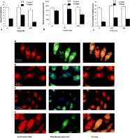
Fig. 2. Effect of drugs on the acidic compartments of 2-h-old schistosomula. (A–C) Effect of wortmannin (W), 3- methyladenine (3-MA) and N-ethylmaleimide (NEM) on the area (μm2)/compartment, the quantity of fluorescence (pixels)/compartment and the total number of compartments/schistosomulum. Schistosomula were quantified at 590 nm. Measurements were made with the OpenLab. Histograms represent mean±S.E.M. (n=30). (D–G) Schistosomula labelled with the LysoTracker Red (left panel) and monodansylcadaverine (middle panel) in the absence of drugs (D, control) or in the presence of wortmannin (E), 3-methyladenine (F) or N-ethylmaleimide (G). Some of the compartments overlapped in the merged images (right panel).

Fig. 3. Effect of starvation on the acidic compartments in 24-h-old schistosomula and adult worms. (A–B) Schistosomula were incubated in the amino acid-rich medium GMEM (A) or in the starvation medium EBSS (B) for 24 h at 37 °C. Following this incubation, schistosomula were labelled with the LysoTracker Red (LTR) (left panel) and monodansylcadaverine (MDC) (middle panel) and then washed and analysed by fluorescence microscopy. Some of the acidic compartments were overlapped in the merged images (right panel). (C–D) Adult worms were incubated in GMEM (C) or in EBSS (D) for 24 h at 37 °C and treated as indicated above for the schistosomula. The left, middle and right panels represent adult worms labelled with the LTR, MDC and the overlay of the merged images respectively. (E) Schistosomula were transformed in GMEM (control) or in EBSS (starved) and incubated in the corresponding medium in the presence or the absence of wortmannin (10 μM) for 24 h at 37 °C. After incubation, schistosomula were washed and labelled with MDC. Histograms represent mean±S.E.M. fluorescence/schistosomulum. Error bars are standard errors (n=30).
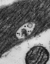
Fig. 4. Section through the muscle layer of 2-h-old schistosomula prepared by mechanical transformation showing a vacuole containing degraded materials and membranes (arrow). (×25000.)
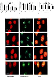
Fig. 5. Effect of irradiation on the acidic compartments in schistosomula of Schistosoma mansoni. (A–C) Effect of exposure to the UV light for 90 sec, 3 min or 5 min on the development of acidic compartments in the transformed schistosomula. The irradiated cercariae were transformed by the mechanical method and then labelled with the LysoTracker Red (LTR) and monodansylcadaverine (MDC) 2 h after transformation (2 h). The second group of irradiated cercariae were incubated for 24 h and then labelled with the LTR and MDC (24 h). Measurements of the area (μm2)/compartment (A), the quantity of fluorescence/compartment (pixels) (B) and the total number of compartments/schistosomula (C) were made with the OpenLab. Histograms represent mean± S.E.M. (n=30). All groups were quantified at 590 nm. (D–G) Control (D) and irradiated (E–G) schistosomula labelled with the LTR (left panel) and MDC (middle panel). The irradiated schistosomula were exposed to UV light for 90 sec (E), 3 min (F) or 5 min (G). The right panel shows the overlapping of some of the acidic compartments in the merged images.
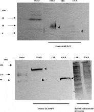
Fig. 6. Western blot analysis of schistosome extracts using anti-microtubule associated protein antibodies (anti-MAP-LC3) and anti-lysosome membrane associated proteins (anti-LAMP) antibodies. (A) The lysate of human embryo kidney cells (HEK29), cercarial extract (CER) and the extract of cultured schistosomula (CSCH) were subjected to immunoblot analysis with the anti-microtubule associated antibodies (goat anti-MAP-LC3). Bands of approximately 18 kDa and 6 kDa were obtained with the HEK29 and CSCH respectively (arrowheads). (B) Each of the extracts mentioned above in A (HEK29, CER and CSCH) was subjected to immunoblot analysis using anti-lysosomal membrane associated proteins (mouse anti-LAMP-1) antibodies or anti-schistosome glycocalyx antibodies (rabbit anti-schistosome glycocalyx). Single bands of approximately 110 kDa and 66 kDa were revealed with the HEK29, CER or CSCH extracts. Multiple bands were recognized by the anti-schistosome glycocalyx antibodies in both the CER and CSCH extracts. 10% SDS-polyacrylamide gels were used in A and B.
 You have
Access
- 14
- Cited by