A non-contained aortic rupture is defined as a complete disruption of the aortic wall, and shows angiographic extravasation of contrast medium beyond the confines of the aortic wall. Caused by catheter interventional stenting or balloon dilation of the aorta for re-coarctation/coarctation, it results in severe bleeding into the pleural space and into the mediastinumReference Qureshi, Lock, Landzberg, Lang and Marshall 1 and is an acute emergency situation. If the bleeding cannot be controlled instantly, the patient is at risk for immediate death due to circulatory collapse. We report a case of aortic rupture at stenting for re-coarctation in an adult patient. The bleeding was successfully controlled by rapid covered stent implantation with the NuDEL (all-in-one) catheter system (NuMED Canada Inc., Cornwall, Ontario, Canada).
Case report
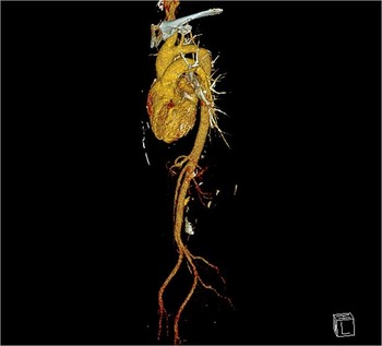
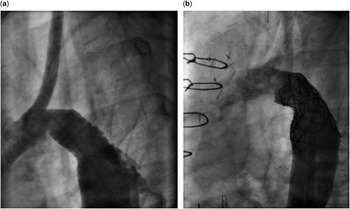
A 32-year-old woman (172 cm, 68 kg) was treated surgically with resection of an aortic coarctation and end-to-end anastomosis in the neonatal period. A re-coarctation resection was necessary 1 year later. In April 2016, re-coarctation and a stenotic bicuspid aortic valve were diagnosed. First, the aortic valve was replaced surgically by a 19-mm Edwards Perimount bioprosthesis. Next, a cardiac catheterisation was scheduled for treatment of the re-coarctation. Her blood pressure was 129/66 mmHg in the right arm, 101/78 mmHg in the left arm, and 100/73 mmHg in the legs (clinical gradient right arm – legs 28 mmHg). The catheter intervention was performed with sedation using propofol. A peak invasive systolic pressure gradient of 20 mmHg was assessed between the ascending and the descending aorta. Angiography in multiple planes showed a transverse aortic arch of 15-mm diameter, a re-coarctation with a minimal diameter of 7 mm, and an enlarged descending aortic diameter of up to 28 mm. An ultra-stiff, 0.035-inch guide wire (Amplatz ultrastiff; Cook Medical Inc., Bloomington, Indiana, United States of America) was placed into the ascending aorta. As the left subclavian artery originated in the stenotic segment (Fig 1), a 36-mm bare-metal stent (MaxLD EV3; Covidien/Medtronic, Meerbusch, Germany) was mounted on a 16×40-mm balloon-in-balloon (NuMed, Numed Canada Inc., Cornwall, Ontario, Canada), and was then advanced through a 12-French-long sheath (Flexor Cook Inc., Bloomington, Indiana, United States of America) into the coarcted segment. The stent was deployed successfully. The distal part of the stent was then shaped with a 22×40-mm low-pressure balloon catheter (Tyshak; NuMed) for better adherence to the vessel wall. The narrowest vessel diameter at this point was 13 mm. It was then decided to dilate the narrow stent segment with a 18×20-mm high-pressure balloon (Atlas; Bard, Tempe, Arizona, United States of America). The stent expanded at 10-atm inflation pressure; however, soon after balloon deflation, the patient became uneasy, and the aortic pressures dropped to critically low levels (systolic pressures <50 mmHg). The patient was intubated instantly, and a central venous line was placed in the groin through which saline (500 ml 0.9%) was infused by hand injection. An angiogram showed significant extravasation of contrast medium at the distal part of the bare-metal stent (Fig 2a). Rapidly, the 22-mm Tyshak balloon was re-advanced into the stent and inflated with diluted contrast at low pressure (1–2 atm) for temporary vessel blocking. A NuDEL catheter (45-mm, covered 8 zig Cheatham Platinum stent pre-mounted on a 24×50-mm balloon-in-balloon; NuMed) was prepared. After removal of the Tyshak balloon and the 12-French-long sheath, the NuDEL system was directly advanced percutaneously over the wire into the bare-metal stent, and the outer balloon was inflated. This resulted in instant stabilisation of blood pressures (systolic pressures >100 mmHg, mean arterial pressure >70 mmHg). On a pullback manoeuver, there was a residual gradient of 5-mmHg between the ascending and the descending aorta. Repeated angiography showed complete sealing of the aortic rupture (Fig 2b); however, a considerable effusion was noted in the left pleural space. For possible re-canalisation of the left subclavian artery, the left brachial artery was canulated with a 5-French sheath, and a 5-French multipurpose catheter was placed into the left axillary artery, demonstrating blood pressures 10-mmHg lower than in the ascending aorta, which was thought to be tolerable. Re-canalisation of the left subclavian artery by puncture of the GoreTex was thought to be dangerous and unpredictable in this situation. A pleural tab drained 1000 ml of blood from the left pleural space. A CT scan performed 2 hours later still showed a significant left pleural effusion, and a second drain was placed more apically into the left pleural space. Another 1500 ml of blood was drained. Her haemoglobin levels dropped from 12.9 to 7.7 g/dl, and two units of blood were transfused. The patient was extubated the next morning, showed no abnormal neurological symptoms, and was discharged home 6 days later in good clinical condition. Her blood pressures in the right arm were normal under medication with losartan 12.5 mg and bisoprolol 2×2.5 mg.
CT scan before stent implantation: the small left subclavian artery originates from the descending aorta just at the site of re-coarctation. The transverse aortic arch diameter is 15–16 mm, the stenosis 7 mm, and the descending aorta has a diameter of 22 mm.

( a ) After dilatation with a high-pressure balloon (Atlas 18×20 mm), the patient became uneasy. The patient’s aortic pressures dropped, and rapid angiography showed extravasation of contrast outside the aortic wall. ( b ) After implantation of a 45-mm, covered cheatham platinum stent premounted on a 24×50-mm balloon-in-balloon through the all-in-one NuDEL system, the aortic rupture was sealed off.

Discussion
Aortic rupture in a patient with re-coarctation during catheter interventional balloon-expandable stent implantation is a true nightmare for every catheter interventionalist. The largest study on stenting of coarctation so far (n=565) published by Forbes et al reported two procedure-related deaths. Both patients were adults with aortic rupture.Reference Forbes, Amin and Zahn 2 , Reference Forbes, Pedra and Zahn 3 Hence, the incidence of aortic rupture during balloon angioplasty or stenting for aortic coarctation is low (<1%) as Tretter et al report in a recent review,Reference Tretter and McElhinney 4 because in general stent implantation for coarctation of the aorta is safe and effective, and many centres use this intervention today as first-line treatment;Reference Eicken, Sebening and Hager 5 – Reference Ewert, Peters, Abdul-Khaliq, Nagdyman and Lange 8 however, not every case of fatal aortic rupture is reported in the literature. The true incidence of aortic rupture may be under-reported. Tretter et al have found a documented number of 23 fatal aortic ruptures in the literature.Reference Tretter and McElhinney 4
In our patient, the NuDEL system enabled rapid sealing of the aorta with a covered stent. If the NuDEL system had not been available, the sheath would have needed to be upsized from 12 French to 14 French. Time-related uncontrolled bleeding could have occurred after removal of the Tyshak balloon. In this situation, it is extremely important to keep a stable guide wire position. The exchange of the sheath to the NuDEL system was possible very rapidly, and the all-in-one system enabled instant sealing of the aortic wall. Haemodynamic stabilisation occurred instantly, after the inflated, covered Cheatham Platinum stent was in place. The NuDEL system is Conformite Europeene marked and available since late 2015 in Europe. It includes a triaxial balloon-in-balloon designed catheter with a covered Cheatham Platinum stent mounted on it, which is then covered by a sheath – an all-in-one system. The catheter tip is tapered, permitting safe entry of the access vessel.
In retrospect, it could be argued that a smaller-sized high-pressure balloon (16 mm) and sequential widening of the coarcted segment could have been safer, or the final enlargement to 18 mm could have been postponed to a second catheterisation later on. It remains speculative, however, whether then the aortic rupture could have been avoided. A covered stent was not used primarily, as the left subclavian artery originated in the coarcted segment. Obviously, the stenotic origin of this vessel led to an efficient perfusion of the left arm via the circulus of Willisi, which was documented after covered stent implantation. Hence, knowing this and after the aortic rupture following bare-metal stenting, primary, covered stent treatment would have been the more adequate therapy.
It is known for years that aortic medial abnormalities occur in patients with coarctation of the aorta and may predispose to dilatation, aneurysm, and aortic rupture.Reference Niwa, Perloff and Bhuta 9 In patients with re-coarctation after surgical treatment, scar tissue may be present, but the adjacent vessel tissue may have the above-mentioned pathological vessel features and may be more fragile.
In conclusion, we report on successful treatment of aortic rupture with the NuDEL – all-in-one – covered stent during bare-metal stenting for residual aortic coarctation in an adult patient.
Acknowledgements
The authors thank Katarzyna Gendera for the help in preparing the manuscript.
Financial Support
This research received no specific grant from any funding agency or from commercial or not-for-profit sectors.
Conflicts of Interest
None.
Ethical Standards
The authors assert that the procedure described in this study complies with the ethical standards of the relevant national guidelines on human experimentation and with the Helsinki Declaration of 1975, revised in 2008, and has been approved by the CCTIRS (national advisory committee for data processing in health research) on 18 April, 2012 (registration number 12.101).