I. INTRODUCTION
Breast cancer is the second leading cause of cancer-related deaths among women in U.S.Reference Jemal, Siegel, Ward, Hao, Xu and Thun1 The National Cancer Institute estimates that, in 2010, there were over 207,000 new cases, and nearly 40,000 deaths, from breast cancer among women in the United States.Reference Jemal, Siegel, Ward, Hao, Xu and Thun1 There is, therefore, a great need for advancements in the early detection of breast cancer and other cancers. Such advancements could both increase survival rates and improve other clinical outcomes.Reference Meng, Fan, Galiana, Branca, Clasen, Ma, Zhou, Leuschner, Kumar, Hormes, Otiti, Beye, Harmer, Kiely, Warren, Haataja and Soboyejo2 In addition, localized cancer treatment methods are needed to overcome the limitations and disadvantages of current systemic therapies.Reference Tanaka, Decuzzi, Cristofanilli, Sakamoto, Tasciotti, Robertson and Ferrari3
Recently, there has been interest in targeting cancer cells with gold nanoparticles (AuNPs), which are biocompatible.Reference Huang, El-Sayed, Qian and El-Sayed4–Reference Bhattacharya, Patra, Earl, Wang, Katarya, Lu, Kizhakkedathu, Yaszemski, Greipp, Mukhopadhyay and Mukherjee6 One motivation is that AuNPs display a strong plasmon resonance (SPR) in the near-infrared (NIR) region. This property is extremely sensitive to the size, shape, and aggregation state of the nanoparticles.Reference Mulvaney, Perera, Biggs, Grieser and Stevens7 In the NIR region, the high tissue transmissivity, caused by low scattering and absorption from intrinsic chromophores, can be exploited for imaging as well as photothermal therapy of cancer cells.Reference Weissleder8–Reference Rajadhyaksha, Grossman, Esterowitz, Webb and Anderson10
Gold nanorodsReference El-Sayed, Huang and El-Sayed5 and nanoshells,Reference Hirsch, Stafford, Bankson, Sershen, Rivera, Price, Hazle, Halas and West11 conjugated with antibodies, are being developed as photothermal therapy agents. For example, Loo et al.Reference Loo, Lowery, Halas, West and Drezek9 conjugated nanoshells to antibodies that are specific to the human epidermal growth factor receptor 2 (HER2). After incubating SKBr3 breast adenocarcinoma cells, which overexpress HER2 receptors, with anti-HER2-targeted nanoshells, the cells were exposed to NIR irradiation at the SPR of the nanoshells. This treatment led to heating of the nanoshells and, thereby, resulted in cell death.
AuNP-Ab conjugates are also being developed that combine photothermal therapy with NIR imaging capabilities for cancer cells.Reference Loo, Lowery, Halas, West and Drezek9, Reference El-Sayed, Huang and El-Sayed12 In one study, El-Sayed et al.Reference El-Sayed, Huang and El-Sayed12 incubated AuNPs-Ab conjugates specific to the epithelial growth factor receptor with cancerous and noncancerous oral epithelial cells. The SPR imaging experiments showed that the AuNP-Ab conjugates bound to cancerous cells with an affinity that was six times greater than the affinity to the noncancerous control cells. Such an increase in binding affinity could be very useful in cancer detection.
AuNPs can also scatter visible light. Reflectance confocal microscopyReference Smithpeter, Dunn, Welch and Richards-Kortum13 and optical coherence tomographyReference Tearney, Brezinski, Bouma, Boppart, Pitris, Southern and Fujimoto14, Reference Huang, Swanson, Lin, Schuman, Stinson, Chang, Hee, Flotte, Gregory and Puliafito15 use reflected light to provide detailed three-dimensional images of tissue, thus eliminating tissue sectioning. These techniques provide spatial resolutions of ∼1–10 μm and a penetration depth ranging from ∼300 μm to 1–2 mm.Reference Sokolov, Follen, Aaron, Pavlova, Malpica, Lotan and Richards-Kortum16 Sokolov et al.Reference Sokolov, Follen, Aaron, Pavlova, Malpica, Lotan and Richards-Kortum16 have demonstrated the use of gold bioconjugates for such types of reflective imaging.
The potential advantages of using AuNPs in cancer therapies have resulted in increased interest in their synthesis.Reference Huang, Jain, El-Sayed and El-Sayed17–Reference Daniel and Astruc20 However, most of the common methods that are used to synthesize AuNPs involve the use of toxic chemical substances,Reference Brust, Walker, Bethell, Schiffrin and Whyman21–Reference Okitsu, Yue, Tanabe, Matsumoto and Yobiko24 high temperatures,Reference Fleming and Williams25, Reference Frens26 and high pressures.Reference Wu, Bai, Sun, Haddad, Boye, Wang, Huang and Fan27, Reference Zhu, Lines, Ganton, Kerr and Workentin28 In contrast, the biosynthesis of AuNPs provides a novel approach to the development of nontoxic environmentally benign methodsReference Shukla, Nune, Chanda, Katti, Mekapothula, Kulkarni, Welshons, Kannan and Katti29, Reference Han, Ghosh and Rotello30 that have the potential to replace conventional chemical and physical synthesis techniques that are normally used.Reference He, Guo, Zhang, Zhang, Wang and Gu31–Reference Shankar, Rai, Ahmad and Sastry33 Furthermore, the biosynthesis of AuNPs can also be achieved through the use of natural bacteria, fungi, and plants.Reference Shukla, Nune, Chanda, Katti, Mekapothula, Kulkarni, Welshons, Kannan and Katti29, Reference He, Guo, Zhang, Zhang, Wang and Gu31–Reference Shankar, Ahmad, Pasricha and Sastry44 These organisms provide useful new pathways for the synthesis of AuNPs with a range of shapes and sizes.Reference Mandal, Bolander, Mukhopadhyay, Sarkar and Mukherjee42, Reference Grzelczak, Perez-Juste, Mulvaney and Liz-Marzan43
Prior work on the bacterial synthesis of AuNPs has shown that the particles may be formed intracellularly or extracellularly.Reference He, Guo, Zhang, Zhang, Wang and Gu31 The shapes and sizes of the resulting AuNPs have also been shown to vary with pH and the type of bacteria.Reference He, Guo, Zhang, Zhang, Wang and Gu31, Reference He, Zhang, Guo and Gu32, Reference Mandal, Bolander, Mukhopadhyay, Sarkar and Mukherjee42 In an earlier study, Bacillus subtilis 168Reference Beveridge and Murray36 was used to reduce Au3+ions to AuNPs with sizes between 5 and 25 nm. In addition to bacteria, many other microbes have the ability to synthesize AuNPs intracellularly and extracellularly, including yeasts, algae, and fungi.Reference Mukherjee, Ahmad, Mandal, Senapati, Sainkar, Khan, Ramani, Parischa, Ajayakumar, Alam, Sastry and Kumar37, Reference Wen, Lin, Gu, Zhou, Yao, Chen and Fu39, Reference Mandal, Bolander, Mukhopadhyay, Sarkar and Mukherjee42
Microorganisms are known to protect themselves from metal ion stress, either by segregating the ions intracellularly or by secreting them into the external environment.Reference Suresh, Pelletier, Wang, Broich, Moon, Gu, Allison, Joy, Phelps and Doktycz45, Reference Bruins, Kapil and Oehme46 Microorganisms have also developed the following six mechanisms for metal resistance: exclusion by permeability barriers, intra- and extracellular sequestration, active transport efflux pumps, enzymatic detoxification, and increased resistance of cellular targets to metal ions.Reference Thakkar, Mhatre and Parikh47 These resistance mechanisms can be exploited for the bacterial synthesis of nanoparticles, which is advantageous over current chemical synthesis routes.
However, the basic mechanisms of nanoparticle synthesis by biological systems are not fully understood. Furthermore, the potential for scaling-up biosynthesis is yet to be demonstrated. There is, therefore, a need to explore potentially scalable biologically based methods for the synthesis of AuNPs. In addition, the basic biosynthetic mechanisms of AuNP formation should be established. This understanding could guide the future scale-up and applications in niche areas, such as nanomedicine.Reference Thakkar, Mhatre and Parikh47, Reference Badwaik, Bartonojo, Evans, Sahi, Willis and Dakshinamurthy48 Furthermore, since nanomedicine requires the specific targeting of diseased cells, a basic understanding of the adhesion of AuNPs to diseased cells, such as cancer cells, should be developed. Also, since basic studies of adhesion have not been conducted on biosynthesized AuNPs or AuNP-Ab conjugates, the adhesion of biosynthesized AuNPs and specific AuNPs-Ab conjugates, which have the potential for future applications in breast cancer detection and treatment, should be explored.
The present study explores the mechanisms by which AuNPs are formed by reduction of chloroauric acid (HAuCl4) by Bacillus megaterium (BM), a common nonpathogenic soil bacterium. The adhesion of the biosynthesized AuNPs to cancer cells is also demonstrated and compared to the adhesion of chemically synthesized AuNPs. The potential for the specific targeting of breast cancer cells is explored by comparing the adhesion of breast-specific AuNP-Ab conjugates to that of uncoated AuNPs. The implications of the results are then discussed for potential clinical applications of biosynthesized AuNPs.
II. EXPERIMENTAL PROCEDURES
A. Biosynthesis of AuNPs
BM [Strain #B-14308, ARS (NRRL) Culture Collection] cells were cultured in enriched nutrient broth [ENB; 1.3% nutrient broth (Remel #R454202, Lenexa, KS), 1% glucose]. After aerated growth for 48 h at 30 °C, the bacterial cells were harvested by centrifugation and washed five times with sterile purified water (EASYpure UV/UF Compact Reagent Grade Water System; Thermo Fisher Scientific Inc., Waltham, MA). Both the washed bacterial cells and the ENB obtained from the 48-h culture (conditioned media or CM) were used in AuNP synthesis reactions, as described below. One gram (wet weight) of washed BM cells was suspended in 10 mL of sterile aqueous solutions of 1 mM HAuCl4(Sigma-Aldrich, St. Louis, MO) at either pH 4 or pH 7.
To determine whether BM cells were required for the formation of nanoparticles, the CM was aseptically filtered using 0.22 μm syringe filters (SLMP025SS; Millex, Billerica, MA) to remove the cells. Solutions of 1 mM HAuCl4 in 10 mL of filtered CM were then prepared at pH 4 and 7. In addition, the following reactions served as controls: 1 g (wet weight) of BM cells in water at pH 4 and 7, 1 mM HAuCl4 at pH 4 and 7, CM at pH 4 and 7, and 1 mM HAuCl4in ENB at pH 4 and 7. The pH of each starting solution was adjusted using 0.1 M NaOH or 0.1 M HCl. All the experiments were conducted at 30 °C for durations of 24–96 h, without agitation, in a covered water bath.
B. Antibody adsorption
To remove any surface-bound capping proteins, biosynthesized AuNPs were treated with 1% sodium dodecyl sulfate (SDS, #71736; Sigma-Aldrich, St. Louis, MO) and heated for 30 min at 95 °C in a vacuum oven (Model 280A; Fisher Scientific Isotemp Vacuum Oven, Waltham, MA) under ambient pressure. The AuNPs and antibodies were washed with HEPES buffer (Dojindo Laboratory, Kumamoto, Japan) at pH 7 and centrifuged for 10 min at 10,000 rpm. This washing procedure was repeated five times to remove residual proteins and SDS.
Antibodies were adsorbed onto the AuNPs using techniques developed by Geoghegan and AckermanReference Geoghegan and Ackerman49 and used by Jiang et al.Reference Jiang, KimBetty, Rutka and ChanWarren50 to produce Herceptin–AuNP complexes. Briefly, AuNPs were diluted in HEPES buffer (pH 8.5),Reference Yamashita, Katsumata and Okada51, Reference Luo, Huang, Wu and Zare52 and anti-EphA2 antibodies (Invitrogen, Carlsbad, CA) were reconstituted in the same buffer at 100 μg/mL. The solutions were then mixed and allowed to react for 20 min at room temperature. The solution was centrifuged at 10,000 rpm for 1 h to remove any unbound antibodies and then resuspended in HEPES buffer. After the second wash, the pellet was again resuspended in HEPES buffer.
C. Characterization of AuNPs
The ultraviolet–visible (UV–Vis) spectra of the synthesized AuNPs were measured using an Evolution 300 spectrophotometer (Thermo Fisher Scientific Inc., Waltham, MA) operated at a resolution of 0.5 nm with either quartz (UV–Vis) or disposable polystyrene (visible only; Thermo Fisher Scientific Inc., Waltham, MA) reduced volume cells. The products of the different biosynthetic reactions, which were present in the supernatant just above the settled precipitate, were dropped on carbon-coated copper grids (CF200-Cu; Electron Microscopy Sciences, Hatfield, PA) and then imaged by transmission electron microscopy (TEM) using a Philips CM10 TEM (Philips/FEI, Hillsboro, OR). Micrographs of the AuNPs were then analyzed using Word Paint (Redmond, WA) software to determine their respective particle size distributions.
D. Adhesion measurements
MDA-MB-231 and Hs578Bst cell lines, growth media (L-15 and 46-X, respectively), and medium supplements (fetal bovine serum and penicillin/streptomycin) were purchased from American Type Culture Collection (ATCC, Manassas, VA), Sigma-Aldrich (St. Louis, MO), and Thermo Fisher Scientific, Inc. (Waltham, MA). Anti-EphA2 Antibodies were obtained from Invitrogen (Carlsbad, CA).
A dip-coating method, described previously by Hutter and Bechhoefer,Reference Hutter and Bechhoefer53 was used to coat bare phosphorus n-doped silicon atomic force microscopy (AFM) tips (MPP-31100; Veeco, Memphis, TN) with biosynthesized AuNPs, SDS-treated AuNPs, or AuNP-Ab conjugates. Briefly, AFM tips were immersed in solutions containing the particles of interest and then air dried for a few seconds. The tips were dipped and dried twice more in this manner to ensure tip coverage. The tips were then allowed to air dry for a minimum of 24 h before being used for AFM adhesion measurements.
Samples with MDA-MB-231 and Hs578bst cells were prepared for the AFM by fixation in 3.7 wt% formaldehyde (Sigma-Aldrich, St. Louis, MO). Briefly, the cells were cultured in 60 × 15 mm Falcon Petri dishes (Falcon, Franklin Lakes, NJ) for 48 h at high confluence and incubated at 37 °C at normal atmospheric pressure and 5% CO2levels for breast cancer and normal breast cells, respectively. MDA-MB-231 and Hs578bst cells were grown in L-15 or 46-X base medium (ATCC, Manassas, VA), respectively, supplemented with 100 I.U./mL penicillin/100 μg/mL streptomycin and 10% FBS (ATCC, Manassas, VA). The samples were rinsed with phosphate buffered saline (DPBS; Invitrogen, Carlsbad, CA) and fixed with a 3.7 wt% formaldehyde solution for 15 min at room temperature. They were then rinsed again with DPBS before a final rinse with purified water. Finally, the samples were placed in a vacuum desiccator for 2 h to dry at room temperature.
To determine whether the AFM tips were coated with AuNPs, bare (uncoated) AFM tips and the coated AFM tips were imaged using environmental scanning electron microscopy (ESEM). A Phillips FEI Quanta 200 FEG Environmental-SEM (Philips Electronics N.V., Eindhoven, The Netherlands) was used to obtain secondary/backscattered electron images of coated and bare AFM tips. An energy dispersive x-ray spectrometer, EDS (Inca, Oxford Instruments, UK), was used to acquire an elemental spectrum at the apex of the cantilever tip to confirm the presence of AuNPs and antibodies.
Since weak adhesion between the AFM tips and the coatings could result in the detachment of the coatings from the AFM tips during handling or pull-off testing, the AFM tips were checked in the ESEM before and after AFM testing. Thus, the pull-off forces were unrelated to the detachment of the dip coatings from the coated AFM tips.
The spring constants of the coated and uncoated AFM tips were measured experimentally using the thermal tune method.Reference Hutter and Bechhoefer53, Reference Matei, Thoreson, Pratt, Newell and Burnham54 This was done to ensure that the actual spring constants were used to calculate the pull-off forces. Such measurements are also required to account for batch-to-batch variations in the spring constants, as well as the effects of coatings on the cantilever stiffness.
All AFM experiments were carried out using a Dimension 3100 AFM (Bruker Corporation, Billerica, MA) under ambient conditions at room temperature (22–23 °C) and relative humidity of 36–45%. Also, before the adhesion force measurements, the photodetector sensitivity was calibrated using a stiff quartz surface.Reference Bhushan55
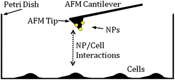
The AuNPs biosynthesized in CM at pH 4 were selected for adhesion measurements with cancer and normal breast cells. AFM tips were used to measure the adhesion to breast cancer cells (MDA-MB-231) and normal breast cells (Hs589Bst), as shown schematically in Fig. 1.
Ligand/receptor interactions. In the experimental setup, ligands on the dip-coated AFM tip interact with surface receptors on breast cancer cells (MDA-MB-231) seeded on a Petri dish.

Adhesive interactions were measured for the following five configurations using biosynthesized AuNPs (i) AuNPs to breast cancer and normal breast cells; (ii) AuNPs after SDS treatment to breast cancer and normal breast cells; (iii) AuNP-Ab conjugate to breast cancer and normal breast cells; (iv) Bare AFM tip on breast cancer and normal breast cells; (v) Ab-coated AFM tip on breast cancer and normal breast cells.
For each pair of interactions, 30 adhesion measurements were obtained, with each AFM tip being tested at six locations on different cells.
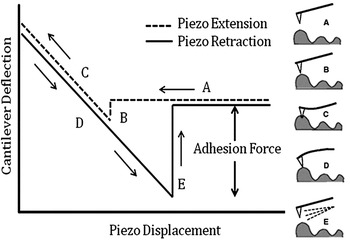
The stages involved in the AFM measurement of adhesion interactions are shown schematically in Fig. 2.Reference Shahin, Ludwig, Schafer, Nikova and Oberleithner56 First, the AFM tip is brought closer to the substrate at a constant velocity (A to B). As the tip approaches the substrate, the adhesive interactions cause the tip to jump to contact at B. The tip then undergoes elastic bending until it is retracted. However, it does not detach from the substrate when the bending force is zero. Hence, the retraction is continued until the applied force is sufficient to pull off the tip from the substrate at point E. The tip then returns to the zero force position. The associated pull-off force is often used as a measure of adhesion from Hooke’s law. This gives:
 $$F = - k{\rm{\delta }}\quad,$$
Schematic of typical force–displacement plot with corresponding stages of force–displacement behavior (Adapted from Ref. Reference Shahin, Ludwig, Schafer, Nikova and Oberleithner56). In one approach-retract cycle, the AFM tip approaches the surface of the sample (A), jumps to contact with the surface when significant van der Waals forces are felt (B), and undergoes elastic bending and is retracted (C, D). Due to adhesive interactions, the tip does not detach from the substrate until a force sufficient to pull the tip off of the surface is achieved (E).

where F is the pull-off force, k is the spring constant of the AFM tip cantilever, and δ is the maximum deflection of the AFM tip cantilever at the onset of pull-off.
III. RESULTS AND DISCUSSION
A. Biosynthesis of AuNPs
In reaction mixtures containing BM cells, aqueous chloroaurate ions were reduced over 24–96 h. The color of the reaction solutions at pH 4 and 7 changed from pale yellow to purple within 24 h, suggesting that AuNPs had formed in the presence of BM as reported in previous studies.Reference He, Zhang, Guo and Gu32, Reference He, Zhang, Guo and Gu32 The formation of AuNPs was also verified using TEM and UV–Vis scanning spectrophotometry, as discussed below.
To determine whether the reduction reaction occurred in the absence of BM cells, reactions consisting of CM with aqueous chloroaurate ions were also monitored. In these reactions, a color change from pale yellow to deep purple occurred at both pH 4 and 7, with the most notable color change occurring at pH 4. These color changes indicated that AuNPs were formed through the possible involvement of extracellular proteins. TEM and UV–Vis scanning spectrophotometry verified that the color change was caused by the formation of AuNPs, as discussed below. In contrast, the control reactions exhibited no change in color, again suggesting that extracellular proteins from BM cells are required for the biosynthesis of AuNPs.
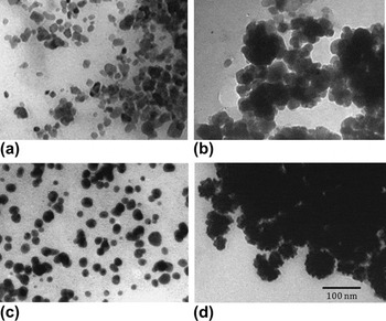
The TEM images obtained from different reactions are presented in Fig. 3. These show the shapes and sizes of AuNPs formed under different reaction conditions. After 72 h of reaction, well-separated mostly spherical AuNPs developed in the reaction containing CM and 1 mM HAuCl4 at pH 4 [Fig. 3(c)]. Figure 3(a) shows that the reaction of BM and 1 mM HAuCl4 at pH 4 produced spherical AuNPs with some minor aggregation. In contrast, reactions containing BM with 1 mM HAuCl4 [Fig. 3(b)] and CM with 1 mM HAuCl4 [Fig. 3(d)] at pH 7 show highly aggregated AuNPs that were not well developed after 72 h. In addition, the reaction containing CM and 1 mM HAuCl4at pH 7 produced AuNPs with branched “nanostar” morphologies.Reference Kumar, Pastoriza-Santos, Rodríguez-González, Javier García de Abajo and Liz-Marzánand57
TEM images of AuNPs produced in reactions after 72 h consisting of BM cells and 1 mM HAuCl4 at (a) pH 4 and (b) pH 7, CM and 1 mM HAuCl4at (c) pH 4 and (d) pH 7.

The reason(s) that the AuNPs synthesized at pH 7 were aggregated, while the AuNPs synthesized at pH 4 were not aggregated, are not currently understood. However, it is possible that the capping protein(s) synthesized at pH 7 differ chemically from the capping protein(s) synthesized at pH 4. Elucidation of the composition of the two types of capping protein(s) is needed and is a potential area for future work.
Since the reactions performed at pH 4 in CM resulted in well-developed mostly spherical AuNPs, the basic mechanisms of AuNP formation were further investigated under these conditions to determine whether the AuNPs were formed by intracellular and/or extracellular processes.
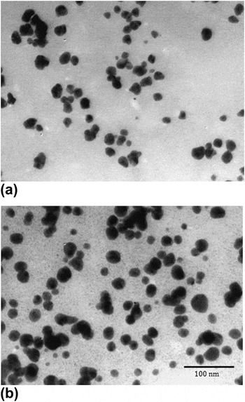
To determine the process of extracellular growth of the AuNPs in CM containing 1 mM HAuCl4 at pH 4, the biosynthesis reaction was monitored at different time points over a period of 96 h using TEM (Fig. 4). The average AuNP radius increased with reaction time, with AuNPs of sizes of 20.2 ± 3.6, 23.5 ± 7.0, and 27.8 ± 5.2 nm produced at durations of 24, 48, and 96 h, respectively. Overall, the TEM images show that the extracellular reaction consisting of CM and 1 mM HAuCl4 at pH 4 produced mostly spherical particles, while reactions consisting of CM and 1 mM HAuCl4at a lower pH, specifically pH 3, produced AuNPs with a variety of shapes and sizes (unpublished data), as previously observed by He et al.Reference He, Guo, Zhang, Zhang, Wang and Gu31, Reference He, Zhang, Guo and Gu32
TEM image (180x) of AuNPs from reactions with CM and 1 mM HAuCl4at pH 4 after (a) 24 and (b) 96 reaction hours.

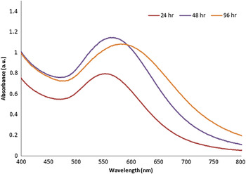
The extracellular reaction consisting of CM containing 1 mM HAuCl4 was also monitored using UV–Vis spectrophotometry at 24, 48, and 96 h (Fig. 5). The results indicate that the reaction containing CM and 1 mM HAuCl4at pH 4 had absorption maxima at 553, 562, and 581 nm, after 24, 48, and 96 h, respectively. These absorption maxima can be attributed to the SPR of the AuNPs.Reference Kumar, Peter and Nadeau58
UV–Vis absorption spectra of AuNPs. UV–Vis absorption spectra of AuNPs produced by reaction of HAuCl4and CM at pH 4.

The spectral redshift of the absorption maxima with reaction time can be attributed to increases in AuNP size,Reference Njoki, Lim, Mott, Park, Khan, Mishra, Sujakumar, Luo and Zhong59 as demonstrated by Njoki et al.Reference Njoki, Lim, Mott, Park, Khan, Mishra, Sujakumar, Luo and Zhong59 Njoki et al.Reference Njoki, Lim, Mott, Park, Khan, Mishra, Sujakumar, Luo and Zhong59 also reported that the redshift is accompanied by a small broadening of the SPR,Reference Njoki, Lim, Mott, Park, Khan, Mishra, Sujakumar, Luo and Zhong59 which is in agreement with the present results. These absorbance peaks were not present in any of the control reactions (data not shown).
Several studies have demonstrated that metal biotransformation by bacteria could involve a complex of capping proteins/peptides that are known to reduce metals.Reference Wen, Lin, Gu, Zhou, Yao, Chen and Fu39, Reference Suresh, Pelletier, Wang, Broich, Moon, Gu, Allison, Joy, Phelps and Doktycz45 To further demonstrate the presence of a possible capping protein(s) bound to the surface of the biosynthesized AuNPs, UV–Vis absorption spectra were captured in lower wave length regions (200–400 nm) for HEPES buffer solutions (pH 7.5) containing antibodies and AuNPs. In all cases, HEPES buffer alone was used as a control.
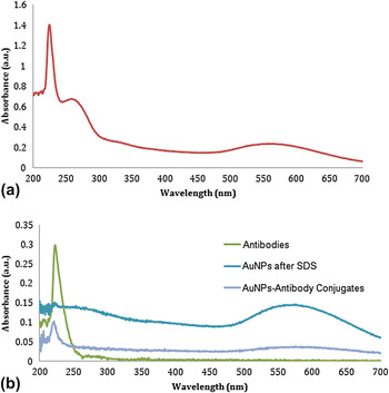
As shown in Fig. 6(a), the biosynthesized AuNPs exhibited two protein absorption bands at wave lengths of ∼220 and 280 nm. Absorption at these wave lengths is expected for proteins since most proteins absorb light at 214 and 280 nm due to peptide bonds and aromatic amino acids, respectively.Reference Landers60 Higher absorbance at ∼214 nm is also expected since the absorbance at any given protein concentration is usually greater at lower UV wave lengths.Reference Landers60 Furthermore, the UV–Vis absorption spectra shown in Fig. 6(a) are similar to those previously reported for bovine serum albumin–AuNP conjugates in 10 mM phosphate at pH 7.0Reference Shang, Wang, Jiang and Dong61 and human serum albumin–AuNP conjugates in phosphate buffer (100 mM at pH 7.4).Reference Gates, Fakayode, Lowry, Ganea, Murugeshu, Robinson, Strongin and Warner62
UV–Vis absorption spectra of the (a) biosynthesized AuNPs and (b) biosynthesized AuNPs with antibodies.

To determine if the surface-bound capping protein(s) contributes to the AuNP integrity, the AuNPs were treated with SDS. This treatment led to protein denaturation, which is supported by the absence of the peak at 214 nm in the UV–Vis absorption spectra for the SDS-treated AuNPs, as shown in Fig. 6(b). The AuNPs, before and after treatment with SDS, still exhibit absorption peaks in the mid-visible region of the spectrum, both at 564 nm. These results support the presence of capping protein(s) on the AuNPs and also demonstrate that these capping proteins can be removed by treatment with SDS. The removal of the protein(s) associated with AuNPs may be necessary for future conjugations with cancer-specific antibodies and, thus, was examined using techniques developed by Geoghegan and Ackerman,Reference Geoghegan and Ackerman49 as described above.
The UV–Vis absorption spectra for the AuNP-Ab conjugates are shown in Fig. 6(b). The synthesized nanoparticles had diameters of ∼20–30 nm that can accommodate about one to two antibodies. Specific optical changes in the UV–Vis spectrum indicate binding of the antibodies: the presence of the antibody absorption band at ∼220 nm after washing and a characteristic redshift in the maximum of the SPR. These optical changes are associated with alterations in the local refractive index around the AuNPs after binding of the antibody.Reference Sokolov, Follen, Aaron, Pavlova, Malpica, Lotan and Richards-Kortum16
B. AFM tip imaging and adhesion measurements
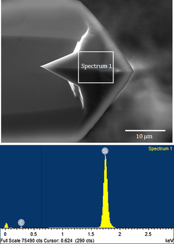
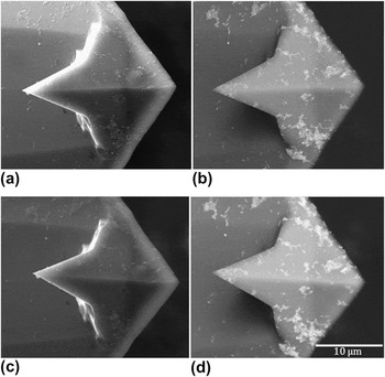
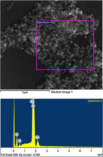
The ESEM image in Fig. 7 shows a typical bare AFM tip with uniformly-smooth surfaces. Figure 7 also shows that silicon and slight traces of carbon are detected in the EDS scan. Figures 8(a) and 8(b) show ESEM images [from secondary electron (SE) and backscattered electron detectors (BSED)] of an AFM tip after dip coating (in CM at pH 4) with AuNPs, respectively. The BSED image of the coated tip clearly shows the presence of AuNPs on the apex of the cantilever [Fig. 8(b)]. The EDS spectrum in Fig. 9 also verifies the presence of the AuNPs. Figures 8(c) and 8(d) show the same coated tip after adhesion experiments. Comparisons of Figs. 8(a) and 8(b) with Figs. 8(c) and 8(d) demonstrate that there is no loss of particles during adhesion testing.
EDS spectrum processing on bare AFM tip showing 99.2 wt% Si.

ESEM image using (a) SE detector and (b) BSED detector on AFM tip after dip coating in CM at pH4 containing AuNPs, before AFM adhesion experiments. ESEM image using (c) SE detector and (d) BSED detector on AFM tip after dip coating in CM at pH4 containing AuNPs, after AFM adhesion experiments.

Spectrum processing on coated AFM tip showing 64.4 wt% Si, 28.6 wt% C, and 5.9 wt% Au.

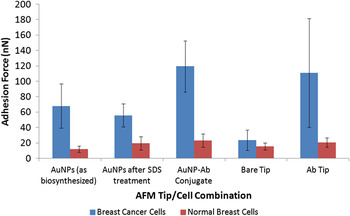
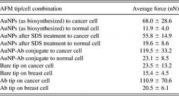
The measured pull-off forces for different AFM tips are presented in Fig. 10 and Table I. The controls show that the interaction forces between the bare tip and the breast cancer cells are in the same range as those between bare tips and normal breast cells. Hence, the adhesion between the bare AFM tips and breast cancer cells is comparable to that between bare AFM tips and normal breast cells. Similarly, there were relatively low levels of adhesion between AuNP-coated AFM tips and normal breast cells.
Summary of pull-off force measurements for breast cancer (MDA-MB-231) and normal breast (Hs578Bst) cells.

Adhesion forces between coated/bare AFM tips and cancer/normal cells.

However, the average adhesion forces between the AuNPs (with an average particle diameter of ∼20.2 ± 3.6 nm) and breast cancer cells are 68.0 ± 28.6 nN and are nearly six times greater than the forces between AuNPs and normal breast cells (11.9 ± 4.0 nN). The higher adhesion values of the AuNPs to breast cancer cells can be attributed to the overexpression of receptors on cancer cells compared to normal cells.Reference Wang, Yang, Chen and Shin63
Table I also shows the average adhesion forces between SDS-treated AuNPs and cancer as well as normal breast cells. The average adhesion forces between the SDS-treated AuNPs and breast cancer cells are 55.8 ± 14.9 nN, which are almost three times greater than the forces between SDS-treated AuNPs and normal breast cells (19.6 ± 8.6 nN). Clearly, without the capping protein(s), the biosynthesized AuNPs have reduced adhesive interactions with cancer cells.
The average adhesion forces between the AuNP-Ab conjugates and cells were also measured. The results shown in Table I demonstrate that the AuNP-Ab conjugates have an adhesion force of 119.5 ± 33.2 nN, which is over five times greater than those forces between AuNP-Ab conjugates and normal breast cells (23.1 ± 8.5 nN). These adhesive forces are in agreement with prior results by Meng et al.,Reference Meng, Paetzell, Bogorad and Soboyejo64 who reported adhesion forces of 111 ± 7 nN between the anti-EphA2 monoclonal antibody and cancer and normal breast cells.
The adhesive forces between AuNPs and cancer cells are attributed to a combination of van der Waals forces and hydrogen bonding. Likewise, the high adhesion forces between the anti-EphA2 antibody and the breast cancer cells are attributed to the van der Waals interactions between the antibody and its particular overexpressed receptor on the surface of the cancer cells.
It is also of interest to compare the results of the current studies on biosynthesized AuNPs with prior work by Oni,Reference Oni65 who investigated the adhesion forces between chemically synthesized AuNPs and breast cancer cells. The AuNPs (Nanopaartx Inc., Loveland, CO) used by OniReference Oni65 had an average diameter of ∼30 nm and a gold colloid composition of <0.1% carboxylic acid functionalized hydrocarbon capping agent and <0.1% hydroxide in distilled water.66 It was found that the average adhesion forces for these chemically synthesized AuNPs to breast cancer cells were less than 20 nN. These forces are over three times smaller than the average adhesion values found between breast cancer cells and biosynthesized AuNPs in the present study. The differences in adhesion values between the chemically synthesized and biosynthesized AuNPs to cancer cells are attributed to the presence of capping protein(s) present on the biosynthesized AuNPs. Further work is clearly needed to determine the composition of the capping proteins.
Future work is also needed to characterize and compare the biocompatibility of the biosynthesized AuNPs to chemically synthesized AuNPs. Toxicity studies are recommended for both types of synthesized AuNPs in breast cancer cells and normal breast cells. These studies constitute further steps in the understanding of the mechanism of the biosynthesis of nanoparticles as well as the physicochemical properties of these AuNPs.
IV. IMPLICATIONS
The implications of the biosynthesis techniques and force microscopy measurements described above are quite significant since biological systems have the potential to synthesize AuNPs for a variety of applications.Reference Mandal, Bolander, Mukhopadhyay, Sarkar and Mukherjee42, Reference Bharde, Kulkarni, Rao, Prabhune and Sastry67, Reference Bhattacharya and Gupta68 This study demonstrates the biosynthesis of mostly spherical AuNPs using BM. This method is a green, simple, and economical process compared to traditional chemical synthesis methods. Although AuNPs are, in general, considered to be biocompatible, chemical synthesis methods may still lead to the presence of some toxic chemical species adsorbed on the surface of the AuNPs, which may have adverse effects in medical applications.Reference Shankar, Rai, Ahmad and Sastry33 Synthesis of AuNPs using microorganisms can potentially eliminate this problem by producing AuNPs that are more biocompatible. Furthermore, since these AuNPs are biosynthesized extracellularly, the production of the AuNPs should be able to be increased for large-scale synthesis.
Greater targeting selectivity is a major goal in the development of therapeutic agents or imaging contrast formulations. Ideally, AuNPs should be selectively enriched in the tumor tissue with minimal damage to normal tissues. Hence, the adhesion techniques presented in this study may be used to screen the effectiveness of potential novel AuNPs that are being developed for the targeting of breast cancer. The high adhesive forces demonstrate the potential to increase the selectivity of AuNPs in the targeting of cancer cells. This should facilitate the attachment, entry, and uptake of AuNPs into cancer cells.
Such improved screening could significantly reduce the cost of identifying new nanotargets through conventional in vitro and in vivo studies. Also, since the average adhesive forces between biosynthesized AuNPs and cancer cells are almost six times greater than those between biosynthesized AuNPs and normal cells, the results suggest that the biosynthesis reaction results in a protein cap that enhances the specific adhesion forces between particles and cancer cells. Such high adhesive forces could improve the selectivity of biosynthesized AuNPs in cancer detection and treatment.
Furthermore, biosynthesized AuNPs alone may be as effective as AuNP-Ab conjugates because the adhesion forces between AuNP-Ab conjugates and cancer cells are five times greater than those to normal cells. In addition, the adhesion forces between biosynthesized AuNPs and cancer cells are nearly six times greater than those to normal cells. These adhesion results, therefore, suggest that biosynthesized AuNPs alone may be effective in breast cancer targeting and treatment.
Finally, it is important to note that the adhesion techniques used in this study may be used to screen the effectiveness of other potential AuNPs that are being developed for the specific targeting of breast cancer. Such screening may enable the rapid identification of particles and/or targeting ligands that are more likely to specifically attach to overexpressed receptors on breast cancer and other types of cancer cells. Further work is clearly needed to explore such possibilities.
V. CONCLUDING REMARKS
This study shows that AuNPs can be synthesized using the common soil bacterium, BM. The successful biosynthesis of the AuNPs was demonstrated through UV–Vis spectrophotometry and TEM. TEM images show that well-developed mostly spherical and homogenous nanoparticles develop from the reaction of CM with aqueous chloroaurate ions at pH 4. This reaction also demonstrates that the AuNPs can be synthesized extracellularly.
Furthermore, the adhesion of biosynthesized AuNPs to breast cancer cells was compared to the adhesion of chemically synthesized AuNPs. The biosynthesized AuNPs were found to exhibit adhesion forces to breast cancer cells that were six times greater than those to normal breast cells. However, chemically synthesized AuNPs had adhesion forces to breast cancer cells that were three times less than those of biosynthesized AuNPs. These results suggest that biosynthesized AuNPs and AuNP-Ab conjugates may be used for the rapid screening of potential ligands for the specific targeting of diseased cells such as cancer cells. Such AuNPs may also be used to design novel types of AuNPs for the early detection and treatment of cancer.
ACKNOWLEDGMENTS
This research was supported by the Princeton Grand Challenges Program (E.H., K.A.M., and W.O.S.), the African Development Bank (R.B. and W.O.S.), and the STEP-B Program of the World Bank (O.S.O., K.A.M., N.A., and W.O.S.).