Secundum atrial septal defects are common and account for 6–10% of congenital heart disease. In general, most patients with even moderate-to-large defects are asymptomatic and are referred for elective closure after 3–4 years of age; however, in premature infants with chronic lung disease, bronchopulmonary dysplasia, or pulmonary hypertension, increased pulmonary blood flow secondary to a left-to-right atrial shunt, may exacerbate their condition. It has been shown that closure of a secundum atrial septal defect either surgicallyReference Lammers, Hager, Eicken, Lange, Hauser and Hess 1 or percutaneouslyReference Thomas, Vincent, Raviele, Diehl, Qian and Kim 2 - Reference Lim and Matherne 4 in these patients results in significant improvement in their respiratory status and clinical condition. The majority of transcatheter procedures in infants described in the literature use the Amplatzer Septal Occluder (St. Jude Medical, St. Paul, Minnesota, United States of America), and very few reports using the Gore HELEX Septal Occluder exist (W. L. Gore & Associates, Flagstaff, Arizona, United States of America). We report the cases of two premature infants with chronic lung disease, who underwent atrial septal defect closure with the HELEX Septal Occluder and discuss the technical aspects of using the device in these patients and their clinical outcomes.
Cases
Patient 1 – was a female infant born at an outside hospital at 24 weeks estimated gestational age with a birth weight of 560 g. She failed multiple attempts at extubation and was transferred to our institution when she was 5 months old for ongoing management of chronic lung disease. An echocardiogram showed a moderate-sized patent arterial duct and a secundum atrial septal defect measuring 6–7 mm in diameter. There was mild right ventricular dilation and normal left ventricular size and function. The morphology of the arterial duct appeared to be a tubular type C, thought to be amenable to transcatheter device occlusion. She was taken to the catheterisation laboratory at 100 days of age weighing 2.6 kg with the goal of decreasing pulmonary blood flow, thereby decreasing left-to-right shunt across her atrial septal defect. This was achieved using a 4-mm Amplatzer Vascular Plug II. Her baseline haemodynamics revealed a pulmonary-to-systemic flow ratio of 1.6, pulmonary artery systolic pressure of 85% of systemic, with an indexed pulmonary vascular resistance of 4.3 Wood Units.
She initially improved clinically following this procedure. She was started on Tadalafil for pulmonary vasodilation by the neonatology team. She was extubated and started on nasogastric feeds 3 days after the catheterisation. Shortly after this, she began to develop increased work of breathing and required re-intubation ten days later. Over the next 4 weeks, she was managed with diuretics and fluid restriction. Despite this, she was unable to separate from the ventilator or tolerate enteral feeds. Serial echocardiograms showed persistence of a moderate secundum atrial septal defect with progressive right ventricular dilation and flattening of the interventricular septum. Her serum beta-natriuretic peptide level was 867 pg/ml (normal range 0–99 pg/ml).
She was referred back to the catheterisation laboratory on the 167th day of life weighing 3.6 kg to re-evaluate her pulmonary artery pressure and perform transcatheter occlusion of the secundum atrial septal defect. Initially, a 5-Fr sheath was placed in the right femoral vein, and a 20-Gauge catheter was placed in the right femoral artery for invasive blood pressure monitoring during the procedure. She was given 100 Units/kg of intravenous Heparin. The baseline catheterisation data, intubated with an inspired oxygen concentration of 30%, revealed a pulmonary-to-systemic flow ratio of 1.5, with pulmonary artery pressure now half systemic with an indexed pulmonary vascular resistance of 3.2 Wood Units. There was no residual shunt across the ductus arteriosus. Repeat right heart catheterisation on 100% oxygen showed an increased pulmonary-to-systemic flow ratio of 2.3, and decreased pulmonary vascular resistance of 2.4 Wood Units. We, therefore, felt that transcatheter occlusion of her atrial septal defect would decrease pulmonary blood flow and right heart volume overload. In addition, we felt that continued management with pulmonary vasodilators in the presence of an intracardiac shunt would likely be detrimental.
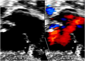
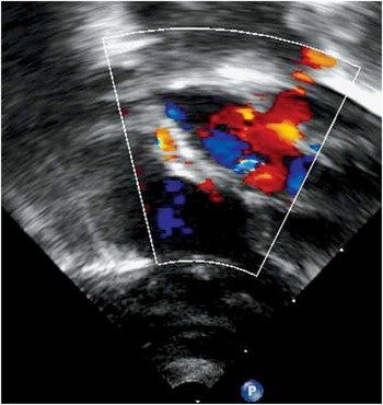
A transthoracic echocardiogram demonstrated a secundum atrial septal defect measuring 6.4 by 7.6 mm in diameter with adequate tissue rims around the defect (Fig 1). The total septal length measured 19–21 mm. The femoral vein sheath was exchanged over a wire for a 9-Fr Terumo sheath. A 15-mm HELEX Septal Occluder and delivery catheter were then advanced through the Terumo sheath and across the atrial septal defect, and subsequently deployed under transthoracic echocardiographic guidance. Once the satisfactory device positioning was confirmed, the device was locked in place. Fluoroscopy showed that the three islets of the device remained well-aligned and the locking loop had deployed properly. Echocardiography demonstrated a well-positioned device with no residual atrial shunt (Fig 2). The total procedure time was 2 hours and 3 minutes, with 17.5 minutes of fluoroscopy. The following day, a repeat echocardiogram showed stable device position and no residual left-to-right shunt. There was evidence of decreased right ventricular volume overload as the interventricular septal wall motion was now normal. She was successfully extubated 3 days after the procedure, and her follow-up chest x-ray showed decreased cardiomegaly. She was continued on Tadalafil and discharged home 6 weeks later on 0.25 L/min of nasal cannula oxygen. The oxygen was discontinued 3 months after discharge, and Tadalafil was discontinued 6 months after atrial septal defect closure. Her most recent outpatient follow-up was 12 months after the procedure, during which her room air saturation was 98%. An echocardiogram demonstrated stable device position with no residual atrial level shunt, normal right ventricular size and systolic function, and no evidence of significant residual pulmonary hypertension. She was tolerating full oral feeds with a weight of 10.1 kg (Fig 2).
Subcostal images of the atrial septal defect in patient 1, demonstrating left-to-right shunt by colour Doppler.

Subcostal images from patient 1 after deployment of the HELEX Septal Occluder. The device is well positioned with the right and left atrial discs well opposed to the atrial septum and no residual left-to-right shunt is seen by colour Doppler.

Patient 2 – was a female infant born at 29 weeks estimated gestational age with a birth weight of 1310 g, who was transferred to our centre at 3 months of age due to chronic lung disease and a persistent oxygen requirement. An initial echocardiogram demonstrated a secundum atrial septal defect measuring 8 mm, mild right ventricular dilation, and mild pulmonary hypertension with estimated right ventricular pressure of half systemic based on tricuspid regurgitation jet velocity. She was initially treated with Diuril and Aldactone and was maintained on nasal cannula oxygen. Over the next month, she developed progressive chronic lung disease with worsening chest x-ray appearance, increased work of breathing, and increased nasal cannula flow to maintain saturation above 88%. A repeat echocardiogram showed evidence of worsening pulmonary hypertension with estimated right ventricular pressure 65% of systemic and progressive right ventricular dilation. She was subsequently brought to the catheterisation laboratory on the 132nd day of life weighing 3.9 kg. She was intubated and placed under general anaesthesia. Baseline haemodynamic data revealed a pulmonary-to-systemic flow ratio of 1.5, pulmonary artery systolic pressure that was 60% of the femoral artery pressure, and an indexed pulmonary vascular resistance of 5.4 Woods Units. There was significant pulmonary venous desaturation with pulmonary vein sats of 79–88%. Repeat haemodynamic assessment on 100% oxygen showed an increased pulmonary-to-systemic flow ratio of 1.9, with decreased pulmonary artery systolic pressure of 40% of systemic, and decreased indexed pulmonary vascular resistance of 3.2 Woods Units. A transthoracic echocardiogram demonstrated a moderate-sized atrial septal defect measuring 8–9 mm with a septal length of 23 mm. Based on these measurements, a 20-mm HELEX Septal Occluder was selected. A 9-Fr Terumo sheath was placed in the right femoral vein, and the device delivery catheter was advanced through the sheath and across the atrial septum. The device was deployed under fluoroscopic and echocardiographic guidance and was subsequently locked. Transthoracic echocardiography showed that the device was in good position with a small (1.2 mm) residual left-to-right shunt seen through the device by colour Doppler. The total procedure time was 2 hours and 20 minutes, with 16.4 minutes of fluoroscopy. She was extubated and brought back to the neonatal intensive care unit on nasal cannula oxygen. Based on her haemodynamic data from the catheterisation laboratory, she was not started on pulmonary vasodilator medication, and was maintained on nasal cannula oxygen to keep her saturation >90%. She was started on enteral feeds by nasogastric tube the following day, and was transitioned to full oral feeds 6 weeks later.
She was discharged home 3 months after the atrial septal defect closure on 0.25 L/min nasal cannula oxygen, and oral Diuril and Aldactone. At her most recent follow-up, at 6 months after the procedure, her nasal cannula oxygen was decreased to 0.1 L/min with a saturation of 99%, and she was on oral diuril only once daily. An echocardiogram showed a trivial (<1 mm) residual shunt through the device with a qualitatively normal right ventricular size and no evidence of significant pulmonary hypertension. He weight was 8.4 kg with an average weight gain of 24 g/day.
Discussion
These cases demonstrate the feasibility of closing a secundum atrial septal defect with the HELEX Septal Occluder in a small infant. In most cases, an atrial septal defect can be closed electively after 4 years of age, as most patients are rarely symptomatic in infancy or childhood. In some cases of genetic syndromes or chronic lung disease from prematurity, however, the additive effect of left-to-right shunt through an atrial septal defect can have a deleterious effect due to increased pulmonary blood flow and volume loading of the right ventricle. Previous reports have shown a benefit of transcatheter atrial septal defect closure in infants with chronic lung disease.Reference Thomas, Vincent, Raviele, Diehl, Qian and Kim 2 - Reference Lim and Matherne 4 Pulmonary hypertension is common in premature infants with chronic lung disease, and the use of pulmonary vasodilators may increase pulmonary blood flow in patients with a significant atrial level shunt. Therefore, closure of an atrial septal defect may allow for improved benefit from pulmonary vasodilator therapy in these patients.
The majority of procedures reported in the literature to date used the Amplatzer Septal Occluder to close the atrial septal defect. We chose to use the HELEX Septal Occluder in our patients because it is a more compliant device and we thought it was potentially less traumatic in a smaller heart with no risk of device erosion reported to date. In patient 1, if we had used an Amplatzer Septal Occluder, we would have likely chosen a 7-mm diameter device, which would have a left atrial disc diameter of 19 mm. Given that our total septal length measured 19–21 mm in multiple planes, we were worried about potential cardiac trauma and erosion with this device. The main potential advantage of the Amplatzer device is that it could be deployed using a 6-Fr delivery sheath; however, the 9-Fr Terumo sheath is hydrophilic and we found that it advanced easily into the femoral vein in these patients. We did not have any difficulty achieving haemostasis from the venous puncture site upon completion of the procedure, and neither patient had any evidence of venous occlusion or venous stasis in the lower extremity during follow-up. The transhepatic approach has also been described for atrial septal defect occlusion in small infants, including using the HELEX Septal Occluder in a 4.9-kg patient.Reference Javois, Van Bergen and Husayni 5 The potential advantage is that the delivery catheter approaches the atrial septal defect in a more perpendicular manner and may allow the left atrial disc to deploy more parallelly to the atrial septum; however, we ultimately felt that using the femoral venous approach was feasible and safe. We did note in both patients that during deployment the atrial septum and device were pulled caudal and repositioned upon setting the locking loop; however, this did not result in any haemodynamic instability or arrhythmias. We did not encounter any difficulties with deploying the left atrial disc in either patient most likely due to the fact that we were able to use the smaller-sized devices in these patients.
Balloon sizing of the atrial septal defect using the “stop flow” technique is often used to determine the appropriate size of the device in older patients.Reference Carlson, Justino and O’Brien 6 We did not use this technique in these cases because we felt that the length and diameter of the available sizing balloons precluded their use in small infants. In the largest series reported for atrial septal defect closure in infants, they used balloon sizing in <25% of their patients.Reference Bishnoi, Everett and Ringel 7 We were able to accurately evaluate the defect size and surrounding tissue rims in multiple planes with transthoracic echocardiogram in both patients. The largest diameter of the defect was 7.4 and 9.1 mm in patients 1 and 2, respectively. We chose to use the 15-mm device in patient 1 and the 20-mm device in patient 2, as these were roughly twice the largest diameter of the defect in each patient. In patient 1, we had a total septal length of 19–21 mm, and thought that the 15-mm device was the safest to use; however, had we not been satisfied with device stability upon deployment, we would have considered using a 20-mm HELEX Septal Occluder in this patient.
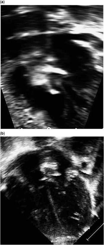
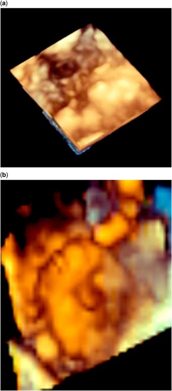
Trans-oesophageal echocardiography is frequently used during transcatheter atrial septal defect occlusion; however, in smaller patients, the trans-oesophageal probe may compress the left atrium and make deployment of the left atrial disc more challenging. Given the size of our patients, we chose to use transthoracic echocardiography and found that this accurately delineated the defect size, septal length, and tissue rims, as well as visualised the device well after deployment (Fig 3a and b). Previous reports have shown that transthoracic echocardiography can be utilised for transcatheter atrial septal defect occlusion.Reference Bartakian, El-Said, Printz and Moore 8 We also found that the use of the three-dimensional imaging to be very helpful in determining the defect diameter in multiple planes, the relationship of the defect to other important structures such as the superior and inferior caval veins, and for evaluating the device after deployment (Fig 4a and b).
( a ) Subcostal and ( b ) apical four-chamber views in patient 2 after deployment of the HELEX Septal Occluder. Both views show the device to be well positioned.

Three-dimensional echocardiogram images from patient 1. ( a ) Demonstrates the atrial septal defect and its relation to the superior and inferior caval veins. ( b ) Shows the HELEX Septal Occluder after deployment. The right atrial disc and locking loop can be seen.

In summary, we have shown that transcatheter occlusion of a secundum atrial septal defect with a HELEX Septal Occluder using the femoral venous approach is technically feasible in a small infant, can be performed safely, and can be beneficial in infants with chronic lung disease. Both patients in our series had significant clinical improvement including improved respiratory status, good weight gain, and ability to wean from medications including diuretics and oral pulmonary vasodilators. Further evaluation with long-term follow-up in a larger cohort of patients is warranted.
Acknowledgements
None
Financial Support
This research received no specific grant from any funding agency, commercial, or not-for-profit sectors
Conflicts of Interest
None.