Introduction
Light microscopic studies (Ishii, Reference Ishii1934; Chen, Reference Chen1937; Rees, Reference Rees1940; Dunn, Reference Dunn1959; Cheng & James, Reference Cheng and James1960; Cheng, Reference Cheng1961; Dobrovolskij, Reference Dobrovolskij1965, Reference Dobrovolskij1971; Cheng & Bier, Reference Cheng and Bier1972; Gerasyov & Dobrovolskij, Reference Gerasyov and Dobrovolskij1977) have revealed a high degree of similarity in the embryonic development of cercariae from different digenean groups. Successive stages of this process were described in most detail by Cheng & Bier (Reference Cheng and Bier1972), whose terminology is used in the present paper. A characteristic feature of cercarial embryogenesis is the formation of an embryonic membrane around early morula-like embryos (called ‘naked cell aggregates’). This transitory envelope has been referred to in various ways (for terminology see Meuleman & Holzmann, Reference Meuleman and Holzmann1975; Dunn et al., Reference Dunn, Dang, Hanna and Nisami1992), the most popular term currently being ‘the primitive epithelium’. After the primitive epithelium has been formed, the embryos acquire a distinctive rounded shape, which has earned them the name ‘germinal balls’. In most light microscope studies, involvement of embryo cells in formation of the primitive epithelium has been noted (Ishii, Reference Ishii1934; Chen, Reference Chen1937; Dunn, Reference Dunn1959; Cheng & James, Reference Cheng and James1960; Cheng, Reference Cheng1961; Dobrovolskij, Reference Dobrovolskij1965, Reference Dobrovolskij1971; James & Bowers, Reference James and Bowers1967; Cheng & Bier, Reference Cheng and Bier1972; Bednarz, Reference Bednarz1973; Gerasyov & Dobrovolskij, Reference Gerasyov and Dobrovolskij1977).
Electron microscopy revealed that the fine structure of the primitive epithelium is fairly similar in various digenean species (Bils & Martin, Reference Bils and Martin1966; Matricon-Gondran, Reference Matricon-Gondran1971; Hockley, Reference Hockley1972; Meuleman & Holzmann, Reference Meuleman and Holzmann1975; Rees & Day, Reference Rees and Day1976; Tihomirov, Reference Tihomirov1980; Göbel & Pan, Reference Göbel and Pan1985; Halton & McCrae, Reference Halton and McCrae1985; Žd'árská & Soboleva, Reference Žd'árská and Soboleva1985; Al-Salman & James, Reference Al-Salman and James1988; Žd'árská, Reference Žd'árská1995; Galaktionov & Dobrovolskij, Reference Galaktionov and Dobrovolskij2003). However, Meuleman & Holzmann (Reference Meuleman and Holzmann1975) observed syncytial connections between the primitive epithelium and tegumental cells of daughter sporocysts in early embryos of Schistosoma mansoni. This observation, combined with similarities in cytoplasm structure of the newly formed primitive epithelium and the tegumental cells of the sporocyst, led Meuleman & Holzmann (Reference Meuleman and Holzmann1975) to the conclusion that the primitive epithelium was a derivative of the sporocyst body wall. This viewpoint was later supported by Halton & McCrae (Reference Halton and McCrae1985), Žd'árská & Soboleva (Reference Žd'árská and Soboleva1985) and Göbel & Pan (Reference Göbel and Pan1985), who studied the developing cercariae of Fellodistomum fellis, Postharmostomum gallinum and Schistosoma japonicum, respectively. However, other ultrastructural studies continued to support the embryonic origin of the primitive epithelium (Rees & Day, Reference Rees and Day1976; Tihomirov, Reference Tihomirov1980; Al-Salman & James, Reference Al-Salman and James1988; Žd'árská, Reference Žd'árská1995; Galaktionov & Dobrovolskij, Reference Galaktionov and Dobrovolskij2003). Significantly, almost all researchers who have studied cercarial development at the electron microscopic level, have illustrated their descriptions with micrographs showing germinal balls with fully formed primitive epithelium. The preceding stage, that of naked cell aggregates, has received relatively little attention using electron microscopy, and the same applies to the process of primitive epithelium formation. To fill this gap and to verify the origin of the primitive epithelium, we have studied cercarial development in embryos preceding the tail bud stage using a bucephalid trematode, Prosorhynchoides borealis. The present work is a continuation of the investigation by Podvyaznaya (Reference Podvyaznaya2007) of germinal development in sporocysts of the same species, which was previously called P. gracilescens. According to a recent revision by Bartoli et al. (Reference Bartoli, Gibson and Bray2006), the specimens studied in both papers should be referred to as P. borealis Bartoli, Gibson et Bray, Reference Bartoli, Gibson and Bray2006.
Materials and methods
Bivalves Abra prismatica infected by P. borealis were collected in the sublittoral zone at the southern coast of Iceland. Small pieces of P. borealis branching sporocysts were dissected out of the molluscs and fixed in 3% glutaraldehyde in 0.1 m cacodylate buffer (pH = 7.4) containing sucrose (760 μosM in total). After rinsing in buffer, the specimens were post-fixed for 3–4 h at 4°C in 1% osmium tetroxide in 0.1 m cacodylate buffer, rapidly dehydrated in ethanol and acetone and embedded in Epon-Araldite mixture. The young embryos were studied in series of alternating ultrathin and semi-thin sections of the terminal swollen portions of sporocyst branches (terminal brood chambers), which contain the largest number of early cercarial developmental stages (Podvyaznaya, Reference Podvyaznaya2007). Ultrathin sections of sporocyst branches were cut on an LKB III ultramicrotome (LKB, Sweden), double stained with uranyl acetate and lead citrate and examined with Leo 900 (Leo, Oberkochen, Germany) and JEM 1200EX (JEOL, Japan) electron microscopes at an accelerating voltage of 80 kV and 60 kV. Semi-thin sections stained with toluidine blue were studied using a Leica DMLS light microscope (Leica, Germany).
Results
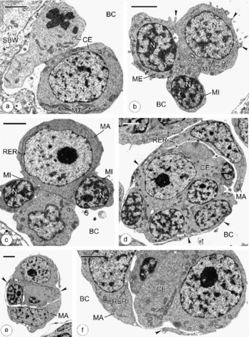
The fine structure of the germinal cells of P. borealis (syn: P. gracilescens) sporocysts was described by Podvyaznaya (Reference Podvyaznaya2007) who showed that, prior to cleavage, germinal cells leave germinal masses and float freely in the terminal brood chambers. The earliest embryos of P. borealis cercariae, corresponding to the stage of naked cell aggregates, also float freely in the sporocyst body cavity (fig. 1a–e). Even when naked cell aggregates are observed close to the body wall of the sporocyst there is no evidence of their entrapment by body wall extensions (fig. 1a) as observed in some other digeneans (Meuleman & Holzmann, Reference Meuleman and Holzmann1975; Tihomirov, Reference Tihomirov1980; Galaktionov & Dobrovolskij, Reference Galaktionov and Dobrovolskij2003). These early embryos are irregular in shape (fig. 1a–c). All their component cells are in direct contact with the contents of the sporocyst body cavity and small irregular outgrowths are often seen on their surfaces (fig. 1b). The cells in each naked cell aggregate differ in size and structure and can be divided conventionally into macro-, meso- and micromeres. Macromeres are similar in appearance to germinal cells (fig. 1c). Each has a characteristically large nucleus with strongly dispersed chromatin and a large nucleolus. Mesomeres are smaller and the proportion of condensed chromatin in their nuclei is larger (fig. 1b). Micromeres, which can be seen in embryos consisting of at least 5–6 cells, are tiny; their small electron-dense nuclei are surrounded by a narrow cytoplasmic rim (fig. 1b and c). In all blastomeres, the cytoplasm is largely filled with free ribosomes and mitochondria, that of macromeres also contains a few large RER cisternae (fig. 1c).
Initial stages of cercarial development in Prosorhynchoides borealis. (a) Section showing the asynchronous division process in a cercarial embryo (CE) composed of two blastomeres. (b) and (c) Sections through cercarial embryos consisting of 5–6 blastomeres. Note macromeres (MA), mesomeres (ME), micromeres (MI) and cytoplasmic outgrowths (arrowheads) on the embryo surface. (d) and (e) Sections showing formation of the primitive epithelium. Note cup-shaped macromeres (MA) and their cytoplasmic extensions (arrowheads). (f) Enlarged fragment of cercarial embryo (CE) showing overlapping extensions (arrowhead) of two superficial macromeres before they become fused to form the primitive epithelium. BC, body cavity; MT, mitochondria; N, nucleus; RER, rough endoplasmic reticulum; SBW, sporocyst body wall. Bars = 2 μm.

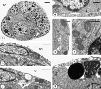
When the number of embryonic cells reaches 12–14, a few (possibly only two) superficial macromeres become concave (cup-like) and start to give rise to flat cytoplasmic extensions that grow around the rest of the embryo (fig. 1d–f). Slightly more mature embryos are already covered with a thin syncytial epithelium, containing at least two large nuclei at the opposite poles of the embryo (fig. 2a). After the superficial macromeres fuse completely, their cytoplasmic structure undergoes noticeable changes (fig. 2b and c). Golgi complexes become distinctly pronounced; large RER cisternae disappear to give way to a few smaller ones (fig. 2c). The number of free ribosomes decreases and the syncytium becomes electron transparent at some places. At the same time, the cytoplasm becomes progressively filled with vesicles of various sizes (fig. 2a–e, g). The bounding membranes of some vesicles appear to be continuous with the inner or outer plasma membranes of the syncytium (fig. 2b and c). Most vesicles are electron-lucent, but some contain particles of electron-dense material (fig. 2c and g). In early germinal balls the primitive epithelium is connected to peripheral embryonic cells by numerous narrow cytoplasmic bridges (fig. 2d and e). The same syncytial connections are also present between neighbouring embryonic cells (fig. 2f) inside the germinal balls (note that this was not observed in the earlier naked cell aggregates). Another feature of the germinal balls is the presence of pyknotic bodies, the result of degeneration of some embryonic cells. In early germinal balls, pyknotic bodies are usually located at the periphery of the embryo (fig. 2g), while in late germinal balls they are distributed throughout the embryo.
Early germinal balls of Prosorhynchoides borealis. (a) Germinal ball (GB) covered by newly formed primitive epithelium (PE); bar = 2 μm. (b) and (c) Micrographs showing developmental changes in the primitive epithelium (PE); bars = 0.5 μm. (d) and (e) Sections showing syncytial connections (arrows) between the primitive epithelium (PE) and peripheral embryonic cells; bars = 0.5 μm. (f) Syncytial connection (arrow) between adjacent embryonic cells inside a germinal ball; bar = 0.5 μm. (g) Micrograph showing pyknotic degeneration (PD) of embryonic cells below the primitive epithelium (PE); bar = 1 μm. BC, body cavity; GC, Golgi complex; N, nucleus; RER, rough endoplasmic reticulum; V, vesicles.

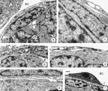
In late germinal balls, small lenticular cells with flattened nuclei become apparent just below the primitive epithelium (fig. 3a). Later these cells progressively flatten, expand and eventually fuse, forming a thin syncytium underlying the primitive epithelium (fig. 3b). This newly formed embryonic tegument has some similarities with the primitive epithelium. Its cytoplasm contains vesicles (fig. 3b), though fewer than in the primitive epithelium, and like the primitive epithelium, the embryonic tegument forms thin syncytial connections with the embryonic cells below (fig. 3c). These connections disappear when the basement lamina, underlying the tegument, is formed.
Initial stages of tegument development in Prosorhynchoides borealis cercariae. (a) Primordial tegumental cell (PTC). (b) Section showing newly formed embryonic tegument underlying primitive epithelium. (c) Section showing syncytial connections (arrows) between the embryonic tegument and peripheral embryonic cell. (d) Micrograph showing detachment of the primitive epithelium from the embryo surface. (e) Section showing two embryos: CE I, germinal ball with primitive epithelium and embryonic tegument; and CE II, young cercaria with tegument, basement lamina (BL) and developing muscles (MU). (f) Section showing degenerating pyknotic nucleus in the tegument. BC, body cavity; N, nucleus; PE, primitive epithelium; T, tegument. Bars = 1 μm.

Germinal balls that possess a newly formed nucleated tegument still retain the primitive epithelium as a closely fitting covering sheath. Progressively the primitive epithelium cytoplasm becomes more electron-dense and its syncytial connections with the underlying cells disappear (fig. 3b). In more mature, elongated germinal balls the primitive epithelium, which has gradually become a thin-walled sac, starts to detach from the embryo (fig. 3d). Early cercariae with a distinct tail bud lack the primitive epithelium completely (fig. 3e and f). At this stage, nuclei that are still present in the embryonic tegument have become pyknotic, usually protruding from the thin syncytial layer as knobs (fig. 3f). At the tail-bud stage, a thin basement lamina and the fibres of the subtegumental musculature become pronounced below the embryonic tegument (fig. 3e) and nuclei can no longer be seen in the tegument of later embryos.
Next, during the tail elongation stage, secretory tegumental cells (cytons) appear to differentiate in the peripheral parenchyma. In late cercariae they become connected by cytoplasmic bridges to the outer cytoplasmic layer of the tegument. The events occurring at this stage have been documented in detail in other trematode species (Matricon-Gondran, Reference Matricon-Gondran1971; Hockley, Reference Hockley1972; Meuleman & Holzmann, Reference Meuleman and Holzmann1975; Rees & Day, Reference Rees and Day1976; Tihomirov, Reference Tihomirov1980; Göbel & Pan, Reference Göbel and Pan1985; Halton & McCrae, Reference Halton and McCrae1985; Dunn et al., Reference Dunn, Dang, Hanna and Nisami1992; Galaktionov & Dobrovolskij, Reference Galaktionov and Dobrovolskij2003). In P. borealis, they follow a similar pattern which will not be described in this study.
Discussion
It has been shown at the light microscopic level (Ishii, Reference Ishii1934; Chen, Reference Chen1937; Rees, Reference Rees1940; Dunn, Reference Dunn1959; Cheng & James, Reference Cheng and James1960; Cheng, Reference Cheng1961; Dobrovolskij, Reference Dobrovolskij1965, Reference Dobrovolskij1971; Cheng & Bier, Reference Cheng and Bier1972; Gerasyov & Dobrovolskij, Reference Gerasyov and Dobrovolskij1977) that earliest cercarial development is achieved by a series of total, unequal, asynchronous divisions. As the result, the germinal cells of the daughter parthenitae form morula-like embryos, consisting of morphologically different cells. The results of our ultrastructural research of the earliest developmental stages of P. borealis cercariae correspond fully with the above light microscopic observations. Later, the early embryos form an embryonic membrane, or primitive epithelium, and become germinal balls. Our investigation shows unambiguously that in P. borealis cercariae the primitive epithelium is formed by the cells of the embryo. In this respect, the obtained results concur with most light microscope descriptions of cercarial embryogenesis (Ishii, Reference Ishii1934; Chen, Reference Chen1937; Dunn, Reference Dunn1959; Cheng & James, Reference Cheng and James1960; Cheng, Reference Cheng1961; Dobrovolskij, Reference Dobrovolskij1965, Reference Dobrovolskij1971; James & Bowers, Reference James and Bowers1967; Cheng & Bier, Reference Cheng and Bier1972; Bednarz, Reference Bednarz1973; Gerasyov & Dobrovolskij, Reference Gerasyov and Dobrovolskij1977), as well as with some ultrastructural data (Hockley, Reference Hockley1972; Rees & Day, Reference Rees and Day1976; Tihomirov, Reference Tihomirov1980; Al-Salman & James, Reference Al-Salman and James1988; Žd'árská, Reference Žd'árská1995; Galaktionov & Dobrovolskij, Reference Galaktionov and Dobrovolskij2003). It is worth noting that the above studies embrace many trematode species from different families (Fasciolidae, Echinostomatidae, Philophthalmidae, Paramphistomidae, Notocotylidae, Bucephalidae, Schistosomatidae, Gymnophallidae, Strigeidae, Troglotrematidae, Allocreadiidae, Brachycoeliidae, Plagiorchiidae, Macroderidae, Heterophyidae, Microphallidae) and orders.
Similarly to other digeneans studied, the primitive epithelium of cercarial embryos in P. borealis is a transitory envelope, which has the following functions: (1) it maintains the integrity of the rapidly growing embryonic mass, (2) protects it, and (3) transports nutrients into the growing embryo until the formation and activation of the true tegument. During the short period of its existence, the primitive epithelium of P. borealis undergoes successive ultrastructural changes. At first, the newly formed syncytium is almost identical in its cytoplasmic and nuclear structure to the macromeres of early embryos. Then the number of ribosomes and the size of RER cisternae decrease, which points to a gradual decrease in protein synthesis. At the same time, the cytoplasm fills with vesicles. Since these vesicles are associated with the inner and the outer plasma membranes of syncytium, they are likely to be involved in endo- or exocytosis. Similar vesicles have been described by Matricon-Gondran (Reference Matricon-Gondran1971) in Bacciger bacciger and by Halton & McCrae (Reference Halton and McCrae1985) in F. fellis. The latter authors consider these structures to be involved in nutrient transport across the primitive epithelium. The emergence of syncytial connections between the primitive epithelium and peripheral embryonic cells in early germinal balls of P. borealis also seems to be associated with an active transport of nutrients to the developing embryos. These connections are observed during active growth of the germinal balls, prior to formation of their outer tegumental layer. After primordial tegumental cells fuse to form a syncytium, the primitive epithelium starts to degenerate: its cytoplasm condenses and then the epithelium detaches from the surface of the embryo. The embryos shed the primitive epithelium completely after the formation of the tail bud; by that time, below the outer tegumental layer there is a distinct basement lamina and the primordial subtegumental musculature.
According to electron microscopic studies (James et al., Reference James, Bowers and Richards1966; Tihomirov, Reference Tihomirov1980; Dunn et al., Reference Dunn, Dang, Hanna and Nisami1992; Galaktionov & Dobrovolskij, Reference Galaktionov and Dobrovolskij2003), early cercarial embryos of some trematodes are surrounded by extensions of redial or sporocyst cells. Examination of these extensions in rediae of Philophthalmus rhionica (Tihomirov, Reference Tihomirov1980) and in daughter sporocysts of Microphallus pirum (Galaktionov & Dobrovolskij, Reference Galaktionov and Dobrovolskij2003) demonstrated that they are not connected to the embryos' primitive epithelium, which is clearly defined in the above species and has a characteristic structure. A different state of affairs was reported in a detailed study of early development of Schistosoma mansoni cercariae (Meuleman & Holzmann, Reference Meuleman and Holzmann1975). According to Meuleman & Holzmann, the germinal cells and very early embryos, consisting of a few cells and located in the body cavity of the daughter sporocysts, are loosely wrapped by thin extensions of the tegumental cells of the sporocyst. At a later stage, the embryos become closely enveloped by a nucleated primitive epithelium, which has cytoplasmic connections with the sporocyst tegumental cells or their extensions. Later, after formation of the outer tegumental layer in the embryos, the syncytial connections between the primitive epithelium and the tegumental cells of the sporocyst are lost and the primitive epithelium gradually degenerates. A similar scenario was reported for daughter sporocysts of S. mansoni, in which embryonic development is also accompanied by primitive epithelium formation (Meuleman et al., Reference Meuleman, Holzmann and Peet1980). Syncytial connections between the primitive epithelium of early cercarial embryos and the ‘subtegumental’ cells of the sporocyst were later revealed in another schistosome species, S. japonicum (Göbel & Pan, Reference Göbel and Pan1985, fig. 2). The authors of all the above studies seem to be confident in their interpretation of these observations, claiming that the primitive epithelium surrounding early schistosome embryos is formed by fusion of extensions from sporocyst cells.
It is our opinion that another interpretation is also possible. In early germinal balls of P. borealis, we observed formation of numerous syncytial connections between the primitive epithelium and the peripheral embryonic cells. Taking into account the genetic identity of the daughter and mother organisms during reproduction of trematode parthenogenetic generations, we propose that the primitive epithelium, formed originally by embryonic cells, may well establish secondary temporary syncytial connections with the sporocyst tegumental cells or their extensions. We suggest that this may be the case in the schistosome species studied. Advantages of the existence of syncytial connections between the primitive epithelium and the sporocyst tegumental cells are evident: they channel nutrients from the mother organism into the developing embryo directly, bypassing the sporocyst body cavity. Our contention would also resolve the apparent discrepancy in the interpretation of data by Rifkin (Reference Rifkin1970), Meuleman & Holzmann (Reference Meuleman and Holzmann1975), Meuleman et al. (Reference Meuleman, Holzmann and Peet1980) and Göbel & Pan (Reference Göbel and Pan1985), on the one hand, and by Maldonado (Reference Maldonado1967), Cheng & Bier (Reference Cheng and Bier1972) and Hockley (Reference Hockley1972), on the other hand. Like us, the latter authors asserted that the primitive epithelium in schistosomes is embryo derived. Furthermore, our hypothesis provides a realistic explanation of the origin of nuclei in the primitive epithelium of schistosomes. According to the ultrastructural data of Hockley (Reference Hockley1972), each cercarial embryo of S. mansoni has several large nuclei in its primitive epithelium. It is hard to imagine how these large nuclei could have migrated there across thin extensions of the sporocyst tegumental cells. The participation of embryonic cells in primitive epithelium formation in schistosomes seems much more likely.
Thus, in most (if not in all) trematode species the primitive epithelium of cercarial embryos is formed as a result of blastomere (macromere) specialization at the naked cell aggregate stage. Moreover, in our opinion this statement can be extended to primitive epithelium formation in parthenogenetic generations – daughter sporocysts and rediae. At the light microscope level no essential differences can be observed between the primitive epithelium formation around daughter sporocyst or redial embryos on the one hand and cercarial embryos on the other (see the review by Galaktionov & Dobrovolskij, Reference Galaktionov and Dobrovolskij2003). At the electron microscope level it has been shown that primitive epithelium was formed from blastomeres (macromeres) in the developing parthenogenetic metacercariae of Parvatrema margaritensis (Irwin et al., Reference Irwin, Galaktionov, Malkova, Saville and Fitzpatrick2003).
Having summarized a vast amount of factual material, Tyler & Tyler (Reference Tyler and Tyler1997) and Tyler & Hooge (Reference Tyler and Hooge2004) showed convincingly that a successive replacement of cover epithelia is a characteristic feature of flatworms in general. In trematodes, this scenario is distinctly expressed in the course of development and metamorphosis of the miracidium (see the review by Galaktionov & Dobrovolskij, Reference Galaktionov and Dobrovolskij2003). Formation of the transitory primitive epithelium in early embryos of cercariae and parthenitae and its subsequent replacement with the definitive tegument is yet another manifestation of the above regularity.
In P. borealis cercariae, the outer tegumental layer formation corresponds fully with the pattern described for most cercariae studied (Matricon-Gondran, Reference Matricon-Gondran1971; Hockley, Reference Hockley1972; Meuleman & Holzmann, Reference Meuleman and Holzmann1975; Rees & Day, Reference Rees and Day1976; Tihomirov, Reference Tihomirov1980; Göbel & Pan, Reference Göbel and Pan1985; Halton & McCrae, Reference Halton and McCrae1985; Dunn et al., Reference Dunn, Dang, Hanna and Nisami1992; Galaktionov & Dobrovolskij, Reference Galaktionov and Dobrovolskij2003). Peripherally located primordial tegumental cells expand and fuse to form a nucleated syncytium underlying the primitive epithelium; the nuclei are later lost. In our species, the nuclei in the embryonic tegument undergo pyknotic degeneration, while perikarya regions protrude outwards. We did not find any indications of perikarya regions submerging into the parenchyma and becoming cytons of tegument, the process described for Acanthoparyphium spinulosum (Bils & Martin, Reference Bils and Martin1966), Meiogymnophallus minutus (Al-Salman & James, Reference Al-Salman and James1988) and Echinostoma revolutum (Žd'árská, Reference Žd'árská1995). At the same time, in P. borealis, thin syncytial connections between the newly formed tegumental layer and the underlying embryonic cells were observed. This arrangement may resemble superficially a sunken epithelium, but careful observation at ultrastructural level reveals that these structures are completely different. Temporary syncytial connections between the embryonic tegument and the underlying embryonic cells seem likely to perform the same function as do the analogous connections between the embryonic cells and the primitive epithelium. We suppose that they are involved in nutrient transport to the developing embryo. Thin syncytial connections between the adjacent embryonic cells inside the germinal balls of P. borealis apparently promote an even distribution of nutrients across the multicellular embryonic mass. The ‘proper’ tegumental cells in P. borealis embryos differentiate later from embryonic cells underlying the layers of developing circular and longitudinal muscles. Projections of cytons pass between the myoblasts and establish syncytial connections with the outer cytoplasmic layer of the tegument. A similar sequence of events has been revealed during tegument formation in cercariae of many trematode species (Matricon-Gondran, Reference Matricon-Gondran1971; Hockley, Reference Hockley1972; Meuleman & Holzmann, Reference Meuleman and Holzmann1975; Rees & Day, Reference Rees and Day1976; Tihomirov, Reference Tihomirov1980; Göbel & Pan, Reference Göbel and Pan1985; Halton & McCrae, Reference Halton and McCrae1985; Dunn et al., Reference Dunn, Dang, Hanna and Nisami1992; Galaktionov & Dobrovolskij, Reference Galaktionov and Dobrovolskij2003). This process appears to be universal.
Acknowledgements
This work was supported by the Russian Foundation for Basic Research (Grant N 07-04-01 675, N 06-04-48 544) and INTAS project (N 05-1000008-8056). We wish to thank Dr M. Eydal (University of Iceland, Reykjavik) for the help in obtaining specimens of molluscs A. prismatica infected by P. borealis and Mr A. K. Levit (Oncological Institute, St. Petersburg) for excellent technical assistance. We are also grateful to Dr S. W. B. Irwin for improvement of the English version of the manuscript.