Congenital sinus of Valsalva aneurysm was first described by Hope in 1839.Reference Magee 1 This is a thin-walled outpouching that develops slowly and remains asymptomatic until it ruptures to form an aorta-to-cardiac fistula.Reference Helmy, Shaaban, Chaikouni and Hajar 2 Rupture can occur into the right ventricle, right atrium, or rarely into the interventricular septum.Reference Abdel Hay 3 When the latter happens, it is also called dissecting aneurysm of the interventricular septum.Reference Lee, Lee, Hsiung, Wei, Chuang and Chang 4 In this study, we describe a patient who was referred with the diagnosis of abscess of the interventricular septum. Echocardiography diagnosed it to be ruptured right sinus of Valsalva into the interventricular septum leading to a huge dissecting aneurysm of the interventricular septum. This case is unusual in that there were no conduction abnormalities either before or after surgery.
Case report
A 17-year-old boy was referred from another tertiary-care facility with a history of low-grade fever, cough, haemoptysis, and shortness of breath. There was no chest pain. He was comfortable, without respiratory distress, and he had an oxygen saturation of 96% on room air. His heart rate was 112 bpm, and his blood pressure was 116/86 mmHg. He had normal volume pulses, a hyperdynamic praecordium, soft heart sounds, and a pansystolic murmur of 2/6 intensity at the cardiac apex. Examination of the respiratory system was unremarkable. His chest X-ray showed cardiomegaly, atelectasis, and infiltrates in the right lower zone. Complete blood counts showed moderate microcytic hypochromic anaemia, with a haemoglobin level of 7.3 mg/dl. The total leucocyte count was 7200/cumm with 60% neutrophils, and the platelet count was 175,000/cumm. C-reactive protein was elevated at 16 mg/dl (normal<0.5 mg/dl).
His echocardiogram showed enlargement of the left atrium and left ventricle and mildly reduced left ventricular systolic function with an ejection fraction of 48%. A fistulous tract was seen from the right sinus of Valsalva dissecting into the interventricular septum along with a “windsock protrusion” of the large aneurysmal sac into the left ventricular cavity (Fig 1). The dissecting aneurysm was, however, intact and did not rupture into the left ventricle. Protrusion into the left ventricular cavity distorted the mitral valve, producing moderate mitral regurgitation (Supplementary Fig 1), and distortion of the right coronary leaflet of the aortic valve caused mild aortic regurgitation. His electrocardiogram did not show any conduction abnormality. The final diagnosis was rupture of the right sinus of Valsalva aneurysm into the interventricular septum producing a large septal dissecting aneurysm.
Parasternal long axis view-Right sinus of Valsalva fistulous connection leading to aneurysm of interventricular septum (lVS) (thin arrow) protruding into left ventricle (LV, broad arrow), hinge point of aortic valve (AV, double arrow). RV=right ventricle, LA=left atrium

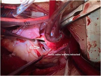
After initial stabilisation, he underwent open-heart surgery (Fig 2) with placement of a bovine pericardial patch to close the ruptured right sinus of Valsalva (Supplementary Fig 2). The ventricular septal end of the dissecting aneurysm was plicated with an over-and-over continuous polypropylene suture bringing the defect edges together, including the “windsock” in the suture, through the aortic valve orifice. The mitral valve was repaired using a 28-mm CG Future Annuloplasty Ring (Medtronic, Minneapolis, Minnesota, United States of America), and the tricuspid valve was repaired with a 32-mm Contour 3D Annuloplasty Ring (Medtronic) with closure of the left atrial appendage. The aortic valve remained competent, and the mitral valve had minimal regurgitation on post-pump transoesophageal echocardiogram.
Sinus of Valsalva aneurysm (SOVA) surgical view (broad arrow) seen after retraction of aortic valve leaflets (thin arrow)

The postoperative course was uneventful, and discharge echocardiography showed near-complete resolution of the aneurysmal cavity in the interventricular septum along with trivial mitral and tricuspid valvar regurgitation. The left ventricular function had returned to normal, with an ejection fraction of 58%. The boy was discharged 8 days after surgery.
Discussion
Aneurysm of the sinus of Valsalva is a rare congenital cardiac defect.Reference Malik, Babar, Khalil, Shah and Laiq 5 Aneurysmal dilatation of the sinus of Valsalva occurs when the media of the aortic wall is defective. The right sinus of Valsalva is most commonly involved (in approximately 65–85% of cases), followed by the non-coronary sinus. Involvement of the left sinus is least common. The dilatation and aneurysmal prolapse of a coronary sinus is a slow process occurring over years and it has been described with other diseases such as ventricular septal defects, bacterial endocarditis, atherosclerosis, and chest trauma.Reference Helmy, Shaaban, Chaikouni and Hajar 2 , Reference Tipoo Sultan, Basir and Fatimi 6
Rupture of the sinus of Valsalva, due to thinning of its wall, can occur into any chamber. A right sinus of Valsalva aneurysm can rupture into the right ventricle,Reference Malik, Babar, Khalil, Shah and Laiq 5 , Reference Lakoumentas, Bonou and Brili 7 and a non-coronary sinus of Valsalva aneurysm can rupture into the right atriumReference López-Velarde, Avila-Vanzzini, López-Velarde, Dabaghi-Richerand, López-Soriano and Espinola-Zavaleta 8 and rarely into the left-sided chambers, pulmonary artery, or pericardium. Rupture of a sinus of Valsalva aneurysm into the interventricular septum can also be described as a dissecting aneurysm into the interventricular septum. It is very rare and is usually associated with aortic valve regurgitation. Owing to its dissecting nature, it can cause conduction abnormalitiesReference Abdel Hay 3 or rupture into the ventricular cavity causing congestive heart failure. Our patient also developed a cystic aneurysmal cavity in the interventricular septum communicating with the right sinus of Valsalva that collapsed in systole and filled with blood during diastole. Preoperative conduction abnormalities were not seen in our case, as have been reported by others.Reference Helmy, Shaaban, Chaikouni and Hajar 2 , Reference Abdel Hay 3
These patients have a potential risk for rupture of the sinus of Valsalva aneurysm, leading to the development of congestive heart failure, endocarditis, conduction abnormalities, stroke, and sudden death, justifying surgical repair even in asymptomatic patients.Reference Malik, Babar, Khalil, Shah and Laiq 5 Surgical management depends on the extent of involvement. Repair of the aortic leaflet may be successful if it is not overtly distorted; however, if there is significant fibrosis and distortion, the aortic valve may require replacement. The condition is usually associated with aortic insufficiency, which was mild in our case. The aortic leaflet was not badly distorted and was successfully repaired with bovine pericardium. Distortion of the mitral valve can also occur, as described by Tomar et al,Reference Tomar, Radhakrishnan, Kausha and Iyer 9 requiring repair or replacement. Our patient had moderate mitral regurgitation, and the valve was successfully repaired.
The most frequently reported complication of surgery is a conduction abnormality due to suture injury. Several techniques have been described to avoid the conduction bundles during surgery.Reference Lee, Lee, Hsiung, Wei, Chuang and Chang 4 , Reference Choudhary, Bhan and Reddy 10 In the case of aortic valve replacement, one technique includes careful placement of sutures in the region of the right coronary artery, using an “outside-in” suture method, wherein the sutures start from outside the aorta and then extend up to the aortic ring. In our case, a continuous polypropylene suture was placed, bringing the defect edges together. The “windsock” was included in the suture through the aortic valve orifice. No postoperative conduction abnormality was observed.
Conclusion
Rupture of the sinus of Valsalva is a rare and serious clinical entity that can occur without coexisting cardiac anomalies, as was in our case. Rupture into the interventricular septum can produce conduction abnormalities, which can also occur because of suture injury during surgery. Distortion of the aortic and mitral valves by dissection into the interventricular septum can lead to insufficiency of these valves. Surgery must be timely with particular care to prevent conduction tissue injury.
Acknowledgements
The authors acknowledge the contribution of Zuhair Syed Ahmed in reviewing the manuscript.
Financial Support
This research received no specific grant from any funding agency, or from commercial or not-for-profit sectors.
Conflicts of Interest
None.
Ethical Standards
The authors assert that all procedures contributing to this work comply with the ethical standards of the relevant national guidelines on human experimentation in our institution, The Aga Khan University, Karachi, Pakistan, and with the Helsinki Declaration of 1975, as revised in 2008.
Supplementary material
To view supplementary material for this article, please visit http://dx.doi.org/10.1017/S1047951116001621