Introduction
Cartilage is currently one of the main materials used in ear surgery. In the last 10 years, different types of tympanoplasty techniques using cartilage have been described. In 1959, Utech was the first author to describe the use of cartilage in ossiculoplasty, as an interposition graft between the eardrum and the stapes.Reference Utech1 In 1963, Salen was the first to use autologous septum cartilage for myringoplasty.Reference Salen2 The first cartilage–perichondrium composite graft was introduced in 1967 by Goodhill.Reference Goodhill3 More recent authors too have preferred to use autologous cartilage in the reconstruction of the middle ear, obtaining excellent results in both the short and long term.Reference Anderson, Cavè-Thomasen and Tos4, Reference Kazikdas, Onal, Boyraz and Karabulut5 In 2008, Tos classified tympanoplasty techniques using cartilage into six main groups.Reference Tos6
Our experience mainly concerns the use of cartilage–perichondrium grafts to repair the tympanic membrane during tympanoplasty. One of the main disadvantages of this type of tympanoplasty is that it is more time-consuming than tympanoplasty with temporalis fascia, due to the need to harvest and model the cartilage graft. The graft is also difficult to fashion in a standard form.
This report proposes two original techniques which may expedite and standardise the preparation of slice-type cartilage–perichondrium composite grafts used in tympanoplasty.
Surgical techniques
Our two proposed techniques relate to tympanoplasty procedures in which a 0.3 mm thick, cartilage–perichondrium composite graft is used to repair the tympanic membrane.
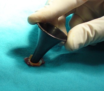
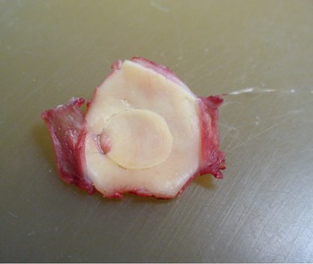
The first technique involves the use of an auricular speculum to isolate a round portion of cartilage, which remains attached to the perichondrium. After evaluating the required graft size, an appropriate auricular speculum is chosen (generally either 8 or 9 mm). The speculum is gently pressed directly onto the cartilage to isolate a round or oval cartilage segment (Figure 1). Holding the speculum in situ, the excess cartilage is then removed with a spatula or a round knife (Figure 2).
Pressure is applied directly to the cartilage via the auricular speculum to isolate a round or oval cartilage graft.

The prepared perichondrium–cartilage island graft, showing central cartilage disc.

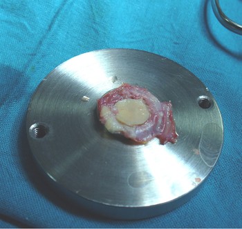
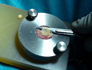
The second technique involves the manufacture of a simple device consisting of two steel discs, used to obtain the required cartilage graft thickness. The first disc is 40 mm in diameter with a raised central cylindrical section 15 mm in diameter and 2.6 mm in height. The second disc has the same overall diameter as the first and can be attached with two screws to the first disc; it has a central hole 17 mm in diameter and 3 mm in thickness. Once the graft is placed on the central section of the first disc (Figure 3), the second, holed disc is fastened tightly to the first disc to hold the excess perichondrium in position. A thin blade can then be used across the surface of the second disc to trim the excess cartilage and obtain the required cartilage graft thickness of 0.3 mm. The cartilage remains attached to the perichondrium (Figure 4).
The perichondrium–cartilage island graft sited on the raised central section of the first steel disc.

The final phase: the excess cartilage is removed to obtain a graft of precisely 3 mm thickness.

In a group of 20 patients with subtotal tympanic perforation undergoing tympanoplasty with perichondrium graft, the use of these two surgical techniques saved an average of 15 minutes per procedure, compared with similar, conventional tympanoplasty procedures.
Discussion
In the last few years, the use of cartilage has been preferred for tympanic membrane reconstruction, particularly in cases of allergy, re-perforation, or total or subtotal perforation. The mechanical characteristics of cartilage offer the advantage of high resistance to retraction and re-perforation, while also permitting acoustic performance comparable to that of temporalis fascia, at least for thicknesses of approximately 0.3 mm.
One of the disadvantages of the tympanoplasty cartilage technique is that it is time-consuming, compared with the use of temporalis fascia. Our two proposed techniques save time, guarantee precision and improve graft standardisation, compared with the conventional procedure. In particular, our proposed cartilage-cutting device enables the creation of cartilage slices of a constant thickness. To date, this is the only reported device enabling creation of cartilage–perichondrium grafts in the described fashion; other devices currently available cut only the cartilage.Reference Murbe, Zahnert, Bornitz and Huttenbrink7
The future use of similar steel discs of different thickness would permit different thicknesses of cartilage–perichondrium graft to be created, and would also facilitate comparison of clinical results.
In conclusion, we propose two new techniques to facilitate the creation of composite cartilage–perichondrium grafts. From our initial experience, these techniques can reduce surgical time by 15 minutes, for tympanoplasty procedures requiring a cartilage–perichondrium composite graft.