Introduction
Muonionalusta is an octahedrite that belongs to group IVA, which accounts for ∼8% of all irons known. The primary mineralogy of the meteorite, with a Ni content of ∼9 wt.% and ∼0.4 wt.% Co (Hidaka et al., Reference Hidaka, Shirai, Yamaguchi and Ebihara2019), has been described in detail elsewhere (Buchwald, Reference Buchwald1975; Holtstam et al., Reference Holtstam, Broman, Söderhielm and Zetterqvist2003). The Muonionalusta meteoroid was disrupted from its parent world ∼0.4 billion years ago in a violent impact event (Blichert-Toft et al., Reference Blichert-Toft, Moynier, Lee, Telouk and Albarède2010). It descended to Earth in Middle Pleistocene, breaking up in the atmosphere and scattering over a large area (Lagerbäck and Wickman, Reference Lagerbäck and Wickman1997). The current distribution of meteorite fragments forms a disturbed strewn field spanning at least 25 × 15 km, with the main concentration found around Lake Kitkiöjärvi (lat. 67°49ʹN, long. 23°8ʹE, 255 m a.s.l.) in the Pajala District, Norrbotten, Sweden. Pieces ranging from 0.1 to 1200 kg can be found from near the surface down to at least 4 m in the soil.
The Quaternary deposits of this area consist primarily of till and postglacial accumulations of peat, though large sections are covered with glaciofluvial/lacustrine sediments (Fagerlind, Reference Fagerlind1981). It is inferred that meteorite fragments were moved by the ice sheet up to 8–20 km from the original impact site, based on the distribution pattern of hundreds of samples (Hättestrand, Reference Hättestrand2009). They have thus endured at least one glaciation, the Weichselian (115–12 k.y.), and thereafter, in a subpolar climate, they have been subjected to seasonal freeze-and-thaw cycles.
Muonionalusta meteorite samples are accompanied by a ‘rust crust’, consisting of various terrestrial alteration products, mainly Fe (±Ni) oxides and oxyhydroxides, and relics of meteoritic minerals plus cemented soil particles (Wickman, Reference Wickman1964; Holtstam and Söderhielm, Reference Holtstam and Söderhielm2004; Gurdziel, Reference Gurdziel2012). Recent finds of muonionalustaite [Ni3(OH)4Cl2⋅4H2O] (Holtstam et al., Reference Holtstam, Bindi, Karlsson, Söderhielm and Zetterqvist2021) and parahibbingite [β-Fe2(OH)3Cl] (Margheri et al., Reference Margheri, Bindi, Bonazzi and Holtstam2022) have demonstrated a more diverse secondary mineralogy, emphasising the significance of chlorine in the terrestrial alteration processes.
In the current contribution, we present results from an investigation of akaganeite, (Fe3+,Ni2+)8(OH,O)16Cl1.25·nH2O, and humboldtine, Fe(C2O4)·2H2O, from meteorite rust samples, including the first single-crystal diffraction study of akaganeite and extended Raman spectroscopic data.
Materials and methods
Samples
Akaganeite crystals were taken from a small meteorite sample (∼2 kg) found at 40 cm depth, 5 km NE of Lake Kitkiöjärvi. A second akaganeite sample (see also Margheri et al., Reference Margheri, Bindi, Bonazzi and Holtstam2022) used in the study is from a rust crust attached to a granitic cobble associated with a 22 kg meteorite specimen recovered from 1.4 m below the ground surface in the nearby area. The rust sample with humboldtine is from an unspecified meteorite find from ∼60 cm depth.
Chemical analysis
The composition of the minerals studied was determined with spot analyses using a FEI Quanta 650 field-emission scanning electron microscope (SEM), fitted with an 80 mm2 X-MaxN Oxford Instruments energy-dispersion (EDS) detector (operated at 20 kV, with beam size ∼1 µm and at a working distance of 10 mm) on carbon-coated, polished samples. The beam current was calibrated on Co metal, with the instrument calibrated against metal and mineral standards (including Fe and Ni metal, NaCl) for each element.
Single-crystal X-ray diffraction study of akaganeite
Several crystals were extracted from a cluster of pseudo-hexagonal, platy crystals and tested with single-crystal X-ray diffraction (SCXRD). Among them, only one exhibited good diffraction quality. On this crystal, unit-cell parameters and intensity data were collected using a Bruker D8 Venture diffractometer equipped with a Photon III detector, with a graphite-monochromatised MoKα radiation (λ = 0.71073 Å). Intensity data were corrected for absorption, using the multi-scan correction method (SADABS), and for Lorentz and polarisation effects with the APEX3 software suite (Bruker, 2016). Details of the data collection are listed in Table 1.
Crystal data and SCXRD experimental details

* w = 1/[σ2(F o2) + (0.0381*P)2] where P = (F o2 + 2F c2)/3
Structure refinement of akaganeite
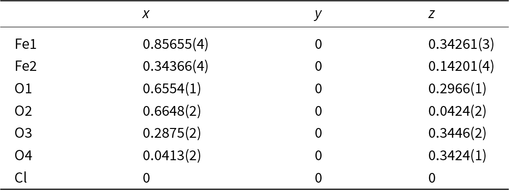
The structure refinement was carried out in the space group I2/m using SHELXL (Sheldrick, Reference Sheldrick2015). Starting atomic positions were taken from Post and Buchwald (Reference Post and Buchwald1991), with two Fe sites and four O sites located on a mirror plane (Wyckoff position 4i) and the Cl site positioned at the origin of the cell (Wyckoff position 2a). Scattering curves for neutral Cl, Fe and O were taken from the International Tables for Crystallography (Wilson, Reference Wilson1992). The Cl-site occupancy was refined and then fixed at the refined value; all the other sites were found to be fully occupied. Notably, the resulting Cl/Fe ratio obtained from the refinement, 0.123(1), is in good agreement with the value from chemical data, 0.13 (see below). Given a still very high R1 parameter (> 20%) at this stage, a non-merohedral twinning was taken into account (matrix: |001/010/100|). The introduction of the twinning dramatically improved the refinement. Convergence was then achieved with an anisotropic model, with a final R1 = 2.23%. Lastly, residues in the ΔF map were examined in order to find the H atoms, but none of the attempts to include known hydrogen positions (see Post et al., Reference Post, Heaney, von Dreele and Hanson2003) led to satisfactory results.
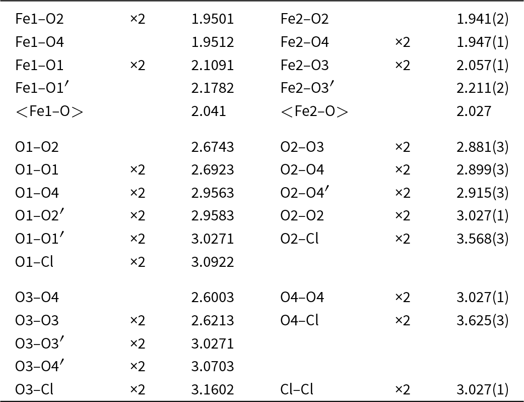
Atomic coordinates and selected bond distances are listed in Tables 2 and 3, respectively. The crystallographic information file has been deposited with the Principal Editor of Mineralogical Magazine and is available as Supplementary material (see below).
Atomic coordinates in the crystal structure of akaganeite

Selected interatomic distances (Å) for akaganeite

Raman spectroscopy
Raman spectra of both akaganeite and humboldtine were collected on a LabRAM HR 800 micro-spectrometer, with a 515 nm continuous-wave single-frequency diode pumped laser (Cobolt Fandango), a Peltier-cooled (−70°C) charge-coupled device detector (Synapse) and an Olympus MPlan N 100×/0.9 objective. The instrument was calibrated against the 521 cm–1 Raman band of silicon before sample measurements. Filters allowing laser throughput of 1% or 10% were employed, with the laser spot on the sample surface being ∼2 µm in diameter. The laser power on the samples for each filter was ∼0.2 and ∼2 mW, respectively. Instrument control and data acquisition (up to 500 s in 2 cycles with a 600 grooves/mm grating) were made with the LabSpec 6 software.
Results
Akaganeite
Akaganeite has been found as (A) large crystals, sometimes grouped in ‘rosettes’ (Figs 1, 2), and (B) as envelopes grown on parahibbingite crystals (Fig. 3; see also Margheri et al., Reference Margheri, Bindi, Bonazzi and Holtstam2022). The single crystals are lustrous reddish brown and euhedral, with a (pseudo)hexagonal platy outline, and up to 1 mm across. They occur in cavities in a crust consisting of mainly fine-grained goethite and maghemite. Relics of unoxidised taenite lamellae and thin crusts of a greenish yellow, unknown Ni-(Fe)-Cl hydrate mineral also occur on the rust sample.
(a–b) Colour images of akaganeite crystals. Sample GEO-NRM #20040065.

Colour image of a cavity with akaganeite crystals and dark brown, botryoidal goethite. Sample GEO-NRM #20040065.

SEM-BSE image of akaganeite forming a rim on parahibbingite. Sample GEO-NRM #20060957.

Chemical analyses (n = 35) of akaganeite (A) gave no detectable Ni or Co and traces of Si, Al and S (< 0.1 wt.% each). Cl ranges from 2.8 to 5.6 wt.% (average 4.4%; 1σ = 0.80). The average mole Fe/Cl ratio is 7.6. The sample (B; n = 6) is more homogeneous, with average Ni 1.3 wt.%, Co 0.6 wt.%; S 0.2 wt.% and Cl 7.5 wt.%; (Fe + Ni + Co)/Cl = 4.7. Whereas the minor Ni, Co, Si and Al are possibly substituting for Fe3+ in akaganeite, SO42– ions probably occupy space in the structural ‘tunnels’ (Peretyazhko et al., Reference Peretyazhko, Pan, Ming, Rampe, Morris and Agresti2019).
For the Raman spectra of akaganeite, sample (B) (Fig. 4) produced the best result (highest peak-to-background and lower noise level). The spectrum was collected with the 1% filter, because the sample transformed with higher laser throughput. Although relative intensities vary (partly due to different excitation wavelengths of the lasers used in different studies), peak positions are in general in good agreement (±7 cm–1) with synthetic akaganeite (Oh et al., Reference Oh, Cook and Townsend1998; Rémazeilles and Refait, Reference Rémazeilles and Refait2007; Li and Hihara, Reference Li and Hihara2015), but previous studies show fewer observed bands in total and do not cover regions above 1000 cm–1 (including possible O–H vibration modes).
Raman spectrum of akaganeite. Sample GEO-NRM #20060957.

For akaganeite, the relatively weak 3510 cm–1 band is assigned to O–H stretching, and in sample (A) this band occurs at a close position, 3522 cm–1. Both are compatible with an O…O–H distance > 3 Å (Libowitzky, Reference Libowitzky1999) as found from structural refinement.
Infra-red (IR) spectra of synthetic akaganeite powder show an absorption band at ∼3485 cm–1, ascribed to O–H stretching modes (Murad and Bishop, Reference Murad and Bishop2000), that may well correspond to the 3510 peak of the Raman spectrum. Another band of approximately equal strength at ∼3390 cm–1 that is also reported from IR spectra (Murad and Bishop, Reference Murad and Bishop2000) has no counterpart in the Raman spectrum, although the background from 3300 to 3500 is slightly elevated here. The 3390 cm–1 band is assigned to OH–Cl interactions (Song and Boily, Reference Song and Boily2012).
Akaganeite is a thermally less stable mineral, known to transform to hematite or goethite depending on physical and chemical conditions (Ståhl et al., Reference Ståhl, Nielsen, Jiang, Lebech, Hanson, Norby and van Lanschot2003; Fu et al., Reference Fu, Jia, Wang, Cao, Ling, Liu, Shi, Wu, Li and Zhang2020). Raman recordings of akaganeite with a lower degree of filtering produced a distinct and reproducible result (Fig. 5) with well-defined peaks. The spectra obtained show no close resemblance to the common phases known in the Fe–O–(H) system. There is a very good match with data ascribed to synthetic nanoparticles of magnetite of ∼20 nm in size (Yew et al., Reference Yew, Shameli, Miyake, Khairudin, Mohamad, Hara, Nordin and Lee2017), with bands at 212, 274, 388, 474, 585, and a broad feature at 1277 cm–1. However, a more probable explanation is that their Fe3O4 oxidised to Fe2O3 under the laser beam, producing a pattern with red-shifted hematite Raman bands at high temperature, a phenomenon described by Shebanova and Lazor (Reference Shebanova and Lazor2003). Our data, then, imply that temperatures at ∼400°C were prevailing during analysis of akaganeite that unintentionally transformed into α-Fe2O3 with higher laser power.
Raman spectrum of laser-transformed akaganeite.

The present material (A) was considered a good candidate for the first SCXRD study of the species, because akaganeite forms exceptional (from outer morphology), large crystals and has a negligible Ni content. The unit-cell parameters observed here (Table 1) are comparable to those found in previous structural studies, some of which employed neutron and synchrotron radiation on synthetic and natural powders (Post et al., Reference Post, Heaney, von Dreele and Hanson2003; Ståhl et al., Reference Ståhl, Nielsen, Jiang, Lebech, Hanson, Norby and van Lanschot2003).
Humboldtine
Humboldtine was identified from powder X-ray diffraction, chemical analyses and Raman spectroscopy. It occurs in clusters, up to 0.5 mm wide (Fig. 6), of small, greenish yellow translucent crystals with complex morphology. They are found in a fragile, porous aggregate of mainly goethite, ferrihydrite and quartz + feldspar sand grains. The chemical data indicate solid solutions between humboldtine and its Ni-analogue, andreybulakhite (IMA no. 2023-037, Vereshchagin et al., Reference Vereshchagin, Britvin, Pankin, Zelenskaya, Krzhizhanovskaya, Kuz’mina, Vlasenko and Frank-Kamenetskaya2025), with a range of atomic Fe/(Fe + Ni) values 0.77–0.51; average from 18 analyses is 0.60. No other metals were detected. The Raman spectrum (Fig. 7) is very similar to published data for humboldtine (Echigo and Kimata, Reference Echigo and Kimata2008) and isotypic M 2+ oxalate dihydrates (M = Mg, Mn, Fe, Ni and Zn; Giester et al., Reference Giester, Rieck, Lengauer, Kolitsch and Nasdala2023). The intense band at 1470 cm–1, related to C–O bending modes, can be compared with the corresponding Raman shift for the synthetic end-member Fe and Ni compounds, at 1463 and 1482 cm–1, respectively. The band at 531 cm–1, assigned to metal–O stretching, is apparently slightly moved (from pure Fe oxalate dihydrate at 523 cm–1), probably related to the shorter average bond lengths in Ni-bearing humboldtine. It may be noted that, whereas the mineral was unstable under the electron beam of the SEM, there was no sample damage from the laser, not even for with the low-density filter (10%). Therefore, signals from vibrations of the H2O molecules, manifesting O–H stretching at 3330 and bending at 1610 cm–1, respectively, are clearly visible in the spectrum.
Colour image of yellowish humboldtine aggregate. Black spherical grains are poorly crystalline goethite. Sample GEO-NRM #20050170.

Raman spectrum of humboldtine. Sample GEO-NRM #20050170.

Discussion
Crystal structure details of akaganeite
The crystal symmetry of akaganeite was considered tetragonal until the early 1990s, when Post and Buchwald (Reference Post and Buchwald1991) proposed the currently accepted monoclinic model. The SCXRD data obtained in the present study are in excellent agreement with the latest literature data (Post and Buchwald, Reference Post and Buchwald1991; Post et al., Reference Post, Heaney, von Dreele and Hanson2003; Ståhl et al., Reference Ståhl, Nielsen, Jiang, Lebech, Hanson, Norby and van Lanschot2003), thus confirming a monoclinic, I-centred crystal lattice.
Akaganeite, also known as β-FeOOH, is isostructural with monoclinic members of the hollandite supergroup (except hollandite sensu stricto and strontiomelane; Biagioni et al., Reference Biagioni, Capalbo and Pasero2013): the structure consists of double chains of edge-linked Fe octahedra that form 2 × 2 tunnels oriented along the b axis (Fig. 8).
Projection of the akaganeite structure approximately down the [010] axis. The green spheres at the centre of the tunnel represent chloride ions. The structure was drawn using VESTA (Momma and Izumi, Reference Momma and Izumi2011).

Unlike hollandite-supergroup minerals, where the octahedrally coordinated sites are occupied by tetravalent cations (i.e. Ti4+ and Mn4+), in the akaganeite structure they mostly host trivalent iron. When akaganeite forms in Ni-rich environments, e.g. when it occurs as a weathering product of metal-bearing meteorites, Ni2+ can enter the octahedra at the expense of Fe3+ (Buchwald and Clarke, Reference Buchwald and Clarke1989; Post and Buchwald, Reference Post and Buchwald1991; Post et al., Reference Post, Heaney, von Dreele and Hanson2003), and the charge neutrality is kept by the addition of OH– groups instead of O2–. The sample (A) examined here seems to be an exception: despite being a secondary phase derived from the alteration of the Muonionalusta iron meteorite, it is devoid of nickel, leaving Fe3+ the only cation to occupy the octahedra (which is the case for ‘terrestrial’ akaganeites, e.g. Mackay, Reference Mackay1962; Holtstam, Reference Holtstam2006). Fe3+ can be found at two distinct, distorted sites (Fe1 and Fe2) positioned slightly off-centre, possibly in order to minimise the cation–cation repulsion as occurs in other akaganeite samples (Post and Buchwald, Reference Post and Buchwald1991; Post et al., Reference Post, Heaney, von Dreele and Hanson2003) as well as in hollandite-supergroup minerals (e.g. Post et al., Reference Post, von Dreele and Buseck1982). The distortion of the Fe2 octahedron is slightly more pronounced than that of Fe1. Both octahedra have three longer and three shorter distances, Fe1–O1 and Fe2–O3 being the longer ones (Table 3). This is expected, because hydrogen is bonded to O1 and O3 (Post et al., Reference Post, Heaney, von Dreele and Hanson2003): the longer Fe–O distances are due to a reduced effective charge for O in the hydroxyl group. The mean distances <Fe1–O> and <Fe2–O> are nearly identical to those presented by Post and Buchwald (Reference Post and Buchwald1991). However, in the crystal studied here the bond distances range from 1.950 to 2.178 Å and from 1.941 to 2.211 Å, respectively. When compared to the values reported by Post and Buchwald (Reference Post and Buchwald1991), these ranges appear fairly different, especially the Fe2–O one (1.80–2.32 Å in Post and Buchwald, Reference Post and Buchwald1991). This discrepancy could be explained by the improved accuracy of our data, considering that, in the aforementioned study, the pronounced difference in regularity between the two octahedra could not be explained and was attributed to a measurement artifact.
The site located at the centre of the tunnels is half-occupied by Cl– anions, corresponding to 1 Cl atom per formula unit in the structural formula. In samples containing larger amounts of Cl (e.g. Post et al., Reference Post, Heaney, von Dreele and Hanson2003; Ståhl et al., Reference Ståhl, Nielsen, Jiang, Lebech, Hanson, Norby and van Lanschot2003), where the tunnel site is occupied for two thirds, it has been observed that in order to have compatible Cl–Cl distances (∼3.7 Å) the position of the site must slightly shift its position along the length of the tunnels. This is not necessary in the case of the crystal studied, because Cl ions could occupy every second site, thus not needing further separation from one another. Such an ordered configuration is in agreement with the relatively low thermal displacement parameter (0.043 Å2), which corresponds to about half of that observed by Post et al. (Reference Post, Heaney, von Dreele and Hanson2003) in the model involving the split Cl site (0.075 Å2).
Formation of akaganeite
Akaganeite is a relatively common corrosion product of iron metal in natural environments, where there is a supply of Cl (Buchwald and Clarke, Reference Buchwald and Clarke1989; Bland et al., Reference Bland, Zolensky, Benedix, Sephton, Lauretta and McSween2006; Holtstam, Reference Holtstam2006). It is also a well-known component on some rusty archaeological artifacts (e.g. Selwyn et al., Reference Selwyn, Sirois and Argyropoulos1999) and as a product of steel corrosion (e.g. de la Fuente et al., Reference de la Fuente, Díaz, Simancas, Chico and Morcillo2011). Gurdziel (Reference Gurdziel2012) has previously reported the occurrence of fine-grained akaganeite from Muonionalusta, but without detailed data. There are pedochemical factors to consider that promote formation of akaganeite in this investigation. The soils in this part of Sweden are exceptionally high in Cl, near 0.1%, which is related to weathering of scapolite- and apatite-group minerals in the regolith (Ladenberger et al., Reference Ladenberger, Andersson, Gonzalez, Lax, Carlsson, Ohlsson and Jelinek2012). Experimental studies show that akaganeite can form at a wide range of pH (2–8) in Cl-rich environments, but is favoured at low pH (Peretyazhko et al., Reference Peretyazhko, Ming, Rampe, Morris and Agresti2018). Values in soil (till) interstitial water of the present region are in the range 5.5–6 (Andersson et al., Reference Andersson, Carlsson, Ladenberger, Morris, Sadeghi and Uhlbäck2014). In the near vicinity of rusting meteoritic iron, pH conditions could, however, be significantly different, depending on the active corrosion mechanism. For example, hydrolysis in the presence of chloride anions promotes production of H+ (Selwyn et al., Reference Selwyn, Sirois and Argyropoulos1999). In sample (B), akaganeite formed from direct oxidation of parahibbingite (similar to what has been observed for hibbingite [γ-Fe2(OH)3Cl] on anthropogenic iron; Simon et al., Reference Simon, Cibin, Robbins, Day, Tang, Freestone and Schofield2018), and it thus has a remarkably high Cl content.
Remarks on the current akaganeite formula
In light of our findings, a brief discussion around the formula of this mineral is necessary. The current International Mineralogical Association (IMA) approved formula of akaganeite is (Fe3+,Ni2+)8(OH,O)16Cl1.25·nH2O. Starting from the octahedral sites, the current formula involves Ni as an essential structural component. While it is true that some Ni can enter at the expense of Fe, it is well known that akaganeite does not require Ni to form: in fact, there are several examples of Ni-free akaganeite, both synthetic (e.g. Cai et al., Reference Cai, Liu, Gao, Navrotsky and Suib2001; Villalba et al., Reference Villalba, Berezoski, de Almeida Cavicchiolli, Galvani and Anaissi2013) and natural (e.g. Mackay, Reference Mackay1962; Bibi et al., Reference Bibi, Singh and Silvester2011). Furthermore, when present, Ni is disordered over Fe-dominant positions; for all the above reasons, it is not fundamental to display nickel in the formula. Even so, the presence of a divalent cation as a substituent for a trivalent cation implies different O/OH ratios to balance the charge; hence, at the end of this section, two formulae (with and without Ni) will be proposed.
Chlorine is located at a specific structural position, and it is then correctly indicated in the formula. However, we suggest that Cl should be reported as a range, because the amount of measured Cl in each sample can vary considerably, also having consequences on the amount of OH– substituting for O2– required to compensate the negative charge introduced by the chloride ion.
Based on all these considerations, the new proposed formulae could be written as FeO1–x(OH)1+xClx (0.01 < x < 0.20), or, taking Ni into account, (Fe1–xNix)O1–x–y(OH)1+x+yCly (0 < x < 0.19 and 0.01 < y < 0.20), and Z = 8.
On the occurrence of humboldtine
Oxalate minerals are quite rare in Nature, but occur in diverse environments (Echigo and Kimata, Reference Echigo and Kimata2010); humboldtine from Muonionalusta could be the first example related to terrestrial weathering of a meteorite (none reported by Rubin and Ma, Reference Rubin and Ma2017). Iron oxalate may form from direct reaction between oxalic acid and Fe minerals (e.g. Vehmaanperä et al., Reference Vehmaanperä, Sihvonen, Salmimies and Häkkinen2022). This is not a likely scenario for Muonionalusta, because there is no obvious source for the high concentrations necessary, and with the reaction kinetics expected in the present climate zone, it is not realistic. The high Ni content of the present mineral is not in line with a simple dissolution–precipitation mechanism either. Instead, interaction with organisms that can produce metal oxalate seems possible, such as lichens or fungi (Gadd et al., Reference Gadd, Bahri-Esfahani, Li, Rhee, Wei, Fomina and Liang2014). For example, experimental work has shown that the ubiquitous fungus Aspergillus niger (black mould) can accumulate both Fe and Ni, and excrete hydrated oxalates corresponding to humboldtine and andreybulakhite (Magyarosy et al., Reference Magyarosy, Laidlaw, Kilaas, Echer, Clark and Keasling2002; Zelenskaya et al., Reference Zelenskaya, Izatulina, Frank-Kamenetskaya and Vlasov2021).
Concluding statement
The conditions under which akaganeite forms commonly favour rapid nucleation, resulting in the simultaneous formation of many small crystals. This leads to competition for available iron, chlorine and oxygen, limiting the resources each crystal can access and thereby restricting their size. Moreover, the environments that facilitate akaganeite formation frequently offer only relatively short periods for growth, such as during specific stages of corrosion, which further prevents the development of large crystals. All these considerations make the discovery of large, well-developed akaganeite crystals unexpected and interesting.
Geochemical and mineralogical studies of mineral rust samples offer fundamental insights into both the terrestrial and extraterrestrial realms. The analysis of composition and transformation of rust in meteorite samples unravel the complex interactions between cosmic materials and their environments.
Supplementary material
The supplementary material for this article can be found at http://dx.doi.org/10.1180/mgm.2024.78.
Acknowledgements
Thanks are due to Andreas Forsberg, Johan Söderhielm and Anders Zetterqvist for providing Muonionalusta specimens over the years to the Swedish Museum of Natural History. Single-crystal X-ray data were collected at CRIST (Centro di Servizi di Cristallografia Strutturale), Università degli Studi di Firenze. The authors acknowledge P. Leverett and two anonymous referees for their feedback and suggestions, which have significantly improved the quality of this manuscript.
Competing interests
The authors declare none.