Catheterisation in paediatric cardiology has developed from a diagnostic to an almost exclusively therapeutic approach.Reference Feltes, Bacha and Beekman 1 There are no standardised procedures. Examinations are rather individual due to the anatomic characteristics in each patient. Transcatheter interventions in CHD are essential for a number of different intracardial and extracardial procedures. Catheterisation can be necessary in the pre-operative evaluation of complex lesions.Reference Feltes, Bacha and Beekman 1 Repetitive contrast administrations in varying C-arm angulations are necessary in order to visualise the complete cardiovascular anatomy by conventional biplane angiography systems, and are attended by repeated radiation exposure and an increased amount of contrast dye.Reference Fagan, Kay, Carroll and Neubauer 2 – Reference Gutleben, Nolker and Ritscher 7
Current methods to overlay two-dimensional fluoroscopic images with three-dimensional volume data sets are used to simplify guidance during interventions. The three-dimensional heart models can be generated from pre-interventional MRI or CT, and are visualised in volume-rendering technique.Reference Tomkowiak, Klein and Vigen 8 , Reference Abu Hazeem, Dori and Whitehead 9 This visualisation technique is not standardised.Reference Glockler, Halbfaß, Koch, Achenbach and Dittrich 4
Registration with live fluoroscopy has been mostly done by acquiring three-dimensional data sets with flat-detector CT intraprocedurally for a three-dimensional–three-dimensional co-registration. Therefore, additional application of contrast dye and radiation dose has always been necessary. Newest developments show that two- to three-dimensional registration is also possible and radiation can be minimised.Reference Goreczny, Dryzek, Morgan, Lukaszewski, Moll and Moszura 10
A novel software solution provides the possibility of merging pre-interventionally created segmented and tessellated three-dimensional models. Their surface is displayed more or less opaque depending on the angle between a specific part and the screen. This ensures that devices are permanently visible throughout the examination. All visualisation properties of the meshes can be changed individually. Heart segments (atrium, ventricle, vessels) can be shown or hidden separately. For registration, the vascular model is moved manually by using the tracheal bifurcation as a fiducial mark, which is permanently visible throughout the examination and displayed according to the selected C-arm angulation and table position.
We analysed and rated feasibility, benefit, and accuracy of the unprecedented technique in 60 consecutive cases over a 10-month time period.
Materials and methods
In our prospective study, we analysed 60 consecutive interventional and diagnostic catheterisations between August 2015 and May 2016. In all cases, pre-procedural acquired data sets from MRI or multi-detector CT were available. These had previously been performed as a part of clinical routine and not primarily for the use in interventions. Radiation used for the mentioned multi-detector CT was therefore not added to our results. All patients were in compliance with our institutional requirements for cardiac catheterisation. All catheter examinations were performed in the usual manner. The registration process of the three-dimensional models did not interfere with the conventional procedure. Radiation was only utilised for the purpose of a classic cardiac catheterisation. Our new technique was exclusively used in order to reduce radiation. The institutional ethics committee approval and written informed consent from each patient or parents were obtained. We merged three-dimensional reconstructions generated from MRI (n=30), multi-detector CT (n=28), and three-dimensional rotational angiography from a former intervention (n=2) with the real-time fluoroscopic image for three-dimensional roadmapping. All examinations were performed on a biplane angiography system, provided with two 20×20 cm2 flat panel detectors, with a 960×906 matrix (Axiom Artis; Siemens Medical Solutions, Forchheim, Germany). The whole workflow is shown in Figure 1.
Workflow of segmentation of 3D heart models and 2D-3D image fusion for catheterization of children with CHD.

Pre-processing and loading
Before the intervention, surface meshes were extracted from the previously acquired MRI, CT, or three-dimensional rotational angiography volumetric data set. For this process, we used a CT Heart Segmentation tool, which is a part of the software Mimics Innovation Suite (Materialise), using both Mimics Medical 18.0 and 3-matic Medical 10.0. This software is medically evaluated. Choosing an optimal threshold value to define the range of voxels of interest, we were capable of creating and editing discrete intracardial and extracardial structures, which were the focus of our respective interventions, including the trachea, which was used later to find a good initial co-registration. The trachea was generated without difficulty from CT and also from three-dimensional angiographic MRI sequences. The “isotropic flash three-dimensional sequence” turned out best for this issue. 3-matic Medical enabled us to further optimise the generated meshes before saving them as stereo-lithography files. Surfaces were smoothed, vessels and heart chambers were hollowed, and overlapping triangles and bad edges were fixed in order to guarantee an ideal display during the intervention. Next, the original MRI or CT volume was loaded into our prototype workstation “Monaco” (Siemens, Forchheim, Germany) along with the extracted surface meshes. Displaying the meshes within the volume allowed for another visual confirmation that the mesh geometry was correct.
Registration
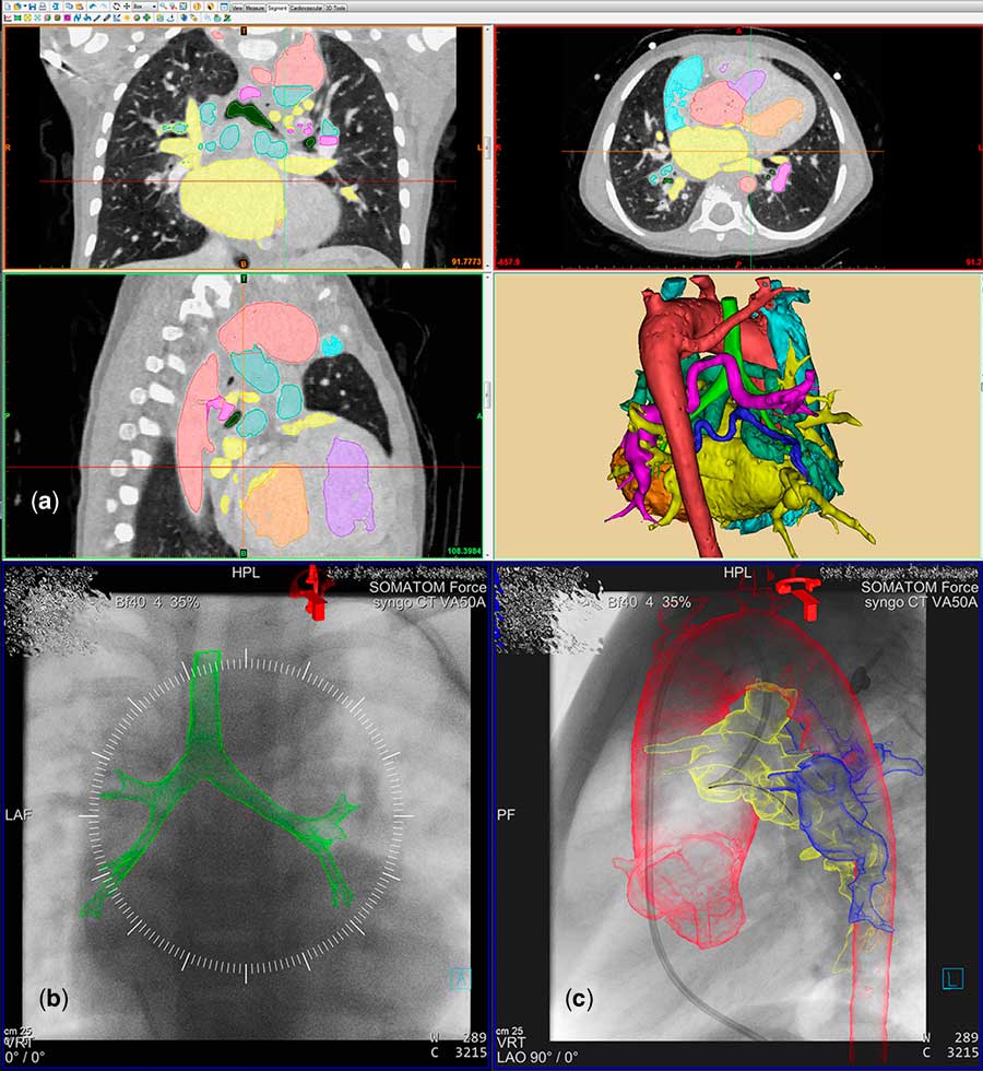
To visualise the meshes on top of fluoroscopic images, a transformation from their coordinate space into the coordinate space of the angiography system is required. This registration is computed in two steps. First, as soon as the first fluoroscopy is performed at roughly the target area of the intervention, a translation is computed that matches the centre of the volume and the iso-centre of the angiography system. This is the starting point for the second step, manual registration, in which the volume’s coordinate system can be translated and rotated to match the meshes with underlying structures. Figure 2 shows the process of segmentation and registration. In this study, a mesh of the trachea was used to match against the outline of the trachea in the fluoroscopic images.
Segmentation and registration of a 3D heart model for the use in catheterization of children with CHD. This figure shows the generation of a 3D-heart model and the following registration process in the cath lab, using the example of a 3-year old male patient with a DORV, atypical anatomy of the aortic arch, pulmonary atresia, stenotic ductus arteriosus and multiple MAPCAs. (a) Segmentation of the different anatomical structures (axial, coronary, sagittal). Generation of a 3D-model on the basis of a CT-dataset. (b) 2D-3D image fusion of the fluoroscopic image and the 3D-model using the tracheal bifurcation as fiducial marker. The outlines of the 3D-trachea (green) are matched with the visible parts of the trachea on the fluoroscopy. (c) During the catheterization, the displayed anatomical parts can be changed (show, hide, change color). In this picture we see the catheter finding its way antegrade through the ostium of the ductus. The model is very helpful in order to navigate correctly without injecting additional contrast medium. Red=aorta, yellow=ductus arteriosus, blue=MAPCA. 2D=two-dimensional; 3D=three-dimensional; CHD=congenital heart disease; DORV=double outlet right ventricle; MAPCA=major aorto-pulmonary collateral artery.

Manual corrections to the coordinate system registration can be performed throughout the procedure. For example, once a contrast injection is performed, the resulting images can be used to match meshes with the outline of their corresponding vessels. As long as the patient does not move relative to the table, the primary registration is completely sufficient and must not be adjusted.
Visualisation
To ensure that none of the devices (catheters, stents) are blocked from view, the Monaco prototype conceives of the meshes like this: If a part of the surface is parallel with the screen, it is transparent, if it is perpendicular, it is completely opaque. Anything in between becomes more or less opaque depending on the angle between the surface and the screen. This produces renderings of the meshes that show only the vessel borders very clearly, while hiding the rest. All visualisation properties of the meshes can be changed individually, such as colour, strength of the outline effect, overall opacity, and so on.
Quantitative and qualitative evaluation
Precision of the initial two- to three-dimensional registration was quantified by measuring the distance between the positions of the trachea-registered models and the subsequently adjusted images. This was performed on the basis of big contrast medium-filled vessels – aortic arch, pulmonary artery, lower hollow vein, upper hollow vein. Deviation was indicated as a percentage of the aforementioned distance in relation to the vessel diameter. This was performed with the software “Adobe Photoshop CS6” (Adobe Systems, Unites States of America) both on the A plane and the B plane, using adequate screenshots.
To assess the relevance of the three-dimensional navigation for each intervention or diagnostic catheterisation, a standardised five-point Likert scaleReference Likert 11 was used by the operator in agreement with a second paediatric cardiologist. If the intervention was not able to be performed appropriately without additional information using the three-dimensional model, the case was rated as “essential”. If three-dimensional navigation provided advantageous information to the already existing one, the case was deemed “very useful”. “Useful” was utilised if secondary information was available but with only marginal advantages compared with classic biplane angiographies. The case was rated as “not useful” if no helpful information was obtained, and if the additional information was deceptive it was rated as “misleading”.
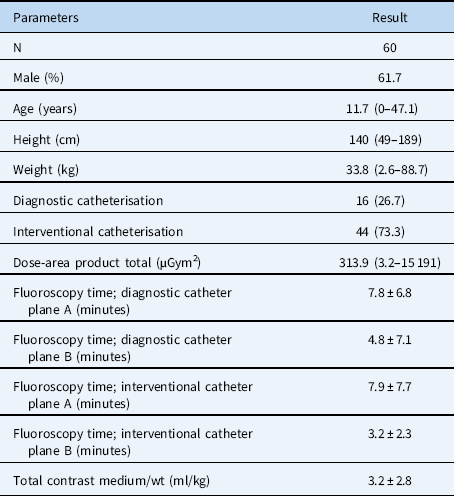
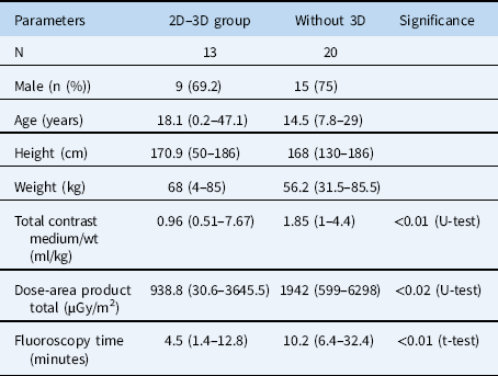
Demographic data and radiation dose (dose-area product) and the amount of used contrast dye and duration of the catheterisations were evaluated (Table 1). In all, 13 of our 60 patients underwent treatment on the aortic arch, and thus these cases were compared with a historical control group – 20 cases with aortic interventions between 2004 and 2011 without three-dimensional navigation. To analyse the collected data, we used the software package IBM® SPSS® Statistics 21.0 (IBM, Germany). We made an Independent Samples t-test (fluoroscopy time) and a non-parametric Mann–Whitney U-test (contrast medium and radiation dose). The level of significance was set at 0.05.
Demographic and procedural data.

Median (with range), mean±standard deviation, or number (percentage of total)
Results
Demographic and procedural data
In the 10-month study period, a total of 354 patients underwent procedures in our catheterisation suite for CHD. In 136 cases, we were able to use three-dimensional roadmapping, of which 60 catheterisations were performed with our prototype workstation. Median patient age was 11.7 years (range 0–47.1 years) and median weight was 33.8 kg (range 2.6–88.7 kg). Median dose-area product in the total investigation was 313.9 µGym2 (3.2–15 191.0) [median; (range)], used contrast dye was 2.0 ml/kg (0.0–8.6), and total fluoroscopy time was 8.2 min (1.4–44.4) (Table 1). No gender-based differences were observed in our evaluation.
In 28 cases (46.7%), we used a CT data set, in 30 cases (50%) an MRI, and in 2 cases (3.3%) a three-dimensional rotational angiography for generating the three-dimensional heart model. A total of 16 patients underwent a diagnostic procedure and 44 patients an interventional procedure. Of the 44, 16 were solely concerning the aorta – e.g. such as stent placement in the aortic isthmus or patent arterial duct. In all, 12 patients were treated on the pulmonary artery by stent implantation, balloon dilation, or transcatheter valve implantation. A total of 10 patients were treated otherwise – e.g. in terms of a myocardial biopsy, coiling of collateral vessels, or dilatation of a ventricular septal defect. A total of six patients received a treatment concerning more than one of these targets. The three-dimensional model was used in 44 cases (73.3%) for precise device positioning without repetitive control angiographies, and in all 60 cases (100%) to define the optimal C-arm angulation. In one case it was possible to completely perform a diagnostic catheterisation without using contrast dye (patient with a small atrial septal defect and a partial anomalous pulmonary venous connection).
Accuracy and benefit
The accuracy of two- to three-dimensional registration was sufficient and three-dimensional guidance was rated superior to conventional biplane imaging in all 60 cases. The value of the overlaid three-dimensional model was classified as “useful” in 10 cases (16.7%), “very useful” in 48 cases (80.0%), and “essential” in two cases (3.3%). Utility was never rated as “misleading” or “not useful”.
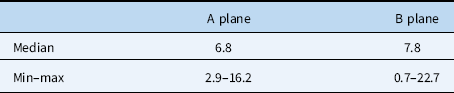
Precision of the initial two- to three-dimensional registration with the trachea as a fiducial marker was highly accurate in all cases. Median deviation was 6.84% on plane A (2.86–16.21) and 7.82% on plane B (0.69–22.74) (Table 2). Furthermore, we were able to register significantly smaller amounts of used contrast dye (p<0.01), lower levels of radiation dose (p<0.02), and less fluoroscopy time (p<0.01) during interventions concerning patients with aortic isthmus anomalies, compared with patients in the control group (Table 3).
Accuracy of the initial two- to three-dimensional registration with the trachea as a fiducial marker.

Deviation of the model given in percentage of the vessel diameter (aorta, pulmonary artery, superior caval vein, inferior caval vein)
Comparison of balloon dilatation or stenting of aortic isthmus anomalies with (two- to three-dimensional (2D–3D) group) and without 3D guidance (control group).

Median (min–max). Results compared with independent samples t-test (fluoroscopy time) and with non-parametric Mann–Whitney U-test. Level of significance 0.05
The expenditure of time cannot be assessed consistently. Generation of a three-dimensional model with our technique takes minutes – aortic arch, right ventricular outflow tract, pulmonary artery – to an hour – complex heart model with illustration of every anatomic structure, e.g. hypoplastic left heart syndrome. It also depends on the quality of the MRI or CT data set. Owing to the higher contrast to noise ratio of CT images, these data sets are easier to handle compared with MRI images.
Discussion
The high potential of three-dimensional guidance during catheter-based interventions to reduce radiation dose, contrast dye, and fluoroscopic time has been demonstrated in former investigations.Reference Stenger, Dittrich and Glockler 12 In our department, about 25% of all interventions are currently performed using three-dimensional overlay.
In our study, we demonstrated that the latest developments in three-dimensional imaging techniques used for catheterisation of children with CHD are highly beneficial and feasible in every respect. Segmented and tessellated heart models can be generated successfully from MRI and CT. All cases were rated “essential”, “very useful”, or “useful”, which shows the huge potential our image overlays possess for making interventions faster and safer and for reducing the amount of contrast dye used and radiation dose given. The comparison of the results of our study with a control group provided evidence supporting our initial hypothesis that especially routine interventions such as the treatment of the aortic arch can achieve a new level of efficiency regarding fluoroscopy time, radiation dose, and used contrast dye. In Stenger et alReference Stenger, Dittrich and Glockler 12 , this development is already portrayed clearly. Our data prove that three-dimensional navigation is about to expand its potential further.Reference Glockler, Halbfaß, Koch, Achenbach and Dittrich 4 , Reference Stangenberg, Shuja, Carelsen, Elenbaas, Wyers and Schermerhorn 13 , Reference Goreczny, Dryzek and Moszura 14 Additionally, other interventions, such as treatment of the pulmonary artery, percutaneous pulmonary valve replacement, or coiling of collateral vessels, will benefit from this new technique.Reference Hinrichs, Renne, Hoeper, Olsson, Wacker and Meyer 15 This is also shown by the rating of our cases. Decreasing numbers of three-dimensional rotational angiographies in our cath lab emphasise this valuable progress. The percentage of three-dimensional rotational angiographies performed in our catheterisation laboratory was reduced from 38% in 2014 to 33% in 2015 and finally to 23% in the first half of 2016 (January 2016–May 2016). Children are particularly sensitive to radiation exposureReference Bacher, Bogaert, Lapere, De Wolf and Thierens 16 , so this is important progress.
When overlaying information on fluoroscopic images, it is crucial that none of the devices (catheters, stents) are blocked from view. In Stenger et alReference Stenger, Dittrich and Glockler 12 , where volumetric data are used as well, consistent visibility is achieved by carefully designing transfer functions and providing an overall opacity to the rendered volume. However, this approach never leaves the devices completely visible. We are the first to use meshes in stereo-lithography format in the clinical routine. This allows for more control over the visualisation. The threshold-based and visually controlled segmentation ensures that the outline of the contrasted vessel correlates exactly with its diameter, while the precision of VRTs depends on the windowing of the fluoroscopy parameters and thus varies more.Reference Cody 17 , Reference Hammon, Rompel and Seuss 18 Being available at the beginning of the catheterisation, the three-dimensional models are able to provide a high-quality three-dimensional impression throughout the whole examination. The accuracy of the models was rated superior to previously utilised volume rendering.Reference Glockler, Halbfaß, Koch, Achenbach and Dittrich 4
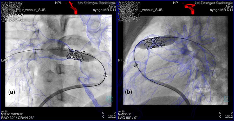
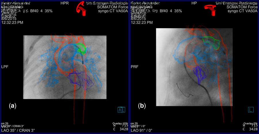
The initial registration with the trachea as fiducial marker was very accurate, with a deviation of <8%. Few manual corrections were sufficient to adjust the meshes to the correct position, so this has the potential to become a recommended procedural method. It has previously been described as a possibility to overlay three-dimensional data sets with exact precision.Reference Fagan, Truong and Jone 19 Furthermore, the segmentation is very helpful in order to depict the structures, which are the focus of the respective intervention, while hiding the rest. On the other hand, the displayed anatomical parts of the heart can be changed during the intervention, depending on the course of the catheterisation. Preliminary work includes not only the generation of the meshes but also contributes to reducing radiation exposure and fluoroscopy time, as the interventionalist is already familiar with the individual anatomy of the patient and can therefore work more safely and faster. Figures 3 and 4 illustrate how the three-dimensional models can be used during interventions concerning the pulmonary artery (Fig 3) and a patent ductus arteriosus (Fig 4).
Dilatation and stenting of the RPA. This figure shows an intervention on the pulmonary artery in a 21-year old male patient with a stenosis of the RPA and LPA. The 3D-model was generated from a previously acquired MRI-dataset. A stent was implanted with the help of the model, which displayed the location of the stenosis exactly. Blue=pulmonary artery, A=A tube, B=B-tube. 3D=three-dimensional; LPA=left pulmonary artery; MRI=magnetic resonance imaging; RPA=right pulmonary artery.

Stenting of PDA in a patient with HLHS. This figure shows the act of trying to find the ostium of the PDA in a patient with HLHS. A stent was implanted in order to complete the Gießen-I-procedure. The catheter with the stent was placed exactly at the right position without repetitive contrast dye injection. The 3D-heart model was generated from a CT dataset. Red=aorta; dark blue=hypoplastic left ventricle; light blue=pulmonary artery; green=patent arterial duct, A=A-tube; B=B-tube 3D=three-dimensional; CT=computed tomography; HLHS=hypoplastic left heart syndrome; PDA=patent arterial duct.

On the basis of our long-standing experience in overlaying three-dimensional heart models during catheterisations, we can confidently state that our new approach to two- to three-dimensional registration with segmented heart models yields many advantages. Overlaying three-dimensional heart models with fluoroscopic images can now be performed without an intraprocedurally acquired three-dimensional rotational angiography. Another solution has been described by Dori et al., where X-ray magnetic resonance fusion allows for use of three-dimensional data during cardiac catheterisation.Reference Dori, Sarmiento and Glatz 20 Moreover, the model is available from the beginning of the intervention. It can be displayed not only on the A plane but also on the B plane, which is highly beneficial to safe interventions and has not been possible so far. Our experience indicates that time and effort of the whole procedure, including the preparation of the meshes, is less than using traditional three-dimensional rotational angiography. In addition, the three-dimensional models help to find an ideal projection angle.Reference Glockler, Halbfaß, Koch, Achenbach and Dittrich 4 They offer a more realistic perception of the anatomical situation during and also before an intervention, which is also beneficial for less experienced interventionalists during their education. Catheterisation time can be reduced consequently.Reference Glockler, Halbfaß, Koch, Achenbach and Dittrich 4 , Reference Goreczny, Dryzek and Moszura 14
The intensive preoccupation with the individual anatomy is advantageous for the interventionalists, as they can already choose suitable equipment. Control angiographies during stent positioning can be reduced. Yet, it is a quite young procedure. Although the models are very exact and provide an accurate description of the position, we do not dispense with a final angiography before stent positioning.
One current limitation lies with the alignment process during a catheterisation. Currently, it must be executed by a third person from an extra workstation. The interventionalist himself can neither move the three-dimensional-volume if necessary nor can he control the displayed or hidden parts of the heart model, which are in the focus of the respective examination. Future software developments will try to overcome this issue and also focus on further intents like electrocardiography-triggered compensation of systole and diastole.
Another limitation of the study lies within the technological progress itself. When comparing our results with the historical control group, we did not consider the improvement of the catheterisation laboratories and thus the reduction of radiation going hand-in-hand with that development.
Conclusion
Segmented three-dimensional heart models are optimal for three-dimensional navigation during catheterisation of CHD. Segmented heart models provide a conspicuous visual advantage. Used devices are perfectly visible throughout the whole catheterisation. At the same time, vessel borders of the model are shown very clearly. The interventionalist receives an optimal three-dimensional impression because of the adjusted opacity. Our study shows that two- to three-dimensional image fusion can be applied successfully using the trachea as fiducial marker. Contrast medium and radiation dose can be reduced effectively.
Financial Support
This research received no specific grant from any funding agency or commercial or not-for-profit sectors.
Conflicts of Interest
None.
Ethical Standards
The authors assert that all procedures contributing to this work comply with the ethical standards of the relevant national guidelines on human experimentation and the Helsinki Declaration of 1975, as revised in 2008, and has been approved by the institutional committees (Ethics Commission of the University Erlangen-Nuremberg).
Acknowledgements
The present work was performed in (partial) fulfilment of the requirements for N.E. to obtain the degree – “Dr med” – from the Friedrich-Alexander University Erlangen-Nürnberg (FAU).