Introduction
Human schistosomiasis is one of the most important tropical diseases (Steinmann et al., Reference Steinmann, Keiser, Bos, Tanner and Utzinger2006) and ranks second to malaria in terms of prevalence, morbidity and mortality rates (WHO Expert Committee, 2002). The disease is caused by any one of six species of the trematode worm Schistosoma; however, infections with Schistosoma haematobium, S. japonicum and S. mansoni account for most cases (Dkhil et al., Reference Dkhil, Abdel Moneim and Al-Quraishy2014). During S. mansoni infection, embolization of eggs occurs from the intestine to the liver through the portal vessels. These eggs stimulate granulomatous inflammatory reactions that heal with liver fibrosis (Colley & Secor, Reference Colley and Secor2014). The inflammatory cells in these granulomas become activated and secrete reactive oxygen species (ROS) (Halliwell & Gutteridge, Reference Halliwell and Gutteridge2015). Failure of the antioxidant enzymes to manage the excess ROS leads to oxidative stress (Othman et al., Reference Othman, Shoheib, Abdel-Aleem and Shareef2008). Additionally, the trapped eggs in hepatic sinusoids initiate a vicious cycle of inflammation, fibrosis, vascular obstruction and hypoxia (Andrade, Reference Andrade2004). Thereafter, hypoxia and ROS together stimulate the process of neoangiogenesis (Coulon et al., Reference Coulon, Heindryckx, Geerts, Van Steenkiste, Colle and Van Vlierberghe2011; Elpek, Reference Elpek2015), which takes place during the repair of injured tissues (Semenza, Reference Semenza2012).
Despite the global burden and seriousness of schistosomiasis, the current treatment of this disease relies only on praziquantel (PZQ), which has been used extensively over the last four decades and is the cornerstone of the current World Health Organization (WHO) control strategy against schistosomiasis (Bergquist et al., Reference Bergquist, Utzinger and Keiser2017). Unfortunately, as a result of this extensive and prolonged dependence on praziquantel, tolerant/resistant strains of S. mansoni have emerged (Melman et al., Reference Melman, Steinauer and Cunningham2009). Moreover, we are still years away from the possible application of a vaccine for schistosomiasis. Thus, the discovery of a novel drug to replace or potentiate PZQ is now an urgent priority. Herbal extracts are considered to be an important source for such discoveries because of their variety of chemical components and biomedical activities (Bergquist et al., Reference Bergquist, Utzinger and Keiser2017).
One plant that has been used in traditional medicine is pumpkin, which belongs to the Cucurbitaceae family. This family includes many members; however, Cucurbita pepo L., C. maxima and C. moschata are the most cultivated throughout the world (Caili et al., Reference Caili, Huan and Quanhong2006). Pumpkin seed oil is a rich source of a variety of bioactive compounds, such as essential fatty acids, tocopherols, sterols, polysaccharides, phenolic glycosides, omega-3 and omega-6 fatty acids, vitamins E, A and C, zinc and other trace minerals (Glew et al., Reference Glew, Glew, Chuang, Huang, Millson, Constans and Vanderjagt2006). Moreover, it has been found to have medical properties, as an antidiabetic, antihypertensive, antibacterial, antioxidant and anti-inflammatory agent (Caili et al., Reference Caili, Huan and Quanhong2006; Bardaa et al., Reference Bardaa, Halima, Aloui, Mansour, Jabeur, Bouaziz and Sahnoun2016).
Therefore, this study was designed to evaluate Cucurbita pepo L. seed oil alone and combined with PZQ against S. mansoni in experimentally infected mice through measurement of parasitological parameters such as worm burden, tissue egg load, oogram pattern, and evaluation of the morphological alterations of S. mansoni adult worms by scanning electron microscopy (SEM). Also, the liver condition was assessed by histopathological examination of hepatic granulomas, fibrosis and neoangiogenesis. Moreover, serum liver enzyme activities were measured and the oxidative stress was evaluated by determination of malondialdehyde amount and superoxide dismutase activity in liver tissue homogenates.
Material and methods
Animals and infection
Pathogen-free CD-1 Swiss albino mice, 6–8 weeks of age and with a mean weight of 20 ± 2 g, were purchased from the Schistosome Biological Supply Center at Theodor Bilharz Research Institute (SBSC, Imbaba, Giza, Egypt). Cercariae were obtained from Biomphalaria alexandrina snails experimentally infected with human S. mansoni (Egyptian strain) at the SBSC. Shedding of cercariae was induced by placing the snails (four weeks after infection) in distilled water and exposing them to artificial light at 28°C for 2 h (Tekwu et al., Reference Tekwu, Bosompem and Anyan2017). Each mouse was infected with 70 ± 5 freshly shed cercariae through subcutaneous injection in the abdominal skin, as described previously (Liang et al., Reference Liang, Bruce and Boyd1987). Mice were then transported to the Experimental Research Unit at the Faculty of Science (Menoufia, Egypt), where they were housed under controlled conditions of temperature and humidity (25 ± 2°C, 70%), with free access to standard food and water.
Praziquantel and Cucurbita pepo seed oil
Praziquantel (Discocide® 600 mg; Egyptian International Pharmaceutical Industries Company (EIPICO), Egypt) was given orally in a full curative dose of 500 mg/kg/day for two successive days, according to Gönnert & Andrews (Reference Gönnert and Andrews1977), commencing six weeks after infection, in the form of an aqueous suspension in 2% Cremophor EL (Sigma-Aldrich, St. Louis, USA). Cucurbita pepo seed oil (NOW® pumpkin seed oil 1000 mg; Health Group Inc., USA) was given orally at a dose of 50 mg/kg/day for four weeks, commencing four weeks post infection, in the form of an aqueous suspension in 2% Cremophor EL.
Experimental design
Forty mice were used in this study; eight healthy non-infected mice were assigned to the control group (GI) and 32 S. mansoni-infected mice were divided randomly into the following four groups of eight mice each (n = 8): GII was the infected non-treated group (infected control), GIII was treated with PZQ, GIV was treated with PSO, and GV was treated with PZQ combined with PSO. Two weeks after administration of the last doses of the tested drugs, the experiment was terminated.
Sampling
Under ether anaesthesia, blood sampling was performed on each mouse via a cardiac puncture, and then mice were sacrificed by cervical dislocation. Sera were separated and stored at −80°C until used. All the infected mice were subjected to perfusion of the portal and mesenteric veins and estimation of the worm burden, and the egg load in liver and ileum tissues, and the oogram pattern of the developmental stages of the eggs was investigated. After recording worm counts, all the male worms recovered from the treated groups, and samples of those retrieved from the control group, were fixed in 4% glutaraldehyde with sodium cacodylate for 24 h for ultrastructural morphological examination by SEM. Immediately after perfusion, a 0.5 g sample from each liver was homogenized in 2.5 volumes (w/v) cold potassium phosphate buffer of pH 6.5 and centrifuged at 3500 rpm for 25 minutes at 4°C; the supernatants were then stored at −80°C for determination of malondialdehyde peroxidase (MAD) content and superoxide dismutase (SOD) activity. Additionally, other samples of liver tissues were fixed in 10% buffered formalin for histopathological and immunohistochemical studies.
Assessment of parasitological criteria
Perfusion of the portal and mesenteric veins with sterile physiological saline (0.9% NaCl, w/v) was performed according to Smithers & Terry (Reference Smithers and Terry1965). The retrieved male, female and coupled worms were counted under a stereomicroscope, and the worm burden for each group was calculated. Samples from the perfused liver and ileum of each mouse were taken, weighed and left for digestion in 5% KOH at 37°C for 16 h. The eggs were counted at a magnification of ×40 and the mean number of eggs/g of liver and intestine was calculated (Herbert et al., Reference Herbert, Orekov and Roloson2010). The oogram pattern was investigated according to Mati & Melo (Reference Mati and Melo2013). Briefly, three fragments of the distal part of the ileum of each animal were washed in 0.9% saline solution and dried on filter paper. Each fragment was pressed between two slides and examined under a light microscope. Eggs were counted and categorized according to their developmental stage as immature (the miracidium occupies less than two-thirds of the shell), mature (the miracidium occupies the whole shell) or dead (opaque, with retracted, granular, dead miracidium).
Scanning electron microscopy
According to Matos-Rocha et al. (Reference Matos-Rocha, Cavalcanti and Veras2016), the adult male worms fixed in 4% glutaraldehyde were washed in 0.1 m sodium cacodylate buffer (pH 7.2) and post-fixed with 1% osmium tetroxide for an hour. Thereafter, the samples were dehydrated in ascending concentrations of ethanol, dried, mounted on metal stubs, and coated with gold vapour using a sputter coater (JEOL TFC-1100, Tokyo, Japan). The samples were examined and photographed using a scanning electron microscope (JEOL JSM 5300, Tokyo, Japan) at the Faculty of Medicine, Tanta University, Egypt.
Histopathological study
The liver tissues fixed in 10% buffered formalin were processed to paraffin blocks, and five liver sections (5 μm thick) from each animal were prepared, cutting the sections every 250 μm to avoid measuring the same granuloma twice. Then the sections were stained with haematoxylin and eosin (H & E) (Carleton et al., Reference Carleton, Drury and Wallington1980). For measurement of the granulomas and detection of other histopathological changes, the sections were examined under a light microscope equipped with an ocular micrometer (Olympus BX41, Olympus Corporation, Tokyo, Japan) and a digital camera (Olympus E420DC7, 4V, Olympus Corporation, Tokyo, Japan). For each mouse, the mean diameter of each granuloma was calculated by measuring two diameters perpendicular to each other, according to Von Lichtenberg (Reference Von Lichtenberg1962). Additionally, serial liver sections, 8 μm thick, were stained for 30 minutes with Picro Sirius red (PSR; Polysciences, Inc., Warrington, PA, USA) according to the manufacturer's instructions, to stain the collagen bundles. The sections were examined under a polarized light microscope (Zeiss Axioplan, Carl Zeiss AG, Oberkochen, Germany) and the results were interpreted according to Drifka et al. (Reference Drifka, Loeffler and Mathewson2016); i.e. thick collagen fibres were stained orange to red, whereas thin fibres were stained green to yellow.
Assessment of angiogenesis by CD34+ immunohistochemical staining
To evaluate the process of angiogenesis, the marker of endothelial cells Anti-CD34+ was used. The procedure was performed as described by Abdel Fattah & Ahmed (Reference Abdel Fattah and Ahmed2012); briefly, paraffin sections (4 μm) were fixed on polylysine-coated slides, dried overnight, deparaffinized and dehydrated. Two drops of hydrogen peroxide blocking serum were added for five minutes at room temperature (RT). Anti-CD34+ primary antibody (Anti-CD34+ antibody [EP373Y] ab81289, Abcam, Cambridge, UK) was added to the sections and the sections were incubated at RT for 2 h, then washed in a phosphate buffer solution (PBS) of pH 7.6. The biotinylated secondary antibody (Dako, Glostrup, Denmark) was added to the sections for 30 minutes at RT, followed by washing in the same buffer. Peroxidase-labelled streptavidin was added for 20 minutes at RT, then the slides were washed, incubated with substrate chromogen (DAB) mixture for 10 minutes, rinsed with distilled water and counterstained with Mayer's haematoxylin (Sigma Aldrich, St. Louis, USA). Finally, the slides were washed in tap water, dehydrated in absolute alcohol, cleared in xylene, mounted with Canada balsam and covered.
Evaluation of the oxidative stress in liver tissue
Malondialdehyde (MDA) concentration was measured in liver homogenates by thiobarbituric acid reaction, according to the method of Ohkawa et al. (Reference Ohkawa, Ohishi and Yagi1979). The optical density of the reaction was measured using a spectrophotometer at 530 nm, and the concentrations of MDA in liver tissues were expressed as nmol/g protein. Superoxide dismutase activity in liver tissue homogenates was estimated by the method of Nishikimi et al. (Reference Nishikimi, Rao and Yagi1972), using nitroblue tetrazolium, nicotinamide adenine dinucleotide (NADH) and sodium pyrophosphate buffer. The reaction was read by a spectrophotometer at 560 nm, and SOD activity was expressed as units/mg protein.
Measurement of serum liver enzymes
The activities of alanine aminotransferase (ALT) and aspartate aminotransferase (AST) were estimated in mice sera using the available commercial kits (Sigma-Aldrich, St. Louis, USA). The reactions were read by a spectrophotometer at 450 nm and the enzyme activities were expressed as units/l. Also, the serum alkaline phosphatase (ALP) level was measured using alkaline phosphatase colorimetric assay kits (Abcam, Cambridge, UK). The reaction was read by a spectrophotometer at 405 nm, and ALP activity was expressed as units/l.
Statistical analysis
The data were collected, tabulated, analysed using SPSS v. 22.0 (IBM, Armonk, USA), and expressed as mean ± SD. The data were tested for normality using the Kolmogorov–Smirnov test. A one-way analysis of variance (ANOVA) test was applied to investigate the significance of normally distributed data, and the Kruskal–Wallis test was used to test the significance of nonparametric data. Each test was followed by a post-hoc test to determine the significance of data of each group in relation to the others. Results with P ≤ 0.05 were considered significant.
Results
Parasitological criteria
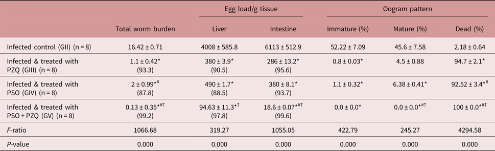
The results revealed significant reductions (P < 0.001) in the total worm burden and liver and intestinal egg load, with significant increases in the percentages of dead ova in all the treated groups in comparison with the infected control group (GII). Treatment of S. mansoni-infected mice with PSO alone (GIV) resulted in significant reductions in total worm burden (87.8%), liver egg load (88.5%) and intestinal egg load (93.7%), and a significant increase in the percentage of dead ova (92.52%). However, treatment with PZQ alone (GIII) resulted in higher percentages than those of PSO (GIV), with a significant difference between both groups. Administration of PSO combined with PZQ (GV) resulted in the highest percentage reduction of worm burden (99.2%), and tissue egg load in liver (97.8%) and intestine (99.6%), with a significant increase in the percentage of dead ova (100%). Moreover, the results for this group were significant (P < 0.05) in comparison with those of PZQ-treated (GIII) and PSO-treated (GIV) groups (table 1 and supplementary table S1).
Effect of PSO treatment with and without PZQ on the total worm burden, egg load/g tissue, and oogram pattern in mice experimentally infected with Schistosoma mansoni.

Data are expressed as mean ± SD.
Numbers in parentheses indicate the percentage of reduction, which was calculated as follows:
% reduction = (value of infected controls−value of treated mice)/(value of infected controls) × 100
* indicates significance (P < 0.05) of the value vs GII, # indicates significance vs GIII and † indicates significance vs GIV by one-way ANOVA followed by a post-hoc test.
Ultrastructure morphological changes in SEM study
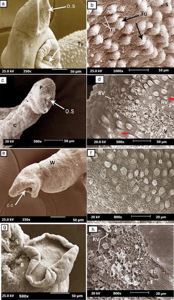
SEM examination of the adult male worms recovered from the infected control group revealed intact oral and ventral suckers (fig. 1a), and intact teguments with numerous large tubercles, with spines and deep tegumental ridges in the areas between the tubercles (fig. 1b). The worms recovered from the PZQ-treated group showed deformity of the oral suckers (fig. 1c), with wide areas of tegumental damage manifested in the loss of integrity of the tubercles and spines, and rupture of several blebs (fig. 1d). Wrinkles (fig. 1e), loss of tubercles, spines and tegumental ridges, and the formation of blebs (fig. 1f) were observed on the adult worms retrieved from the PSO-treated group, whereas the worms retrieved from the combined PSO–PZQ-treated group showed marked deformity of the oral suckers (fig. 1g) and more obvious tegumental damage in the form of extensive peeling and erosion (fig. 1h).
Scanning electron microscopy (SEM) micrographs of adult Schistosoma mansoni worms. (a, b) Micrographs of worms recovered from the untreated control group, showing (a) the anterior end of an adult male with an intact oral sucker (o.s), a ventral sucker (v.s) and tubercles on the dorsal surface (Tu) (×350), and (b) well-developed tubercles (Tu) with spines (S) and apparent tegumental ridges (TR) (×1000). (c, d) Micrographs of worms retrieved from the PZQ-treated group: (c) the anterior end of an adult male with a contracted and deformed oral sucker (o.s), a rough, corrugated tegument (C) and peeling (P) (×500); (d) the tubercles show loss of spines (red arrow), with appearance of vesicles (V), ruptured vesicles (RV) and erosion (E) (×500). (e, f) Micrographs of adult worms recovered from the PSO-treated group: (e) tegumental wrinkles (W) at the anterior region of the worm (×350); (f) the tegument shows loss of the normal architecture and deformity of the tubercles, loss of spines, disappearance of the tegumental ridges, and formation of blebs (B) (×800). (g, h) Micrographs of adult worms recovered from the combined PSO–PZQ-treated group: (g) obvious destruction of the anterior region, sloughing of the tegument and bleb formation (B) (×500); (h) severely destructed tegument, with vesicles (V), ruptured vesicles (RV), erosions (E) and exposure of the tegumental layers (TL) (×800).

Histopathological study
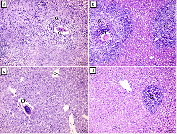
Histopathological examination of liver sections from the infected control group (GII) revealed loss of the normal architecture, disorganization of hepatic cords and the presence of large fibrocellular granulomas, with a mean diameter of 308.63 ± 31.85 µm (table 2). These granulomas comprised activated macrophages, epithelioid cells, lymphocytes, and many fibroblasts encircling the trapped S. mansoni eggs with abundant fibrous tissues (fig. 2a). This fibrous tissue was formed of thick collagen bundles that appeared dark red when stained with PSR (supplementary fig. S1a). Treatment with PZQ (GIII) resulted in a significant reduction (59.7%) in granuloma diameters (table 2); these fibrocellular granulomas were less defined, with degenerated eggs and less fibrosis (fig. 2b), which appeared faint red when stained with PSR (supplementary fig. S1b). Administration of PSO (GIV) reduced the granuloma diameters by 62.6%, with a significant difference in comparison with PZQ (table 2). These granulomas were less defined, with minimal fibrosis and inflammatory reactions, and occasional absence of the inflammatory reaction around the degenerated ova (fig. 2c); moreover, the collagen fibres were markedly decreased and stained greenish to yellow by PSR, denoting their conversion from thick to thin (supplementary fig. S1c). The combined PSO–PZQ treatment (GV) reduced the granuloma diameters by 89.2%, which was significant in comparison with GII, GIII and GIV (table 2). The granulomas of this group were devoid of eggs, less defined and comprised inflammatory cells with minimal fibrosis (fig. 2d), rendered as scanty, greenish thin collagen fibres by PSR stain (supplementary fig. S1d).
Haematoxylin and eosin stained liver sections from S. mansoni-infected mice groups. (a) Infected control group, showing an inflammatory granulomatous (G) lesion with condensed fibrous connective tissue surrounding an S. mansoni ovum (O) with a visible miracidium. (b) PZQ-treated group, showing inflammatory granulomatous lesions with condensed fibrous connective tissue and degenerated ovum. (c) PSO-treated group, showing less defined fibrous and inflammatory granulomatous reaction around the ovum. (d) Combined PSO–PZQ-treated group, showing inflammatory granuloma with minimal fibrosis and absence of egg (×400).

Effect of PSO treatment with and without PZQ on the diameters of hepatic granulomas in mice experimentally infected with S. mansoni. F-ratio = 242.73; P-value = 0.000.

Data are expressed as mean ± SD. Numbers in parentheses indicate the percentage of reduction, which was calculated as follows:
% reduction = (value of infected controls−value of treated mice)/(value of infected controls) × 100
* indicates significance (P < 0.05) of the value vs GII, # indicates significance vs GIII and † indicates significance vs GIV by one-way ANOVA followed by a post-hoc test.
Effect of PSO on angiogenesis in the liver
Examination of liver sections from different groups stained immunohistochemically with anti-CD34+ revealed the almost absence of CD34-positive cells in liver sections of the normal control group (GI) (supplementary fig. S2a). In the infected control (GII) (supplementary fig. S2b), PZQ-treated (GIII) (supplementary fig. S2c) and PSO-treated (GIV) groups (supplementary fig. S2d), angiogenesis was present within the granulomas, forming vascular collars around some granulomas, and in the intergranulomatous spaces. In the combined PSO–PZQ-treated group (GV), there was evident angiogenesis and vascular remodelling associated with a significant reduction of the granuloma size and restoration of normal liver architecture (supplementary fig. S2e).
Effect of PSO on hepatic lipid peroxidation and antioxidant SOD activity
In the infected control group there was a significant increase in the amount of MDA, associated with a significant decrease in SOD activity when compared with the normal control group (GI). In all the treated groups there were significant decreases in MDA and increases in SOD activity, and the values of the combined PSO–PZQ-treated group (GV) were almost normal (fig. 3 and supplementary table S2).
Effect of PSO treatment with and without PZQ on (a) malondialdehyde and (b) superoxide dismutase concentrations in liver homogenates of mice experimentally infected with S. mansoni. Data are expressed as mean ± SD; eight mice per group; ‡ indicates significance (P < 0.05) of the value vs GI (normal control, NC), * indicates significance vs GII (infected control, IC), # indicates significance vs GIII (PZQ-treated, PZQ) and † indicates significance vs GIV (PSO-treated, PSO) by one-way ANOVA followed by a post-hoc test.

Effect of PSO on liver function tests
In the infected control group (GII) there were significant increases (P < 0.001) in the serum levels of ALT, AST and ALP enzymes in comparison with the normal control group (GI). In all the treated groups there were significant reductions in the levels of these enzymes, and the values of the combined PSO–PZQ-treated group were almost normal (fig. 4 and supplementary table S2).
Effect of PSO treatment with and without PZQ on the liver enzymes aspartate aminotransferase (AST), alanine aminotransferase (ALT) and alkaline phosphatase (ALP) of mice experimentally infected with S. mansoni. Data are expressed as mean ± SD, eight mice per group; ‡ indicates significance (P < 0.05) of the value vs GI (normal control, NC), * indicates significance vs GII (infected control, IC), # indicates significance vs GIII (PZQ-treated, PZQ) and † indicates significance vs GIV (PSO-treated, PSO) by one-way ANOVA followed by a post-hoc test.

Discussion
Despite the global importance and seriousness of schistosomiasis, its treatment depends on PZQ only. Unfortunately, because of the extensive use of PZQ, resistant strains of S. mansoni have emerged. Thus the discovery of novel drugs or therapeutic regimens to treat schistosomiasis is an urgent priority. Herbal extracts are important sources for such discoveries because of their bioactive compounds; therefore, this study was designed to evaluate the therapeutic effects of Cucurbita pepo L. seed oil alone or combined with PZQ against S. mansoni in an experimental mouse model.
The administration of PSO to treat S. mansoni-infected mice resulted in significant reductions in worm burden, and liver and intestinal egg load, with a significant increase in the percentage of dead eggs in the oogram pattern; however, the reference drug PZQ was more effective than PSO alone. Combined PSO–PZQ treatment resulted in the highest reduction percentage. This anthelmintic effect of PSO could be attributable to various secondary metabolites, such as cucurbitacin B, cucurbitin, saponins, sterols, curcumosin, berberine and palmatine, present in the seeds of Cucurbita species (Grzybek et al., Reference Grzybek, Kukula-Koch, Strachecka, Jaworska, Phiri, Paleolog and Tomczuk2016). Moreover, berberine has been reported as an antischistosomal agent with the ability to reduce infection-induced liver damage and oxidative stress (Dkhil et al., Reference Dkhil, Abdel Moneim and Al-Quraishy2014). The results of the current study were in agreement with previous studies that described the anthelmintic effect of pumpkin seeds against Heterophyes heterophyes (Mahmoud et al., Reference Mahmoud, Basiouny and Dawoud2002), Fasciolopsis buski, Ascaris lumbricoides and Hymenolepis diminuta (Iqbal et al., Reference Iqbal, Nadeem, Khan, Akhtar and Waraich2001).
As Schistosoma survival depends on the tegument, a vital structure concerned with host immune evasion, nutrient absorption, metabolism and the selective uptake of drugs (Faghiri & Skelly, Reference Faghiri and Skelly2009; Pereira et al., Reference Pereira, Padilha, Lima-Filho and Chaves2011), the ultrastructural alterations in the tegument of S. mansoni adult worms recovered from the treated mice by SEM were studied. Similar to previous studies by Matos-Rocha et al. (Reference Matos-Rocha, Cavalcanti and Veras2016) and El-Sayad et al. (Reference El-Sayad, Abu Helw, El-Taweel and Aziz2017), the worms recovered from PZQ-treated mice showed severe focal damage, deformity of the oral suckers, and wide areas of tegumental damage. Treatment with PSO alone resulted in some sort of tegumental damage, with the loss of some tubercles and spines, which could be attributed to the secondary metabolites in PSO (Dkhil et al., Reference Dkhil, Abdel Moneim and Al-Quraishy2014). The combined PSO–PZQ treatment resulted in marked deformity of the oral suckers, and more obvious tegumental damage that might be attributed to the synergy of PZQ and PSO antiparasitic actions.
These morphological and ultrastructural changes could explain the simultaneous reductions in worm burden and in the egg load in the liver and intestinal tissues of the treated mice, as well as the increases in the percentages of dead eggs. Severe tegumental damage induced by an antischistosomal drug is irreversible and leads to exposure of the Schistosoma antigens (Harnett & Kusel, Reference Harnett and Kusel1986) and disappearance of their immunological mask, making them easily recognized and attacked by the host immune system. Moreover, the morphological alterations of the oral and ventral suckers restrict feeding of the damaged worms, which results in their death (Shaw & Erasmus, Reference Shaw and Erasmus1987).
In this study, administration of PZQ improved the pathological changes, and reduced granuloma sizes and fibrosis. These findings are in agreement with Liang et al. (Reference Liang, Luo and Yuan2011), who reported the reversal of liver fibrosis in murine schistosomiasis after treatment with PZQ, and attributed their results to the removal of adult Schistosoma worms and the subsequent reduction of egg deposition. Moreover, they reported an anti-inflammatory effect of PZQ. Therefore, PZQ seems to exert dual effects on S. mansoni infection by eliminating the parasites and stimulating the immune response of the host (Pinlaor et al., Reference Pinlaor, Hiraku and Yongvanit2006). This dual action may explain the abundance of inflammatory cells and the diminished fibrosis in liver granulomas of the PZQ-treated group observed in this study. Contrary to our results, S. mansoni eggs and hepatic granulomas persisted for one year after treatment with PZQ in a previous study (Cheever et al., Reference Cheever, Macedonia, Deb, Cheever and Mosimann1992). These contradictory results could be explained by the negative correlation between the ability of an antischistosomal treatment to reverse fibrosis and the grade of liver pathology that already exists at the time of commencement of treatment (Chai, Reference Chai2013).
In the current study, treatment of S. mansoni-infected mice with PSO resulted in an obvious reduction in granuloma sizes, and conversion of collagen fibres from thick to thin as determined by PSR stain. These results were significant in comparison with those of the infected control and PZQ-treated groups, although PZQ showed more significant effects regarding worm burden, tissue egg load, and the oogram pattern. This antifibrotic effect of PSO may be attributable to its content of several antioxidant components, such as phenolic compounds (Andjelkovic et al., Reference Andjelkovic, Van Camp, Trawka and Verhé2010), tocopherols (vitamin E) (Rezig et al., Reference Rezig, Chouaibi, Msaada and Hamdi2012), L-arginine and selenium, which protect against free radical-mediated lipid peroxidation (Dhawan et al., Reference Dhawan, Handu, Nain and Ganguly2005). Moreover, a synergy between PZQ and PSO could explain the most significant reductions of granuloma diameters and fibrosis, with complete eradication of the degenerated eggs, which were detected in the combined PSO–PZQ-treated group. This is in accordance with Othman et al. (Reference Othman, Shoheib, Abdel-Aleem and Shareef2008), who reported the amelioration of S. mansoni-induced liver fibrosis by a combined PZQ–antioxidant treatment.
In accordance with previous studies by Dkhil et al. (Reference Dkhil, Abdel Moneim and Al-Quraishy2014) and Al-Olayan et al. (Reference Al-Olayan, El-Khadragy, Alajmi, Othman, Bauomy, Ibrahim and Moneim2016), the results of the infected control group in the present study revealed significant increases in serum ALT, AST and ALP activities, associated with a significant rise of MAD products and reduction of SOD activity in liver homogenate, coinciding with massive granulomatous reactions. This correlation between the degree of oxidative stress and the increased activity of serum ALT may be attributable to the peroxidative damage of the lipid content of the hepatocyte cell membranes, which leads to leakage of the liver enzymes (De Maria et al., Reference De Maria, Colantonl, Fagiuoli, Liu, Rogers, Farinati, Van Thiel and Floyd1996). On the other hand, there were significant improvements in the oxidative stress markers and serum liver enzymes in all treated groups. Furthermore, in the combined PSO–PZQ-treated group there was a restoration of almost normal values concomitant with the resolution of fibrosis.
Generally, hypoxia is the main inducer of angiogenesis (Elpek, Reference Elpek2015); however, other factors existing during schistosomiasis may also contribute to this process, such as the release of ROS by the activated Kupffer cells, and the inflammatory mediators secreted by hepatic stellate cells (Coulon et al., Reference Coulon, Heindryckx, Geerts, Van Steenkiste, Colle and Van Vlierberghe2011). In this study, neoangiogenesis was prominent in the infected control mice because of the persistence of eggs in the hepatic sinusoids, which initiates a vicious cycle of inflammation, healing with fibrosis, vascular obstruction, and hypoxia (Andrade, Reference Andrade2004). Angiogenesis was also obvious in the PZQ, PSO and PSO–PZQ-treated groups, although granuloma sizes, structure, fibrosis, oxidative stress and liver functions were improved; however, in the latter group there was evidence of tissue remodelling and restoration of the normal architecture, with almost disappearance of fibrosis. Accordingly, in agreement with Kanse et al. (Reference Kanse, Liang, Schubert, Haas, Preissner, Doenhoff and Dennis2005), we suggest that angiogenesis takes place during S. mansoni infection to maintain the hepatic blood flow that was decreased by fibrosis, and, in agreement with Abdel Fattah & Ahmed (Reference Abdel Fattah and Ahmed2012), angiogenesis occurs after treatment as part of the tissue repair process.
In conclusion, administration of PSO to treat S. mansoni infection in an experimental mouse model showed promising schistosomicidal effects, with significant reductions in worm burden and tissue egg loads, and significant increases in the percentage of dead eggs. Also, PSO induced ultrastructure morphological changes in adult worms, and improved granuloma size, structure and fibrosis. This improvement was associated with a reduction in lipid peroxidation, an increase in antioxidant activity, and reductions in serum liver enzyme values. However, regarding these criteria, the results obtained with PZQ were significantly better than those with PSO alone, except for fibrosis and oxidative stress. The results obtained with the combined PSO–PZQ treatment were the most significant, because of a synergy of the schistosomicidal effect of both drugs and the potent antioxidant and antifibrotic effects of PSO. Accordingly, during treatment of S. mansoni infection with PZQ, PSO could be considered as an adjuvant therapy to potentiate the antiparasitic effects of PZQ and eliminate infection-induced fibrosis, oxidative stress and liver damage.
Supplementary material
To view supplementary material for this article, please visit https://doi.org/10.1017/S0022149X18000317
Financial support
This research did not receive any specific grant from funding agencies in the public, commercial or not-for-profit sectors.
Conflict of interest
None.
Ethical standards
The authors assert that all procedures contributing to this work comply with the international ethical guidelines on the care of laboratory animals, and the study was approved by the Scientific Research Ethical Committee, Faculty of Medicine, Menoufia University.