Implications
This study describes the effects of a commonly used feed additive on development of mastitis from infusion of four bovine isolates into mouse mammary glands. Results show that the feed additive reduced the amount of bacterial DNA in the glands for three of four pathogens. The results support field observations on commercial farms and published reports that the additive reduces incidence of bovine mastitis.
Introduction
Clinical mastitis is the most prevalent disease reported in dairy cows, and is the second most common cause of death and reason for culling (National Animal Health Monitoring System (NAHMS), 2007). The most commonly isolated groups of bacterial pathogens associated with intramammary infections (IMIs) include streptococci, staphylococci and coliforms (Bradley, Reference Bradley2002).
Antimicrobial therapies are commonly utilized to combat IMI, often with varying results. Recently, there has been considerable pressure to decrease the amount of antimicrobials utilized in food animals, especially dairy cattle. This effort is aimed at reducing the likelihood of producing and transmitting multi-drug resistant bacteria to humans. Therefore, alternative preventative and therapeutic strategies need to be developed and utilized in order to address this important worldwide concern (Gruet et al., Reference Gruet, Maincent, Berthelot and Kaltosatos2001).
OmniGen-AF (Prince-Agri Products Inc. Quincy, IL, USA) is a commercially available feed additive that exerts immunomodulating effects in ruminant species. Wang et al. (Reference Wang, Puntenney, Burton and Forsberg2007 and Reference Wang, Puntenney, Burton and Forsberg2009) examined effects of this additive on markers of neutrophil function in immunosuppressed sheep and in periparturient dairy cattle. The studies revealed that the additive altered expression of neutrophil function markers (including L-selectin (CD62L)) and interleukin-1β (IL-1β)) and that immunomodulatory actions of the product were preserved following pelleting. In peri-parturient dairy cattle (Wang et al., Reference Wang, Puntenney, Burton and Forsberg2009), gene profiling techniques were used to identify a broader spectrum of genes that were differentially expressed in neutrophils of cows fed the additive. The additive altered concentrations of several messenger RNA (mRNA) including IL-1β converting enzyme (ICE), IL-4 receptor-α, IL-8 receptor and thymopoeitin-α. These observations indicated potential for the additive to reduce incidence of infectious diseases in ruminant animals.
Recent studies have shown that the additive reduces incidence of mastitis on dairy farms (Wada et al., Reference Wada, Endo, Takahashi, Yano, Watanabe, Koya, Abe, Katoh and Ogata2008). However, in this study, information on efficacy against specific mastitic pathogens was not collected. The goal of this study, therefore, was to determine the effects of feeding the immunomodulating feed additive (OmniGen-AF) on the experimental infection of the mouse mammary gland using bovine-origin isolates of Streptococcus uberis, Escherichia coli, Staphylococcus aureus and Klebsiella pneumoniae. Specific objectives were to: (i) determine the extent of infection by quantifying bacterial DNA present in the mammary gland; (ii) assess post-infection histological changes in mammary tissue; (iii) evaluate whether the bacteria penetrated the blood–milk barrier by quantifying bacterial DNA present in blood; and (iv) identify potential mechanisms of action by which the additive reduced mammary infections.
Material and methods
General
Effects of a feed additive (OmniGen-AF) on infection of the mouse mammary gland by four bovine pathogens and on immune responses were assessed. OmniGen-AF is a commercially available feed additive that consists of a proprietary mixture of mineral clay, diatomaceous earth, yeast cell wall derived from Saccharomyces cerevisiae and an extract from Trichoderma longibrachiatum. Pathogens included S. uberis, E. coli, S. aureus and K. pneumoniae. Experimental protocols were approved by the OmniGen Research Animal Care and Use Committee.
Experimental animals and diets
Timed-pregnant CD-1 mice (ca. 15 days of gestation) were purchased from Charles River Laboratories (Hollister, CA, USA). Animals were housed under a 12 h : 12 h light : dark cycle at 20°C. Upon receipt, animals were allowed a 3-day adaptation before the application of experimental treatments. During this period, animals were allowed ad libitum access to feed (powdered Teklad 8604 (Harlan Teklad, Madison, WI, USA)) and water. To initiate the study, animals were randomly assigned to one of the three treatments on approximately day 18 of gestation: (i) negative control (ad libitum intake of powdered Teklad 8604); (ii) positive control (ad libitum intake of Teklad 8604); and (iii) OmniGen-AF (ad libitum intake of Teklad 8604 supplemented with 0.5% (w/w) with OmniGen-AF). Eight animals were assigned to each treatment (24 animals per study). In total, four experiments with 24 mice each were completed.
Mouse mastitis model
The mouse model of mastitis was described previously (Chandler, Reference Chandler1970; Brouillette et al. Reference Brouillette, Grondin, Lefebvre, Talbot and Malouin2004). Briefly, 10 days after parturition, pups were removed from their dams 1 to 2 h before inoculation of the mammary glands. Pups were euthanized by carbon dioxide asphyxiation. Lactating dams were anesthetized using an intraperitoneal injection of xylazine (13 mg/kg) and ketamine (87 mg/kg). Once anesthetized, the mammary glands (fourth on the right (R4) and fourth on the left (L4)) were disinfected and approximately 0.5 to 1 mm of the distal teat end was aseptically removed to allow accurate injection into the mammary duct. In all, 50 μl of sterile phosphate-buffered saline (PBS) was injected using a sterile, blunt 33-gauge needle into the L4 and R4 mammary glands of animals placed on treatment 1 (see previous paragraph). The L4 and R4 mammary glands of animals assigned to treatments 2 and 3 were each infused with 50 μl PBS containing a defined amount (50 colony-forming units (CFU)) of pathogen. CFU infused for S. uberis, E. coli, S. aureus and K. pneumoniae were delivered in 50, 50, 25 and 100 μl, respectively. Durations of infections for the four pathogens were 48, 24, 48 and 24 h, respectively. After the desired incubation period, animals were anesthetized via intraperitoneal injection of xylazine (13 mg/kg) and ketamine (87 mg/kg) and exsanguinated via cardiac puncture with 1 ml of blood sample collected into a 1 ml syringe containing 0.1 ml of acid citrate dextrose. A whole blood sample was stored for later DNA extraction. Remaining blood samples were centrifuged and the serum stored at −20°C until analysis (experiment 3). Following euthanasia, the R4 mammary glands were collected and stored at −20°C until analyzed. In the third study, L4 mammary glands were frozen between blocks of dry ice and then stored at −80°C for later extraction of RNA.
Preparation of bacteria and quantitative determination of pathogen DNA
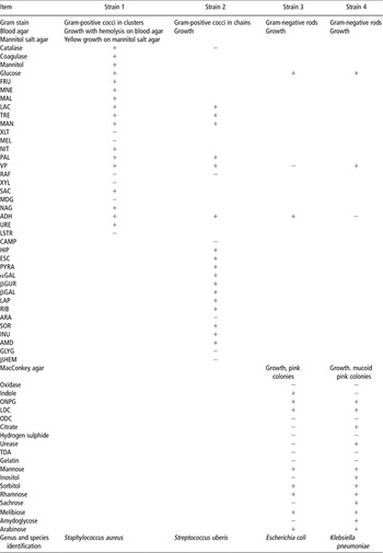
Isolates of S. uberis and E. coli were obtained from Dr. Ken Weston (Postville Veterinary Clinic, Postville, IA). Isolates of S. aureus and K. pneumoniae were obtained from Dr Allan Britten (Udder Health Systems, Bellingham, WA, USA). All strains were fully characterized and identified by Accugen Laboratories (Willowbrook, IL, USA; Table 1) and are available from the authors by request.
Characterization of the bacterial strainsFootnote 1

1 The empty box indicates that the parameter was not assessed.
Isolates of S. uberis, E. coli and S. aureus were grown in Luria–Bertani broth (10 g tryptone, 5 g yeast extract and 10 g NaCl/l). K. pneumoniae was grown in trypticase soy broth (15 g tryptone, 5 g soytone and 5 g NaCl/l). Cultures were grown at 37°C in a shaking water bath. Following culture, bacterial were harvested by centrifugation and diluted into sterile PBS. Serial dilutions of bacterial were then plated onto blood agar plates for determination of the densities (CFU/μL) of individual cultures and to prepare proper dilutions of pathogens for mammary infusions.
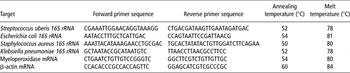
Serial (10-fold) dilutions of pathogens were used to develop quantitative PCR standard curves. DNA from dilutions of the four pathogens was extracted using a Qiagen Qiamp DNA mini kit (Qiagen, Valencia, CA, USA; Catalog no. 51306). Standard curves were developed using quantitative Sybr-green PCR (Bio-Rad Laboratories, Hercules, CA, USA). When samples were run and compared to standard curves, they were diluted and re-run until signal lay within the standard curve. Pathogen-specific primers were obtained by searching the database of the National Center for Biotechnology Information for unique sequences using Beacon Designer 5.1 (Premie Biosoft International, Palo Alto, CA, USA). Sequences were generated against the 16S ribosomal RNA internal transcribed spacer region and were ordered from Invitrogen (Carlsbad, CA, USA). Sequences, annealing and melt temperatures used for quantitative determination of pathogen DNA are given in Table 2. Pathogen DNA concentrations were determined by Sybr-green PCR using a BioRad MyIQ thermocycler (BioRad Laboratories, Hercules, CA, USA). Initial denaturation was carried out for 3 min at 95°C. This was followed by 45 cycles of denaturation at 95°C (30 s), annealing (Table 2; 30 s) and extension at the melt temperature (Table 2; 15 s). Following the 45 cycles, samples were denatured at 95°C for 1 min then cooled to 55°C. Then, melt analysis was performed using a linear gradient of 55°C to 95°C using 80 cycles (0.5°C/cycle). Detection of DNA product was determined at the melt temperature to ensure that no contribution of Sybr-green fluorescence originated from primer-dimer. Standard curves were then constructed for each pathogen such that CFU equivalents could be determined by Sybr-green fluorescence.
Primer sequences and reactions conditions for quantitative PCR and quantitative reverse Transcriptase PCR

To prepare mammary and blood samples for quantitative determination of pathogen, DNA was extracted from tissue and blood sample using the Qiagen DNA mini kit extraction kit. Whole mammary glands were homogenized with a Polytron (Kinematica Inc., Bohemia, NY, USA) and 25 μl of homogenate were then used for DNA extraction. In all, 25 μl of whole blood sample were processed for DNA extraction. Concentrations of pathogen DNA in mammary gland and blood sample were then expressed as CFU equivalents/μl of blood or per microgram of mammary gland homogenate where 1 CFU equivalent equals the amount of target DNA found in one bacterium.
Quantitative reverse transcriptase PCR (QRT-PCR)
Effects of the additive on mRNA concentrations encoding various mammary immune markers and on serum C3 concentration were assessed in the third study. RNA was extracted from mammary tissues using a Qiagen RNeasy Protect Mini kit (Qiagen catalog no. 74124) as per manufacturer’s recommendations. Concentrations of three mRNAs (myeloperoxidase (MPO) and β-actin) in mammary tissue were assessed using Sybr-green QRT-PCR. Unique primer sequences for amplification of MPO and β-actin mRNAs were developed using the Beacon Designer 5.1 software. Sequences, annealing and melt temperatures for these are given in Table 2. Concentrations of MPO mRNA was standardized with housekeeping mRNA (β-actin) using the 2−ΔΔCT method (Livak and Schmittgen, Reference Livak and Schmittgen2001).
Concentrations of macrophage inflammatory protein 1α (MIP) and major histocompatibility complex 2, class II, locus Mb1 (MHC) were assessed using the Applied Biosystems (Foster City, CA, USA) TaqMan kits. Assay IDs for MIP and MHC mRNA assays were Mm9999057_ml and Mm01704827-sl, respectively. Sequence information for the Applied Biosystems TaqMan kits is not available. The TaqMan protocol used for determination of both mRNA concentrations was 48°C for 30 min and 95°C for 10 min then followed by 45 cycles of denaturation (95°C, 15 s) and annealing/extension (60°C, 1 min). This was followed by a final denaturation step of 95°C (1 min) and 55°C for 1 min. Standardization (2−ΔΔCT method) of Fam signals from Taqman MIP and MHC analyses was completed using analytical values for β-actin mRNA.
Analysis of plasma C3 concentration
Concentrations of serum C3 were assessed in the third study using a Mouse Complement C3 ELISA assay (Kamiya Biomedical Co., Seattle, WA, USA) as per manufacturer’s recommendations.
Histology
Samples of mammary tissue from the first study (S. uberis infection) were placed into formalin at time of euthanasia and processed for histological examination. Samples were embedded in paraffin, sectioned and stained with hematoxylin and eosin (H + E). Slides were assessed by a veterinary pathologist who was unaware of treatments.
Statistical analyses
Differences between treatments were assessed using the analysis of variance with S-Plus (Lucent Technologies Inc., Murray Hill, NJ, USA). Studies were conducted as completely randomized designs with three treatments in each study. The independent variable in each study was represented by the treatment effect. The dependent variable in each study was pathogen DNA concentration (in either mammary tissue or blood). Data were log-transformed before statistical analyses. Where differences among treatment means were detected, a Fisher’s multiple range test was used to examine differences between individual treatment means. A P-value of 0.05 was adopted for all comparisons.
Results
Four experiments were completed to assess the effect of feeding a commercial feed additive (OmniGen-AF) on the development of mammary infections and on immune responses in mice intentionally infected with four bovine mastitic strains. Eight mice were assigned to each treatment.
Following intramammary infusion of the four pathogens, animals were monitored closely to follow the course of infection. Signs of inflammation included swelling and reddening of the mammary gland and changes in mouse behavior. Infections with E. coli and K. pneumoniae progressed quickly and, as a result, animals infected with these two pathogens were processed earlier (24 h following infection) than those in other experiments. In general, however, external signs of inflammation bore little relationship to the extent of mammary damage noted following euthanasia and examination of the tissues both grossly and histologically.
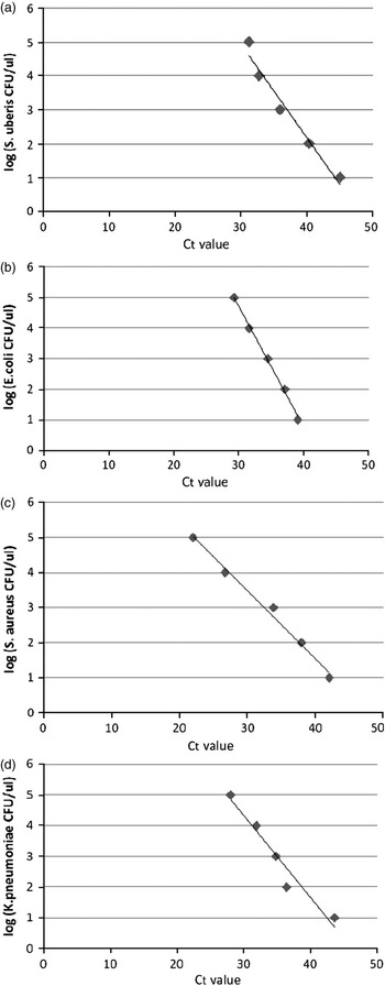
Standard curves relating CFU equivalents for the four pathogens to C t values derived from quantitative PCR are shown in Figure 1. R 2 values for the four standard curves were 0.966, 0.997, 0.989 and 0.947 for S. uberis, E. coli, S. aureus and K. pneumoniae assays, respectively. Regression equations derived for each standard curve were y = −0.2745x + 13.162, y = −0.3971x + 16.629, y = −0.1932x + 9.2757 and y = −0.2653x + 12.297, respectively, where ‘y’ equaled log CFU equivalents and ‘x’ equaled the derived C t value.
Standard curves describing relationships between quantitative PCR and colony-forming units (CFU equivalents) for the four pathogens used in this study. Streptococcus uberis, Escherichia coli, Staphylococcus aureus and Klebsiella pneumoniae are represented (a to d, respectively). R 2 values and regression equations for each relationship are provided in the text.

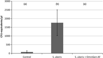
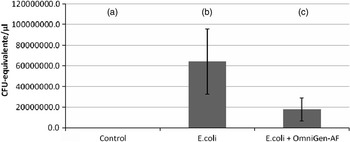
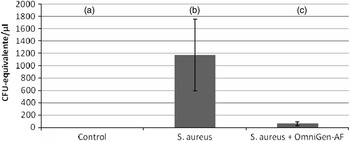
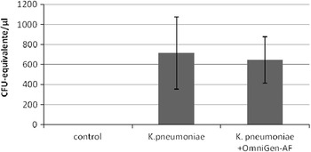
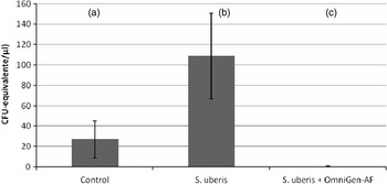
Effects of feeding the additive on mammary concentrations of S. uberis, E. coli, S. aureus and K. pneumoniae DNA are shown in Figures 2 to 5, respectively. Intramammary infusion of these pathogens caused significant (P < 0.05) elevations in mammary pathogen DNA concentrations. S. uberis DNA was also detected in control (sterile PBS-infused) animals in the first study (Figure 2).
Effects of the three experimental treatments on accumulation of Streptococcus uberis DNA in mammary tissue. Duration of infection was 48 h and eight mice were used for each treatment. Values (y axis) are expressed as colony-forming units (CFU)-equivalents/μl mammary gland homogenate ± s.e. Values are arithmetic means, whereas the statistical analysis was performed on log-transformed data. Treatments with differing superscripts (a, b) differ significantly (P < 0.05).

Effects of the three experimental treatments on accumulation of Escherichia coli in the mouse mammary gland. Duration of infection was 24 h and eight mice were used for each treatment. Values on the y axis are expressed as Escherichia coli colony-forming units (CFU) equivalents ± s.e. Differences were detected among the three treatments (P < 0.05). Values are arithmetic means, whereas the statistical analysis was performed on log-transformed data. Treatments with differing superscripts (a, b, c) differ significantly (P < 0.05).

Effects of the three experimental treatments on accumulation of Staphylococcus aureus DNA in mammary tissue. Duration of infection was 48 h and eight mice were used for each treatment. Values (y axis) are expressed as colony-forming units (CFU) equivalents/μl mammary gland homogenate ± s.e. Values are arithmetic means, whereas the statistical analysis was performed on log-transformed data. Differences were detected among the three treatments (P = 0.0017). Treatments with differing superscripts (a, b, c) differ significantly (P < 0.05).

Effects of three experimental treatments on accumulation of Klebsiella pneumoniae DNA in mammary tissue. Duration of infection was 24 h and eight mice were used for each treatment. Values are arithmetic means, whereas the statistical analysis was performed on log-transformed data. Values (y axis) are expressed as colony-forming units (CFU)-equivalents/μl mammary gland homogenate ± s.e.

Feeding OmniGen-AF for approximately 2 weeks before the infusion of pathogen and during early lactation caused significant (P < 0.05) reductions in concentrations of mammary S. uberis, E. coli and S. aureus DNA concentrations. Specifically, feeding OmniGen-AF caused >99%, 71% and 94% reductions in mammary S. uberis, E. coli and S. aureus DNA concentrations (Figures 2 to 4, respectively). Although infusion of glands with K. pneumoniae caused infection, the additive had no effect (P > 0.05) on K. pneumoniae infection progression (Figure 5).
Abilities of pathogens in an established mammary infection to disseminate from the gland were assessed by quantifying pathogen DNA concentration in whole blood sample. Concentrations of whole blood S. uberis DNA were significantly elevated (P < 0.05; Figure 6) in mice infected with S. uberis. S. uberis DNA concentrations in blood sample were approximately 10% of that noted for mammary tissue. In addition, similar to the data shown in Figure 2, feeding OmniGen-AF reduced (P < 0.05) blood S. uberis DNA concentrations. Concentrations of E. coli, S. aureus and K. pneumoniae DNA in blood sample were also assessed following IMI. Only trace concentrations of the DNA from E. coli and K. pneumoniae (0.004% and 0.01% of the concentrations detected in mammary tissue, respectively) were detected in blood sample, whereas no S. aureus DNA was detected. No differences in blood pathogen DNA concentrations (P > 0.05) were detected among treatments in the E. coli, S. aureus and K. pneumoniae studies (experiments 2 through 4, data not shown).
Effects of the three experimental treatments on accumulation of Streptococcus uberis DNA in serum. Values (y axis) are expressed as colony-forming units (CFU) equivalents/μl serum ± s.e. Values are arithmetic means, whereas the statistical analysis was performed on log-transformed data. Differences were detected among the three treatments (P = 0.020). Treatments with differing superscripts (a, b, c) differ significantly (P < 0.05).

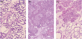
Mammary tissue samples from the first (S. uberis) study were processed for histological examination. Representative sections are presented in Figure 7a to c. Mammary tissue from control animals appeared normal (Figure 7a). Infection of the mammary gland of control-fed animals with S. uberis caused loss of tissue integrity (Figure 7b). Epithelium was largely degraded and alveoli were filled with cellular infiltrate (primarily neutrophils) and cellular debris. Mammary tissue from OmniGen-AF-fed animals which received an S. uberis infusion appeared normal (Figure 7c).
Hematoxylin and eosin staining to show the effects of Streptococcus uberis infusion on mammary histology. Animals which were fed a control diet and received an infusion of sterile phosphate-buffered saline (a), animals fed the control diet but infused with S. uberis (b) and animals fed with OmniGen-AF (0.5% w/w) and infused with S. uberis (c) are represented (magnification = ×20).

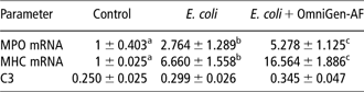
Effects of infection of the mammary gland with E. coli and of feeding OmniGen-AF during a mammary E. coli infection on mammary MPO and MHC mRNA concentrations are shown in Table 3. Infusion of E. coli into the mammary gland caused significant elevations (P < 0.05) in both mammary MPO and MHC mRNA concentrations. Feeding OmniGen-AF caused an even greater (P < 0.05) increase in mammary MPO and MHC concentrations. E. coli infusion and feeding of OmniGen-AF to E. coli-infused mice had no effect (P > 0.05) on mammary MIP mRNA concentrations (data not shown).
Effects of three treatments on relative mammary MPO and MHC mRNAs and on serum C3 concentrations in animals challenged with Escherichia coli
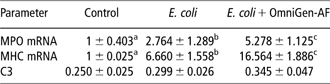
MPO = myeloperoxidase; MHC = major histocompatibility complex.
Values are means ± s.e. Values for MPO and MHC mRNAs are expressed as a proportion of control (control = 1.0) according to the 2−ΔΔCT method. C3 values are means ± s.e. and are expressed in arbitrary densitometry units. Treatments with differing superscripts (a, b, c) within a parameter differ significantly (P < 0.05).
Effects of infection of the mammary gland with E. coli and of feeding OmniGen-AF on serum C3 concentrations are shown in Table 3. No differences were detected among the three treatment means (P = 0.26). However, E. coli infusion tended to increase serum C3 concentration. Furthermore, feeding OmniGen-AF tended to increase the C3 response to E. coli infusion.
Discussion
Previous studies (Wada et al., Reference Wada, Endo, Takahashi, Yano, Watanabe, Koya, Abe, Katoh and Ogata2008) have shown that OmniGen-AF reduces incidence of mastitis on commercial dairies. In that study, mastitis species were not identified. In this study, therefore, ability of the additive to prevent new IMI resulting from four known pathogens was tested. Pathogens utilized in this study were isolated from clinical cases of bovine mastitis and included S. uberis (a Gram-positive facultative anaerobe), E. coli (a Gram-negative facultative anaerobe), S. aureus (a Gram-positive facultative anaerobe) and K. pneumoniae (a Gram-negative, lactose-fermenting, facultative anaerobe; see Table 1) – some of the most commonly isolated bacteria causing mastitis (Barkema et al., Reference Barkema, Schukken, Lam, Beiboer, Wilmink, Benedictus and Brand1998).
Using lactating mice as a model for bovine mastitis was first developed and characterized by Chandler (Reference Chandler1969 and Reference Chandler1970). Since then, this model has been utilized to investigate numerous pathogens causing bovine mastitis (e.g. Sordillo and Nickerson, Reference Sordillo and Nickerson1986). The murine model for the study of bovine mastitis has also been utilized to evaluate the efficacy of numerous vaccines (Mamo et al., Reference Mamo, Froman and Muller2000; Gomez et al., Reference Gomez, Sordelli, Buzzola and Garcia2002), antimicrobials (Brouillette et al., Reference Brouillette, Grondin, Lefebvre, Talbot and Malouin2004) and immunomodulatory compounds (Sordillo and Nickerson, Reference Sordillo and Nickerson1986; Lee et al., Reference Lee, Paape and Zhao2003; Boulanger et al., Reference Boulanger, Brouillette, Jasper, Malouin, Mainil, Bureau and Lekeux2007) in order to elucidate agents that might prevent or treat bovine mastitis.
Quantitative PCR assays were developed and used to quantify pathogen DNA. PCR was used in lieu of culture methods for three reasons: (i) the specific pathogen under investigation could be quantified (i.e. culture methods may not adequately distinguish between the pathogen under investigation from other bacteria); (ii) PCR-based assays are more rapid than conventional assays; and (iii) results obtained from quantitative PCR-based assays correlate highly with bacteriological assays and have been termed ‘synonymous’ (Studer et al., Reference Studer, Schaeren, Naskova, Pfaeffli, Kaufmann, Kirchhofer, Steiner and Graber2008). Our assays are not the first quantitative PCR assays used for S. aureus, E. coli and K. pneumoniae. Previous reports on the use of quantitative PCR for these pathogens include Studer et al. (Reference Studer, Schaeren, Naskova, Pfaeffli, Kaufmann, Kirchhofer, Steiner and Graber2008; S. aureus), Li and Drake (Reference Li and Drake2001), Shannon et al. (Reference Shannon, Lee, Trevors and Beaudette2007; E. coli) and Shannon et al. (Reference Shannon, Lee, Trevors and Beaudette2007; K. pneumoniae). Methods for quantitative PCR-based detection of S. uberis have not been published; however, methods for other streptococcal species are available (e.g. del Rio et al., Reference del Rio, Martin, Martinez, Magadan and Alvarez2008). Most studies that utilize PCR to quantify the degree of bacterial colonization or infection report DNA accumulation in terms of ‘CFU’. However, the limitation of the PCR assays is that they detect DNA whether it resides within a viable CFU or not. As a result, quantitative PCR may overestimate viable CFU. To address this limitation, we have reported accumulations of bacterial DNA as ‘CFU equivalents’ where 1 CFU equivalent equals the amount of DNA present in a CFU.
As expected, infusion of the mammary gland with mastitis pathogens resulted in accumulations of pathogen DNA. Provision of the additive for 2 weeks before intentional infection with three of the pathogens (S. uberis, E. coli and S. aureus) successfully reduced the extent of pathogen DNA accumulation. Quantitative molecular and histological analyses support these observations. The additive was unsuccessful in reducing accumulation of K. pneumoniae DNA.
Non-infected control-fed mice harbored lower levels of S. uberis DNA in mammary tissue and blood. We attribute this to an error in technique; possibly to contamination of some mice by re-use of the same warming pad during recovery from anesthesia.
Mechanisms by which the additive reduced mammary accumulations of pathogen DNA were examined in this and in other studies (Wang et al., Reference Wang, Puntenney, Burton and Forsberg2007 and Reference Wang, Puntenney, Burton and Forsberg2009). In this study, the additive increased mammary concentrations of MPO and MHC mRNAs. Myeloperoxidase is involved in generation of reactive oxygen species in neutrophils and therefore serves as a reliable marker of neutrophil infiltration into a tissue and inflammation (Faith et al., Reference Faith, Sukumaran, Pulimood and Jacob2008). An increase in mammary MPO mRNA implies that the additive may have enhanced the inflammatory response of mammary tissue to the E. coli challenge. These data are supported by earlier studies (e.g. Wang et al., Reference Wang, Puntenney, Burton and Forsberg2007 and Reference Wang, Puntenney, Burton and Forsberg2009) in which the additive increased the expression of neutrophil markers in sheep and dairy cattle.
Antigens in infected tissues may be phagocytosed by macrophages and dendritic cells and presented on the surfaces of antigen-presenting cells in association with MHC molecules (Murphy et al., Reference Murphy, Travers and Walport2008). Enhanced expression of an MHC molecule in mammary tissue of OmniGen-AF-fed animals implies that the additive increased either the total number of antigen-presenting cells in mammary tissue or increased the abilities of individual antigen-presenting cells to present antigens on their surfaces. Either response may explain the ability of the additive to reduce the amount of bacterial DNA in the mammary glands following experimental infection with S. uberis, E. coli or S. aureus.
We do not fully understand why the additive successfully reduced mammary S. uberis, E. coli and S. aureus DNA concentrations but had no effect on K. pneumoniae. We propose three reasons for this observation. First, the strain of K. pneumoniae used in this study was extremely fast growing. It is possible that the introduction of a lactose-fermenting, rapidly growing bacterium (Walia et al., Reference Walia, Madhavan, Chugh and Sharma1987) overwhelmed the natural and augmented aspects of mammary gland immunity. Second, K. pneumoniae is surrounded by a large bio-protective mucoid polysaccharide (Pan et al., Reference Pan, Fang, Yang, Lin, Hsieh, Tsai, Keynan and Wang2008) capsule that protects it from phagocytosis. This capsule may have protected the organism from neutrophil-mediated killing by the host’s natural and augmented immune systems. Finally, some strains of K. pneumoniae produce an extracellular toxic complex that is lethal and produces extensive lung pathology in mice (Straus, Reference Straus1987). The presence of this complex may have also reduced ability of the host’s immune system to slow its growth.
In this study, only S. uberis infection disseminated from the mammary gland to the blood. Although it has been reported that E. coli infection can lead to bacteremia in up to 32% of cases (Cebra et al., Reference Cebra, Garry and Dinsmore1996), that was not confirmed in this study using mice.
Enhancement of a cow’s natural defense mechanisms, particularly during times of stress, may prevent new cases of mastitis thereby decreasing or possibly eliminating the need for antimicrobials. Despite the similarities that exist between the two species, there are critical differences between the bovine and murine mammary glands, especially on a cellular and/or molecular level. Therefore, even though results obtained using the mouse mastitis model are suggestive for similar findings in cows, final conclusions await confirmation in a bovine model. Furthermore, analyses of the effects of the additive on colonization of the mammary gland with additional strains of the pathogens used in this study are clearly warranted.
Acknowledgements
ADR and Y-QW contributed equally to this study. The authors are grateful to Dr Ken Weston (Postville, IA, USA) for provision of S. uberis and E. coli and to Dr Allan Britten (Bellingham, WA, USA) for provision of S. aureus and K. pneumoniae. They also thank Dr C. Loehr (Oregon State University College of Veterinary Medicine, Corvallis, OR, USA) for histology. Finally, The authors are grateful to Dr Eric Brouillette (University of Sherbrooke, Québec, Canada) for his assistance and advice in the development of the murine mastitis model and to Dr. Steve Nickerson (University of Georgia, Athens, GA, USA) for critical reading of this manuscript and for his helpful comments.