




No CrossRef data available.
Article contents
Clustering of filarial infection in an age-graded study: genetic, household and environmental influences
Published online by Cambridge University Press: 03 March 2004
Abstract
A statistical method that analyses correlation structures in families to delineate the contribution of genetic, household and environmental factors on clustering of infection, has been applied to data collected in an area endemic for brugian filariasis in South Sulawesi, Indonesia. Infection was assessed both by microfilaraemia and by anti-filarial IgG4. The results confirmed earlier findings that genetic factors play an important role in clustering of infection. When clustering of infection was analysed in children (<10 years of age) and adults (>20 years of age) separately, it was found that the genetic factors influence clustering of infection in children more profoundly than environmental or household effects. In contrast, genetic factors could not fully explain the clustering of infection seen in adults, which seemed to be mainly determined by household and environmental effects. The data have implications for genotyping studies in brugian filariasis; they indicate that it may be important to concentrate on the younger age groups where individual environmental effects have not yet overruled the genetic influences on gain/loss of infection.
Information
- Type
- Research Article
- Information
- Copyright
- 2004 Cambridge University Press
INTRODUCTION
Lymphatic filariasis is a vector-borne disease affecting approximately 120 million people in tropical and subtropical areas of the world and resulting in a burden of disease that has been estimated by WHO to be 4 918 000 disability-adjusted life years (http://www.filariasis.org/index.pl?iid=2392), primarily in people of reproduction age. In a community where lymphatic filariasis is endemic host–parasite interaction leads to a spectrum of clinical and immunological outcomes ranging from asymptomatic amicrofilaraemia through asymptomatic microfilaraemia, symptomatic microfilaraemia to elephantiasis or other chronic lesions (Maizels et al. 1995). Epidemiological surveys in endemic areas have documented some degree of specific clustering of filarial infection and disease within families/households in affected communities (Walter, 1974; Ottesen et al. 1981; Terhell et al. 2000). Studies of such patterns of clustering within populations may help shed light on factors that control acquisition of pathogens and subsequent outcomes of host–pathogen interaction and may help to design appropriate control strategies.
Contribution of household-related factors to clustering of filarial infection was first documented by Walter in 1974 who demonstrated a highly significant household clustering in brugian filariasis in India and suggested that genetic and/or environmental factors may regulate gain of filarial infection. Later Ottesen and coworkers (1981) studied clustering of filarial disease in Mauke Island and, by applying segregation analysis, compared the influence of genetic and environmental factors and showed a strong genetic effect on disease susceptibility. Since then, several studies have investigated the association between genes and filarial disease with no consistent outcome (Chan et al. 1984; Yazdanbakhsh et al. 1995; Choi et al. 2001). Other risk factors that may contribute towards susceptibility to infection are intrauterine events. Lammie et al. (1991) reported that children born to microfilaraemic mothers had higher prevalence of microfilaraemia than children whose mothers were amicrofilaraemic. Although they carefully analysed the paternal influence, the study design did not allow a thorough analysis of genetic or environmental factors that may have affected their outcome.
To start analysing the possible contribution of host genetics to susceptibility or resistance to filarial infection, we collected family data in Karondang, South Sulawesi (Terhell et al. 2000) where infection status was not only determined by the presence of microfilariae in blood but also by anti-filarial IgG4. Anti-filarial IgG4 is a sensitive marker of filarial infection which is highly suitable for immunoepidemiological studies and detects low level or amicrofilaraemic active infections within a population (Ottesen et al. 1985; Kwan-Lim, Forsyth & Maizels, 1990; Kurniawan et al. 1993; Mahanty et al. 1994; Haarbrink et al. 1995). The family data were analysed by a statistical method developed by Houwing-Duistermaat et al. (1995) where correlation structures are compared corresponding to a genetic, environmental or household model. The correlation structures are based on information available about the genetic distance, household distance and spatial distance between two subjects. When applied to filariasis, the results showed significant evidence for a genetic (for Mf status as well as anti-filarial IgG4), household (anti-filarial IgG4), and environmental (anti-filarial IgG4) model that could explain clustering of filarial infection within the community (Houwing-Duistermaat et al. 1998; Terhell et al. 2000). Since the correlation structures are partially overlapping whereby genetic effects could influence the result of household and environmental models or vice versa, the question arises whether by using different age groups it will be possible to gain better insight into the relative importance of the three models in a more dynamic manner. To confirm our earlier findings (Terhell et al. 2000) in an independent study and to assess whether the model could be applied to different age groups, we repeated the study in a larger population.
The present study was undertaken in Salubarana, another area endemic for lymphatic filariasis, which was 50 km away from our previous study area, Karondang (Terhell et al. 2000). The population was studied as a whole but also after separating into adults and children to provide additional information on potential risk factors specific to children and adults. This knowledge is important when deciding which subjects should be used for DNA analysis to identify genes that link to a particular phenotype.
MATERIALS AND METHODS
Study population
Salubarana is a rural village in the Mamuju district, South Sulawesi, Indonesia with 600 inhabitants and endemic for Brugia malayi infections. The filarial parasites have a nocturnal periodicity and are transmitted by the vectors Anopheles barbirostris and Mansonia uniformis (Slee, 1930; Partono et al. 1977). The study was performed with the cooperation of the head of the village, local medical doctor, local nurses and key persons in villages, such as elementary school teachers and spiritual teachers. Written informed consent was obtained from all study participants or parents of underage children before blood withdrawal and the study was approved by the ethics committee of the Faculty of Medicine, Hasanuddin University, Indonesia.
Study design
Salubarana village is located 3 km from the seaside and most of the houses are situated along the main road and spread over a distance of approximately 1·2 km. The major economic activity of the population is subsistence farming which mainly involves the growing of cacao and corn. The first step in this study was to visit households one by one and obtain information about the members of the households; name, age, sex and the interrelationship of each. To maximize information of the link among individuals, multiple- and cross-checks were performed before constructing pedigrees. Information on children was obtained from parents, and when both mother and father were not available, an adult who lived with the child provided the information. The second step was measuring the distance between the houses and their neighbours. The map of the village including all houses was drawn first, x and y coordinates were put on it and finally the real distances were measured using the stride of one investigator. One stride was assumed to be 1 m. The third step was collecting blood samples, where invitation for blood withdrawal was distributed 2 days prior to blood collection to a few households per night to ensure maximal coverage.
Blood collection
Blood samples were collected from all study participants between 7 p.m. and 2 a.m. by finger prick and were absorbed on discs of Whatman no. 3 filter paper (Maidstone, UK). Care was taken to ensure that the blood penetrated to the reverse side of the paper and that the spots were at least 1 cm in diameter. Approximately 25 μl of blood were absorbed on each disc and assumed to be equivalent to 12·5 μl of serum. The papers were air-dried, placed in self-sealing plastic bags with silica gel, transported to the Netherlands and stored at 4 °C until eluted.
Parasitological examination
For the presence of microfilariae, thick blood smears were made, fixed with methanol, stained with Giemsa's stain and examined microscopically.
Parasite antigen
Adult B. malayi worms were purchased from TRS laboratories, Athens, Georgia, USA. A total of 100 female worms were freeze-dried, ground to powder on ice, dissolved in PBS (phosphate buffered saline) and slowly stirred at 4 °C. The protein concentration was determined by the BCA (2,2′-biquinoline-4,4′-dicarboxylic acid disodium salt hydrate) method before storage at −20 °C (Smith et al. 1985).
Dried blood spot elution
Blood-impregnated filter paper was punched out in 1 cm diameter disks and placed in an Eppendorf tube, followed by 250 μl of assay buffer (PBS containing 5% foetal calf serum (FCS) and 0·05% Tween-20). The dilution was assumed to be 1/20. The tubes were shaken overnight at 4 °C to allow the elution of antibodies from the paper disc.
Elisa for detection of IgG4
Microtitre plates (Nunc-Immuno Plate, Maxisorp, Inter Med, Denmark) were coated overnight at 4 °C with 100 μl/well of B. malayi antigen (5 μg/ml) in 0·1 M carbonate buffer (pH 9·6) and blocked for 1 h at 37 °C with 120 μl of PBS containing 5% bovine serum albumin (BSA, Organon, Teknika). Patient plasma was diluted 1/100 and 1/1000 in assay buffer (PBS containing 5% FCS and 0·05% Tween-20) and incubated at 100 μl per well. A positive standard plasma containing 1×10⁶ arbitrary units (A.U.) parasite-specific IgG4 from a patient in Central Sulawesi, was incubated on each plate in a starting concentration of 1/30 and diluted down 1/√10 to a final concentration of 1/30000. Plates were incubated overnight at 4 °C. After washing, 100 μl of mouse anti-human IgG4 monoclonal antibodies were added (1[ratio ]20000 diluted in PBS containing 5% Tween-20 (Sigma, St Louis, USA)) followed by alkaline phosphatase (AP)-conjugated goat anti-mouse IgG (1/1000 diluted in PBS containing 5% Tween-20 and 4% BSA (Sigma)). Both incubations were for 1 h at 37 °C. The colour was developed by addition of substrate para-nitrophenylphosphate (p-NPP, Boehringer Mannheim, Germany), diluted in diethanolamine buffer (DEA, 0·5 mM MgCl2, 0·1 M DEA, pH 9·6) (Merck, Germany) and absorbency (OD) was measured at 405 nm after 30 min. Absorbency of patient plasma was converted into A.U. by drawing a standard curve of the positive control plasma. A cut-off value for BmA-specific IgG4 antibodies was determined by taking the mean IgG4 reactivity (A.U.) plus 3 times standard deviations of 20 Indonesian student donors who had been living in Leiden, the Netherlands for at least 1 year.
Statistical analysis
Specific IgG4 levels were transformed to log 10 to obtain a normally distributed outcome. Logistic and linear regression models were fitted to model the effect of age and gender on the microfilaraemia and anti-filarial IgG4 respectively. Just as in Terhell et al. (2000) age was incorporated as the natural logarithm of age [ln(age)]. Interactions between age and gender were examined. In the children group, the relation between the IgG4 levels of children and of their mothers was studied by adding the IgG4 level of the mother as independent variable to the linear regression model. Analogously the relation between the IgG4 levels of children and their fathers was estimated. The regression analyses were performed in SPSS for windows version 10.
In the total, children and adults groups, genetic, household and environmental clustering of filarial infection were tested by use of the statistic test of Houwing-Duistermaat et al. (1995) using SPLUS 2000 for Windows. This test has been applied to test clustering of filarial infection due to genetic, household and environmental effects (Houwing-Duistermaat et al. 1995; Terhell et al. 2000). Briefly, the genetic, the household and the environmental correlation structures were based on distances between individuals. The correlation between genetic effects of two individuals equals 2−dg in which dg is the genetic distance in the pedigree. Thus parents and offspring have dg=1, pairs of siblings have dg=1, grandparents and grandchildren have dg=2, aunts/uncles and nephews have dg=2, cousins have dg=3, great-grandparents and great-grandchildren have dg=3, great-uncles/aunts and nephews have dg=3, and grand-nephews can have dg=4 or dg=5 depending on the degree of kinship. Individuals who are not related have infinity as distance. The distances for the household model were defined by dh, with dh=0 for individuals sharing the same house (correlation 1) and distance infinity for inhabitants of different households (correlation 0). The distance for the environmental model de equals the distance between two households in meters. The correlation follows from the formula:
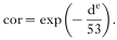
The number 53 is the mean distance between neighbouring houses. For de=53 the correlation is about 0·37 and for de=4*53 the correlation is only 0·02. Thus the correlation decreases if the distance between two households becomes larger. The hypotheses of no correlation due to genetic, household and environmental effect were tested versus the alternatives using the test statistic of Houwing-Duistermaat et al. (1995). The outcomes for anti-filarial IgG4 level and microfilaraemia were adjusted for the covariates age and sex.
For children and adults, variance components for a genetic and for a household effect were estimated by fitting a linear mixed-effects model (Laird & Ware, 1982; Houwing-Duistermaat et al. 1998). As a measure of the effect of genes on clustering of IgG4 levels within families, we used the part of clustering explained by genes for monozygotic twins living in the same house. Since these monozygotic twins share all genes and households, the variance of the total cluster effect is the sum of the genetic variance and the household variance. Hence the part of clustering explained by genes equals the genetic variance divided by this total cluster variance. Finally, we tested whether the difference between the variance components for adults and children was significant using a likelihood ratio statistic. Also in these analyses the anti-filarial IgG4 levels are adjusted for age and sex.
RESULTS
Microfilariae prevalence and anti-filarial IgG4
A total of 583 individuals from Salubarana village were involved in this study (Table 1). The age ranged from 2 months to 80 years (mean 23·7 years±17·5) and was comprised of more males (n=303) than females (n=280). The microfilariae prevalence increased with age starting from 19 years whereas anti-filarial IgG4 production started early in life; in the 0–9 year age group 50% (78/156) of the children were already positive for IgG4 reaching a plateau in subjects older than 10 years, these results are in agreement with our previous findings (Terhell et al. 2000). Therefore the children group was defined as children of age less than 10 years (n=157) and the adult group was defined as subjects of age 20 years and older (n=313). Because of the small number of microfilaraemics, age stratified analyses were only performed for anti-filarial IgG4 levels.

Logistic regression analysis showed that in the total group the prevalence of microfilaraemics was significantly higher in males than in females (b=1·03, CI=0·278–1·786, P=0·006) and increased significantly with age (b=0·66, CI=0·216–1·100, P=0·003). No significant interaction between age and sex was found. For anti-filarial IgG4 interaction between age and sex was significant (P<0·001). The relation between age and IgG4 levels was more pronounced in males (b=0·36, CI=0·28–0·44, P<0·001) than in females (b=0·12, CI=0·00–0·20, P=0·004). Furthermore, in children the effect of the IgG4 levels of mothers on their children was significant (b=0·20, CI=0·05–0·35, P=0·007). The effect of the level of the father was smaller and not significant (b=0·10, CI=0·04–0·25, P=0·149).
Clustering
The data comprise 35 families which ranged in size from 2 to 243 individuals, the mean number of members per family was 16·7. The 583 inhabitants examined were spread over 133 households, the households ranged in size from 1 to 11 individuals and the mean number of members per household was 4·4. Also for the children group we were able to distinguish between genetic and household effects, because this group comprises genetically related pairs who did not share households; for example there were 232 cousin pairs.
For the outcome, filarial infection as determined by microfilariae status, the statistic of Houwing-Duistermaat et al. (1998) showed that in the whole population both genetic and household factors were important (P=0·019 and P=0·003, respectively). No significant effect for environmental factors was found (P=0·58). When as marker of infection the anti-filarial IgG4 was studied, the genetic and household effects were statistically significant for the total population (P<0·001 and P=0·002, respectively) and for environmental factors reached borderline significance (P=0·075) (Table 2), a finding in agreement with our previous study (Terhell et al. 2000).
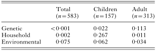
When the analysis was applied to adults and children separately, both household and environmental effects were significant for adults whereas the clustering could not be fully explained by genetic effects (Table 2). In children, the analysis showed that genetic factors were significant for clustering of infection whereas no household or environmental effect was seen (Table 2).
For children, the genetic variance was 0·07 (standard error (S.E.) 0·06) and the household variance was 0 (S.E. could not be estimated) indicating that 100% of the clustering was explained by genes, while for adults the genetic variance was 0·07 (S.E. 0·09) and the household variance was 0·09 (S.E. 0·04) showing that 44% of the clustering was explained by genes. However, the difference between the variance components of household effects for children (0) and adults (0·09) was not statistically significant (P=0·11).
DISCUSSION
The risk of filarial infections within families can be caused by genetic or unknown effects that also aggregate within families. A new statistical method developed by Houwing-Duistermaat et al. (1995), exploits the fact that genetic and environmental effects give different correlation structures in family studies and therefore is able to estimate what are the important causes of aggregation of a medical trait within families. For example, the correlation due to genetic effects decreases with the distance in the pedigree, whereas an environmental factor affecting one area would influence relatives from one area and not relatives from a distant area. When in a population residing in an area endemic for filarial infection family data are collected with information on pedigree structure, household structure and distances between houses, the statistics based on the correlation structure can be applied. For previous data collected in another village with endemic B. malayi infections, the statistical test was successfully applied (Houwing-Duistermaat et al. 1998) and the results showed that genetic effects regulated the microfilaraemic state of an individual (Terhell et al. 2000) whereas both genetic and environmental effects accounted for the clustering of infections when anti-filarial IgG4 was taken as an indicator of active infection. One way to explain the different outcomes when using microfilaria count versus anti-filarial IgG4 as an indicator of infection may be that microfilariae-positive individuals carry a relatively high burden of infection and this is governed by genetic factors, whereas light infections that can be detected by anti-filarial IgG4 positivity (Haarbrink et al. 1995) are influenced not only by genetic factors but also by household and environmental factors. Needless to say, the genetic regulation of IgG4 itself could also be a contributing factor when infection is measured indirectly.
In the present report we have studied the data collected in a nearby village with lower prevalence of microfilariae prevalence (6·8% in Salubarana, the present study, compared with 33·3% in Karondang, our previous study (Terhell et al. 2000)). For the whole population clustering of infection as determined by microfilariae, was influenced by genetic and household factors whereas in our previous study which consisted of 171 participants, belonging to 35 pedigrees and residing in 57 households (Terhell et al. 2000) the genetic effect was the only significant outcome. This might reflect the impact of larger sample size in the present study. When using anti-filarial IgG4, we showed that genetic factors play an important role in controlling filarial infection. Both household and environmental effects could also explain the clustering of anti-filarial IgG4 levels as a marker of filarial infection within families, but their effect was less significant than the genetic effect. In a study of another filarial infection, Loa loa, the importance of the genetic control of microfilaraemic infection has been documented by segregation analysis of the familial data collected in Cameroon (Garcia et al. 1999). Interestingly, in the latter study there was a significant association between microfilaraemic status of a mother and child whereas no such association was found for father and child. However, when a major gene was included in their model the maternal influence disappeared, vouching for the ability of a major gene effect to explain the association. In our study, we also observed that the level of anti-filarial IgG4 in children was significantly correlated with that of mothers but no correlation was found with fathers. Although the strong correlation between infection status of mother and child may be the consequence of in utero exposure to filarial antigen, our data also provide evidence for a genetic effect. Future studies should unravel the contribution of each of these components to the mother/child relationship.
Very few family studies in filariasis have analysed the data in an age-stratified manner. The question of what the relative importance of genetic, household and environmental factors are in clustering of infection in children versus adults has been addressed in the present study. The prevalence of microfilariae was too low to allow reliable analysis of the correlation structure for this trait in children and adult groups separately, therefore anti-filarial IgG4 only was used. The analysis indicated that for children below 10 years of age, the genetic model best explained the clustering of active infection whereas environmental factors had a borderline influence and household effects were not significant. Interestingly, this was different from what was seen in adults aged 20 years or more, where genetic effects could not fully explain the clustering of infection in this population. By calculating the variance, in order to estimate the relative contribution of the genetic versus household effects (the outcome where significant effects were seen), we observed that whereas 100% of clustering in children in the model was explained by genetic factors, only 44% of the clustering could be accounted for by genetic factors in adult. These results suggest that individual behavioural or environmental factors overrule the genetic predisposition to infection in the adult population whereas children with a relatively more homogeneous behaviour/environment allow the expression of the genetic effects. Interestingly, a study carried out in Haiti (Eberhard et al. 1993) where the effects of in utero exposure to filarial antigens was investigated reported that in children up to 9 years of age the influence of maternal infection status was more profound than in older children. Indeed the profound influence of environmental factors in adults may explain the weak and non-reproducible associations between genes and filarial disease, which were studied in adults (Yazdanbakhsh et al. 1995, 1997).
In conclusion, our present findings have important implications for the DNA genotyping when specific genes are sought for their role in resistance/susceptibility to infection. If confirmed in independent studies, it may be important to concentrate on genotyping subjects that are below the age of 20 years and reside in filaria-endemic areas. This will reduce background variability and would cut down on costs.
Grant support is acknowledged from The Netherlands Foundation for the Advancement of Tropical Research (W93-325); Netherlands Organization for International Cooperation in Higher Education (NUFFIC); European Commission, INCO programme, contract no: ICA4-CT-2001-10081.
References
REFERENCES
CHAN, S. H.,
DISSANAYAKE, S.,
MAK, J. W.,
ISMAIL, M. M.,
WEE, G. B.,
SRINIVASAN, N.,
SOO, B. H. &
ZAMAN, V.
(1984).
HLA and filariasis in Sri Lankans and Indians.
Southeast Asian Journal of Tropical Medicine and Public Health 15, 281–286.Google Scholar
CHOI, E. H.,
ZIMMERMAN, P. A.,
FOSTER, C. B.,
ZHU, S.,
KUMARASWAMI, V.,
NUTMAN, T. B. &
CHANOCK, S. J.
(2001).
Genetic polymorphisms in molecules of innate immunity and susceptibility to infection with Wuchereria bancrofti in South India.
Genes and Immunity 2, 248–253.CrossRefGoogle Scholar
EBERHARD, M. L.,
HITCH, W. L., McNEELEY, D. F. &
LAMMIE, P. J.
(1993).
Transplacental transmission of Wuchereria bancrofti in Haitian women.
Journal of Parasitology 79, 62–66.CrossRefGoogle Scholar
GARCIA, A.,
ABEL, L.,
COT, M.,
RICHARD, P.,
RANQUE, S.,
FEINGOLD, J.,
DEMENAIS, F.,
BOUSSINESQ, M. &
CHIPPAUX, J.-P.
(1999).
Genetic epidemiology of host predisposition microfilaraemia in human loiasis.
Tropical Medicine and International Health 8, 565–574.CrossRefGoogle Scholar
HAARBRINK, M.,
TERHELL, A.,
ABADI, K.,
VAN BEERS, S.,
ASRI, M. &
YAZDANBAKHSH, M.
(1995).
IgG4 antibody assay in detection of filariasis.
Lancet 346, 853–854.CrossRefGoogle Scholar
HOUWING-DUISTERMAAT, J. J.,
DERKX, B. H. F.,
ROSENDAAL, F. R. &
VAN HOUWELINGEN, H. C.
(1995).
Testing familial aggregation.
Biometrics 51, 1292–1301.CrossRefGoogle Scholar
HOUWING-DUISTERMAAT, J. J.,
VAN HOUWELINGEN, H. C. &
TERHELL, A. J.
(1998).
Modelling the cause of dependence with application to filarial infection.
Statistics in Medicine 17, 2939–2954.3.0.CO;2-U>CrossRefGoogle Scholar
KURNIAWAN, A.,
YAZDANBAKHSH, M.,
VAN REE, R.,
AALBERSE, R.,
SELKIRK, M. E.,
PARTONO, F. &
MAIZELS, R. M.
(1993).
Differential expression of IgE and IgG4 specific antibody responses in asymptomatic and chronic human filariasis.
Journal of Immunology 150, 3941–3950.Google Scholar
KWAN-LIM, G. E.,
FORSYTH, K. P. &
MAIZELS, R. M.
(1990).
Filarial-specific IgG4 correlates with active Wuchereria bancrofti infection.
Journal of Immunology 145, 4298–4305.Google Scholar
LAIRD, N. M. &
WARE, J. H.
(1982).
Random-effects models for longitudinal data.
Biometrics 38, 963–974.CrossRefGoogle Scholar
LAMMIE, P. J.,
HITCH, W. L.,
WALKER ALLEN, E. M.,
HIGHTOWER, W. &
EBERHARD, M. L.
(1991).
Maternal filarial infection as risk factor for infection in children.
Lancet 337, 1005–1006.CrossRefGoogle Scholar
MAHANTY, S.,
DAY, K. P.,
ALPERS, M. P. &
KAZURA, J. W.
(1994).
Antifilarial IgG4 antibodies in children from filaria-endemic areas correlated with duration of infection and are dissociated antifilarial IgE antibodies.
Journal of Infectious Diseases 170, 1339–1343.CrossRefGoogle Scholar
MAIZELS, R. M.,
SARTONO, E.,
KURNIAWAN, A.,
PARTONO, F.,
SELKIRK, M. E. &
YAZDANBAKHSH, M.
(1995).
T cell activation and the balance of antibody isotypes in human lymphatic filariasis.
Parasitology Today 11, 50–55.CrossRefGoogle Scholar
OTTESEN, E. A.,
MENDELL, N. R.,
McQUEEN, J. M.,
WELLER, P. F.,
AMOS, D. B. &
WARD, F. E.
(1981).
Familial predisposition to filarial infection-not linked to HLA-A or locus specificities.
Acta Tropica 38, 205–216.Google Scholar
OTTESEN, E. A.,
SKVARIL, F.,
TRIPATHY, S. P.,
POINDEXTER, R. W. &
HUSSAIN, R.
(1985).
Prominence of IgG4 in the IgG antibody response to human filariasis.
Journal of Immunology 134, 2707–2712.Google Scholar
PARTONO, F.,
OEMIJATI, S.,
HUDOJO, J. A.,
SAJIDIMAN, H.,
PUTRALI, J.,
CLARKE, M. D.,
CARNEY, W. P. &
CROSS, J. H.
(1977).
Malayan filariasis in Central Sulawesi (Celebes), Indonesia.
Southeast Asian Journal of Tropical Medicine and Public Health 8, 452–458.Google Scholar
SLEE, W. VAN.
(1930).
Onderzoek naar het voorkomen van filaria te Mamoedjoe.
Geneeskundig Tijdschrift van Nederlands-Indië 70, 444–450.Google Scholar
SMITH, P. K.,
KROHN, R. I.,
HERMANSON, G. T.,
MALLIA, A. K.,
GATRNER, F. H.,
PROVENZANO, M. D.,
FUJIMOTO, E. K.,
GOEKE, N. M.,
OLSOM, B. J. &
KLENK, D. C.
(1985).
Measurement of protein using bicinchoninic acid.
Analytical Biochemistry 150, 67–85.CrossRefGoogle Scholar
TERHELL, A. J.,
HOUWING-DUISTERMAAT, J. J.,
RUITERMAN, Y.,
HAARBRINK, M.,
ABADI, K. &
YAZDANBAKHSH, M.
(2000).
Clustering of Brugia malayi infection in a community in South-Sulawesi, Indonesia.
Parasitology 120, 23–29.CrossRefGoogle Scholar
WALTER, S. D.
(1974).
On the detection of household aggregation of disease.
Biometrics 30, 525–538.CrossRefGoogle Scholar
WORLD HEALTH ORGANIZATION
(http://www.filariasis.org/index.pl?iid=2392).
YAZDANBAKHSH, M.,
SARTONO, E.,
KRUIZE, Y. C. M.,
KURNIAWAN, A.,
PARTONO, F.,
MAIZELS, R. M.,
SCHREUDER, G. M. TH,
SCHIPPER, R. &
DE VRIES, R. R. P.
(1995).
HLA and elephantiasis in lymphatic filariasis.
Human Immunology 44, 58–61.CrossRefGoogle Scholar
YAZDANBAKHSH, M.,
ABADI, K.,
DE ROO, M.,
VAN WOUWE, L.,
DENHAM, D.,
MEDEIROS, F.,
VERDUIJN, W.,
SCHREUDER, G. M. TH,
SCHIPPER, R.,
GIPHART, M. J. &
DE VRIES, R. R. P.
(1997).
HLA and elephantiasis revisited.
European Journal of Immunogenetics 24, 439–442.CrossRefGoogle Scholar
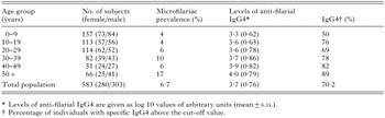
Table 1. Description of the study population (number of subjects, female/male ratio, microfilariae prevalence, mean specific IgG4 level and prevalence of specific IgG4 are given per age-group)
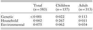
Table 2. The P values for testing the null hypothesis of no clustering of IgG4 levels, adjusted for ln age and sex due to genetic, household and environmental effects using the score statistic in total, adult and children population
 You have
Access
- 13
- Cited by