Introduction
The biological cycle of fish includes several phases, i.e. eggs, embryos, larvae, and adults. Initial development is characterized by morphogenetic events that begin after the egg is fertilized until the larvae hatch (Kimmel et al., Reference Kimmel, Ballard, Kimmel, Ullmann and Schilling1995; Perini et al., Reference Perini, Sato, Rizzo and Bazzoli2010). After hatching, the main internal organs and scales are formed during larval development. Furthermore, their weight and length increase, and skeleton ossification and tissue pigmentation occur. The larval phase ends when total yolk sac resorption and mouth opening occur. This shows the need for exogenous feeding (Perrotti et al., Reference Perrotti, Abdo, Meireles, Marcon and Bazzoli2019).
Development of systems and organs has specific patterns for each species. These show when fish will swim, escape, and capture food (Hall et al., Reference Hall, Smith and Johnston2004). When the formation of the mouth is incomplete, the larva feeds on the yolk through endocytosis from the vitelline syncytial layer (Abdo et al., Reference Abdo, Perrotti, Meireles and Bazzoli2016). This absorption of the yolk sac is essential for maintaining larval health. Indeed, the digestive tract is still developing and cannot digest exogenous feeding (Maria et al., Reference Maria, Ninhaus-Silveira, Orfão and Viveiros2017).
Several environmental factors can influence larval ontogenesis, especially temperature. Studies show that higher temperatures, in the tolerable limits of the species, accelerate metabolism and, consequently, its development. Additionally, temperature can influence other important fish processes such as gonadal maturation, spawning, embryogenesis, fertility, and hatching rates (Sato et al., Reference Sato, Fenerich-Verani, Verani, Vieira and Godinho2000; Borçato et al., Reference Borçato, Bazzoli and Sato2004; Pankhurst and Munday, Reference Pankhurst and Munday2011; Uji et al., Reference Uji, Suzuki, Iwasaki, Teruya, Hirasawa, Shirakashi, Onoue, Yamashita, Tsuji, Tsuchihashi and Okuzawa2015).
G. brasiliensis, known as ‘Cará’ or ‘Acará’, inhabits lentic environments and streams. It adapts well to artificial environments, such as reservoirs, dams, and tanks. This species is found in several South American watersheds. It is active during the day, with detritivorous or omnivorous feeding habits and parental care for its offspring (Abelha and Goulart, Reference Abelha and Goulart2004). The Acará presents sexual dimorphism similar to other cichlids. Males develop more distinct characteristics compared with females in the reproductive season, showing variations in colour and development of a bulged head region called the ‘nuptial callus’(Oliveira and Almada, Reference Oliveira and Almada1995). This is an economically important neotropical species that presents great potential for aquaculture as a result of its striking colour and capacity to reproduce in captivity. However, despite its potential for ornamental aquaculture, there have been few studies related to its reproductive biology and initial development. Given this scenario, this study aimed to increase knowledge about larval development and the influence of temperature in this process, fundamental information for fish farming.
Materials and Methods
Study location and sampling
Adults of G. brasiliensis were captured from a lake in the São Francisco River basin (19°55′14.37′′S, 43°59′31.48′′W), Minas Gerais, Brazil, in the first quarter of 2018. They were caught by means of a mesh net with 3.0 cm between opposing nodes. The captured fish were transferred to a 750-litre aquarium at the Wet Laboratory of the Postgraduate Programme in Vertebrate Biology. The water temperature was maintained at 26°C. All individuals were fed daily with commercial pelleted feed with 36% crude protein at the rate of 1.5–2.0% live weight/day. Breeding and spawning/spermiation of the broodstock occurred spontaneously without induction techniques. Wooden substrates were placed in the aquarium to allow the oviposition and fixation of adhesive eggs. The eggs deposited on the substrate were observed daily and hatched after 72 h.
Experimental conditions of the larvae
After hatching in the 750-litre aquarium, 40 larvae were transferred to each 5-litre aquarium with temperatures of 22°C, 26°C or 30°C. To avoid thermal stress, the 5-litre aquaria were initially heated to the same temperature as the 750-litre aquarium, i.e. 26°C. In the coldest aquarium, the thermostat was turned off until the temperature gradually reached 22°C. In the 30°C aquarium, the water temperature was gradually increased. The acclimatization period lasted 4 h. Water parameters, such as temperature, dissolved oxygen, toxic ammonia, and pH (Table 1), were monitored daily throughout the experiment. Five larvae from each aquarium were collected daily until the complete resorption of the yolk sac. The mortality rate in each treatment was calculated as follows:
 $${\ Dead \ larvae \times 100\over Total \ number \ of \ larvae}.$$
Mean values and standard error of water parameters recorded during larval ontogenesis of G. brasiliensis at temperatures 22, 26 and 30°C

a,b,c Different letters mean statistical difference between treatments (P < 0.05).
The research was approved by the Ethics Committee on the Use of Animals (CEUA PUC Minas protocol no. 021/2017).
Histological and biometric analyses
For microscopic analysis, the larvae collected were euthanized by means of a eugenol overdose, 180 mg/l (Vidal et al., Reference Vidal, Albinati, Albinati, Lira, Almeida and Santos2008), then fixed in Bouin’s liquid for 8–12 h and embedded in historesin. Larvae were sectioned at 3-µm thickness and stained with toluidine blue sodium borate (TB).
Biometric analysis was performed using a stereomicroscope and a light microscope (Olympus BX50 coupled with an Olympus SC-30 camera.) The following measurements were taken in Olympus CELL software: total length (TL), ocular diameter (ED), body height (BH), length of the yolk sac (YSL), and height of the yolk sac (YSH).
Statistical analysis
The values for larval biometry are presented as mean ± standard error. Values were calculated using STATISTICA v.7.0 software. Due to the absence of a normal data distribution, biometric data were compared using non-parametric analysis of variance, Kruskal–Wallis test and Dunn’s post-test. Results were considered significant at a P-value < 0.05.
Results
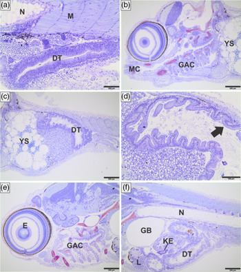
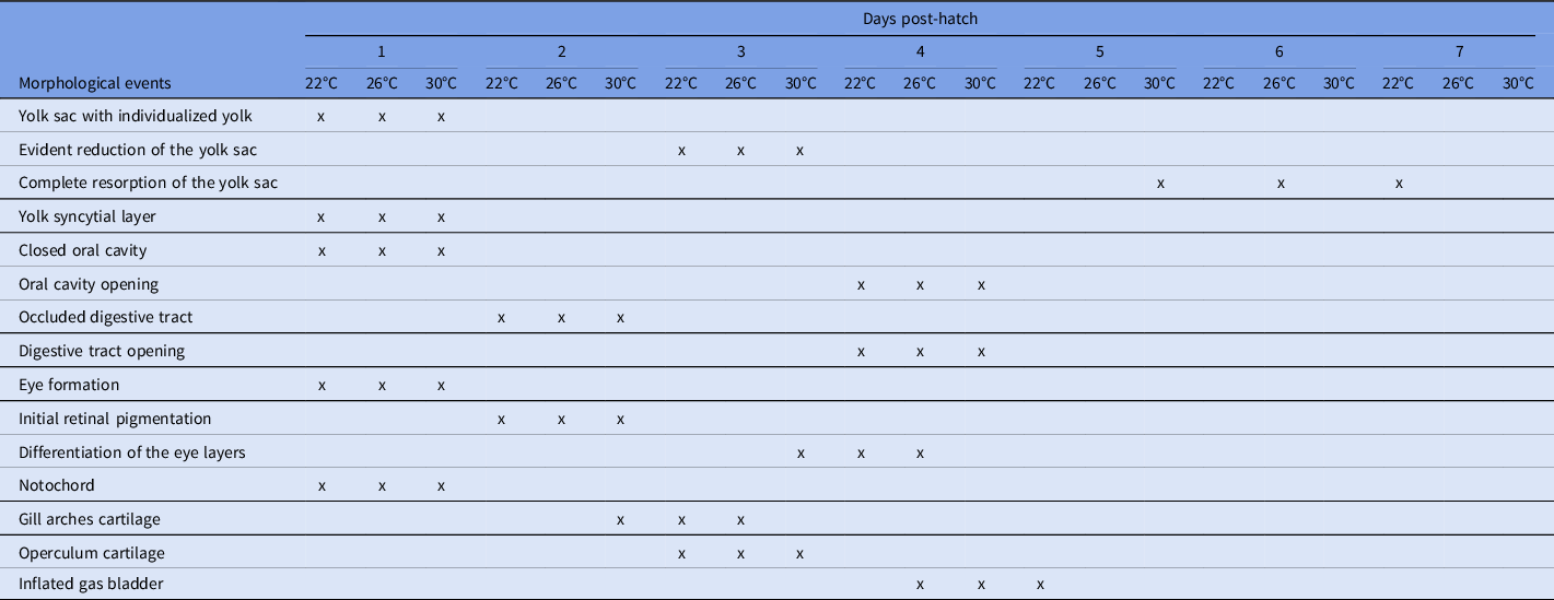
Development of the main structures and the morphological events observed during larval ontogenesis of G. brasiliensis are shown in Figs 1 and 2 and Table 2, and were observed in the following chronological order:
-
First day, the following structures were observed: large yolk sac with individualized yolk granules, eyes, notochord, myomeres, heart, olfactory vesicle, oral cavity, and occluded digestive tube.
-
Second day: optic vesicle, the beginning of pigmentation of retinal epithelial cells, and the beginning of the formation of mandibular cartilages. The formation of the gill arches cartilage was also observed at 30°C.
-
Third day: At 22 and 26°C, the gill arches cartilage was observed. At 30°C, the differentiation of the eye layers was observed. Opercular cartilage, chondrocranial cartilage, renal epithelium, and clearly reduction of the yolk sac were also observed at the three temperatures.
-
Fourth day: differentiation of the eye layers at 22 and 26°C, and the opening of the digestive tract and mouth at the three temperatures.
-
Fifth, sixth, and seventh days: there was total reabsorption of the yolk sac in the three aquariums at temperatures of 30°C, 26°C. and 22°C, respectively.
Main structures observed during larval ontogenesis of G. brasiliensis, in histological slides stained by TB, from the first to the fourth days: (a) yolk sac (YS), myomeres (M), optic vesicle (OV), and brain (B). (b) YS with yolk syncytial layer (YSLa) and clear vesicles protruding (*). (c) Detail of the YSLa and elliptical yolk granules (arrow) in the yolk sac. On the fourth day: (d) gill arches cartilage (GAC), notochord (N), YS, heart (H), and differentiation of the layers of the eye into the inner plexiform layer (IP), the outer plexiform layer (OP), inner nuclear layer (IN), and outer nuclear layer (ON). Scale bars: 200 µm (a, d); 100 µm (b); and 50 µm (c).

Main structures observed during the larval ontogenesis of G. brasiliensis, in histological slides stained by BT, from the fourth to the seventh days: (a) notochord (N), myomeres (M), the initial opening of the digestive tract (DT). On the fifth day: (b) yolk sac (YS), mandibular cartilage (MC), and gill arch cartilage (GAC); (c) complete opening of the DT and reduction of the YS. On the sixth day: (d) detail of the anterior intestine covered by a simple prismatic epithelium with a striated border (arrow). On the seventh day: (e) eye layers (E), GAC; (f) N, gas bladder (GB), DT, liver (L), and renal epithelium (KE). Bars: 200 µm (a–c, e, f); 100 µm (d).

The main morphological events of larval ontogenesis of G. brasiliensis, with the respective day of occurrence, at 22, 26, and 30°C

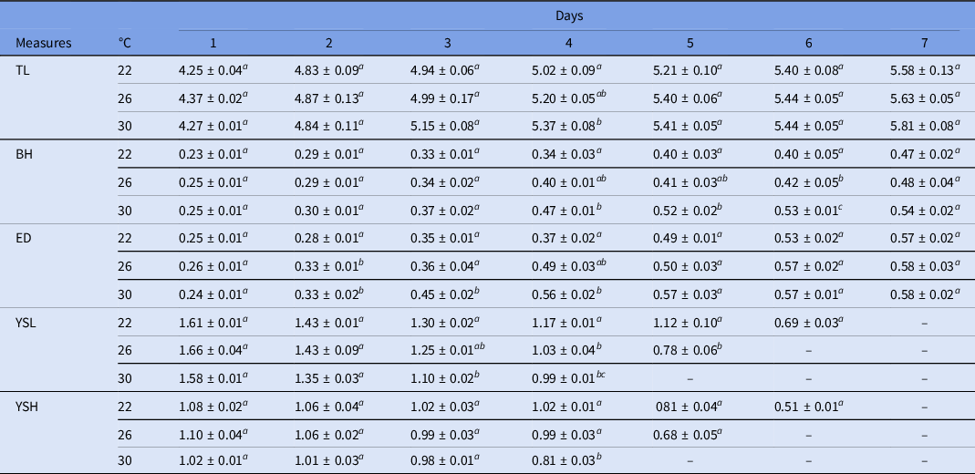
The larvae biometrics from the first to the seventh days at temperatures of 22, 26, and 30°C are shown in Table 3. In general, the yolk sac gradually decreased, while the other structures increased gradually until the seventh day in a temperature-dependent manner.
Mean values (mm) and standard error of the morphometry recorded during the larval ontogenesis of G. brasiliensis at 22, 26, and 30°C

a,b,c Different letters indicate significant differences between treatments (P < 0.05).
Total length (TL), body height (BH), eye diameter (ED), yolk sac length (YSL) and yolk sac height (YSH).
Regarding the TL, the averages showed statistical difference only on the fourth day between temperatures of 22°C and 30°C. On the seventh day it was observed that the larvae in all treatments showed TL without significant variations. BH showed statistical differences on days 4 and 5 between temperatures 22°C and 30°C, on the sixth the significant difference was observed in the three temperatures tested. Eye diameter showed a significantly smaller size from the second to the fourth days at 22°C. At 30°C, the ED showed significantly higher values until day 4, after this day no further statistical differences were observed between treatments. The yolk sac length (YSL) and yolk sac height (YSH) showed similar trends, in which the higher the temperature, the faster the decrease in yolk sac measurements was observed. YSL from the third day showed values statistically higher at 22°C and statistically lower at 30°C. For YSH, statistical difference was observed only on the fourth day at 30°C. Complete resorption of the yolk sac was observed on the fifth day at 30°C, sixth day at 26°C and seventh day at 22°C.
Mortality rate was calculated and was similar in all treatments, presenting a rate of 5% (n = 2) at a temperature of 26°C, and 7.5% (n = 3) at 22°C and 30°C.
Discussion
Studies on larval ontogenesis and its morphological and biometric characteristics (such as eye diameter, pigmentation, organ development and mouth opening) as well as on the evolutionary relationships between species, are of great relevance for fish farming (Perrotti et al., Reference Perrotti, Abdo, Meireles, Marcon and Bazzoli2019). This type of approach increases knowledge on the identification of eggs, larvae and spawning sites (Marques et al., Reference Marques, Nakaghi, Faustino, Ganeco and Senhorini2008; Gomes et al., Reference Gomes, Sato, Rizzo and Bazzoli2013). In this study, the development of the main organs and structures of G. brasiliensis presents similar characteristics to those observed in other Cichlidae fish (Saemi-Komsari et al., Reference Saemi-Komsari, Mousavi-Sabet, Kratochwil, Sattari, Eagderi and Meyer2018a and 2018b). In addition, our data showed that increasing the water temperature to 30°C reduced the duration of larval ontogenesis, which may reflect in cost savings for producers.
Water temperature is a main factor that influences egg hatching. This factor is observed in Prochilodus lineatus in which hatching of eggs at 28°C occurred within 14 h, while at 22°C it occurred within 23 h (Ninhaus-Silveira et al., Reference Ninhaus-Silveira, Foresti and de Azevedo2006). High temperatures accelerate hatching of eggs and the rupture of the corium. This effect is observed in other neotropical species, such as Rhamdia quelen (Rodrigues-Galdino et al., Reference Rodrigues-Galdino, Maiolino, Forgati, Donatti, Mikos, Carneiro and Rios2010), Astyanax altiparanae (Dos Santos et al., Reference Dos Santos, Yasui, Xavier, de Macedo Adamov, do Nascimento, Fujimoto, Senhorini and Nakaghi2016), and Brycon amazonicus (Cristina da Silva et al., Reference Cristina da Silva, Pereira Dos Santos, Senhorini, Paes, Valentin, Fujimoto, do Nascimento, Yasui and Nakaghi2017). Temperature also influences the larval ontogenesis process. The higher the temperature, the shorter the development time of the species (Green and Fisher, Reference Green and Fisher2004; Martell et al., Reference Martell, Kieffer and Trippel2005; Prakoso et al., Reference Prakoso, Pouil, Prabowo, Sundari, Arifin, Subagja, Affandi, Kristanto and Slembrouck2019). This resembles results observed in G. brasiliensis, which showed a faster larval development at 30°C compared with temperatures of 22°C and 26°C.
The tolerance to different temperatures can vary substantially between tropical and phylogenetically close species (Nur et al., Reference Nur, Batubara, Eriani, Tang, Muhammadar, Siti-Azizah, Wilkes, Fadli, Rizal and Muchlisin2020). Although our data showed similar mortality rates at different temperatures, this difference has already been observed in other tropical species such as Trichogaster leeri, which presented a lower mortality rate at temperatures between 26–28°C (Pereira et al., Reference Pereira, De Andrade, Radael, Fosse Filho, De Azevedo, Mattos and Vidal Junior2016). In Oreochromis niloticus, Pandit and Nakamura (Reference Pandit and Nakamura2010) observed that very high temperatures (>35°C) could drastically decrease larvae survival.
The reabsorption of the yolk in fish is carried out through the yolk syncytial layer (Ninhaus-Silveira et al., Reference Ninhaus-Silveira, Foresti, de Azevedo, Agostinho and Veríssimo-Silveira2007). This was observed in histological slides of G. brasiliensis larvae from the second day after hatching. The total resorption time of the yolk sac can vary in interspecific and intraspecific ways, depending on the water temperature. This fact can be confirmed in this study. Indeed, in the G. brasiliensis larvae, total reabsorption of the yolk sac occurred on the seventh day at 22°C, the sixth day at 26°C and the fifth day at 30°C. For other cichlids that were reared at 26–29°C, total yolk resorption occurred in 6 days for O. niloticus (Fujimura and Okada, Reference Fujimura and Okada2007), 13 days for Labidochromis caeruleus (Saemi-Komsari et al., Reference Saemi-Komsari, Mousavi-Sabet, Kratochwil, Sattari, Eagderi and Meyer2018a) and 15 days for Nimbochromis venustus (Saemi-Komsari et al., Reference Saemi-Komsari, Salehi, Mansouri-Chorehi, Eagderi and Mousavi-Sabet2018b).
Determining the mouth opening in teleost larvae is an important event in aquaculture. It indicates the need for exogenous feeding (Perini et al., Reference Perini, Sato, Rizzo and Bazzoli2010; Olaniyi and Omitogun, Reference Olaniyi and Omitogun2014; Perrotti et al., Reference Perrotti, Abdo, Meireles, Marcon and Bazzoli2019). The transition between endogenous and exogenous feeding is critical for larvae survival, constituting a limitation in fish farming (Zaniboni Filho et al., Reference Zaniboni Filho, Reynalte-Tataje and Weingartner2006; Lima et al., Reference Lima, Andrade, Pini, Makrakis and Makrakis2017). The time that the mouth opens varies between species (Ninhaus-Silveira et al., Reference Ninhaus-Silveira, Foresti and de Azevedo2006; Oka and Higashiji, Reference Oka and Higashiji2012; Olaniyi and Omitogun, Reference Olaniyi and Omitogun2014). In G. brasiliensis this event occurred on the fourth day for all temperatures, aligning with the opening of the digestive tract. This finding resembles that observed for other Perciform such as Dicentrarchus labrax (Koumoundouros et al., Reference Koumoundouros, Divanach, Anezaki and Kentouri2001), and in other neotropical fish, such as Megaleporinus elongatus (Perrotti et al., Reference Perrotti, Abdo, Meireles, Marcon and Bazzoli2019) and Prochilodus hartii (Abdo et al., Reference Abdo, Perrotti, Meireles and Bazzoli2016). As shown by Koumoundouros et al. (Reference Koumoundouros, Divanach, Anezaki and Kentouri2001), although mouth opening can occur at the same time under different temperatures, the mouth can becomes functional at different times in a temperature-dependent manner.
Conversely, studies with species phylogenetically close to G. brasiliensis, such as O. niloticus (Morrison et al., Reference Morrison, Miyake and Wright2001) and T. leeri (Pereira et al., Reference Pereira, De Andrade, Radael, Fosse Filho, De Azevedo, Mattos and Vidal Junior2016), showed that the mouth opening time occurred in a temperature-dependent manner, i.e. decreasing the opening time as the temperature was raised.
The time required for the pigmentation of the retinal epithelium is adaptive and may be related to the predation or development of the visual system before the first exogenous feeding (Hall et al., Reference Hall, Smith and Johnston2004). An eye pigmentation analysis is important for taxonomic determination and species identification (Meijide and Guerrero, Reference Meijide and Guerrero2000; Santos et al., Reference Santos, Soares and Bialetzki2020). In G. brasiliensis, retinal pigmentation started on the second day and was completed on the fourth day at 22°C and 26°C, respectively. Similar values were observed in Pterophyllum scalare (Korzelecka-Orkisz et al., Reference Korzelecka-Orkisz, Szalast, Pawlos, Smaruj, Tañski, Szulc and Formicki2012).
The process of inflating the gas bladder in G. brasiliensis occurred between the fourth and fifth days. It was faster at 26°C and 30°C. In other species of Chiclidae this step occurred in approximately 5 days, such as Cichlasoma dimerus (Meijide and Guerrero, Reference Meijide and Guerrero2000) and P. scalare (Korzelecka-Orkisz et al., Reference Korzelecka-Orkisz, Szalast, Pawlos, Smaruj, Tañski, Szulc and Formicki2012).
Morphometric analyses compared different stages of development, which were associated with morphological characteristics. This is an important tool in the identification of species (Abdo et al., Reference Abdo, Perrotti, Meireles and Bazzoli2016). In our data, the mean of the TL of the newly hatched G. brasiliensis larvae was 4.33 ± 0.34 mm. When compared with other cichlids, this value was close to that for O. niloticus (4.8 ± 0.3 mm) (Morrison et al., Reference Morrison, Miyake and Wright2001), bigger compared with L. caeruleus (3.4 mm) (Saemi-Komsari et al., Reference Saemi-Komsari, Mousavi-Sabet, Kratochwil, Sattari, Eagderi and Meyer2018a) and Nannacara anômala (3.74 ± 0.07 mm) (Kupren et al., Reference Kupren, Prusińska, Żarski, Krejszeff and Kucharczyk2014), and smaller compared with N. venustus (8.28 ± 0.04 mm) (Saemi-Komsari et al., Reference Saemi-Komsari, Salehi, Mansouri-Chorehi, Eagderi and Mousavi-Sabet2018b). In Cichlid Symphysodon aequifasciatus, the morphometric parameters increased gradually from the first to the seventh days, except for the length and height of the yolk sac (Satoh et al., Reference Satoh, Tanoue, Ruitton, Mohri and Komatsu2017). This finding resembled our observations in G. brasiliensis; the yolk sac gradually decreased from the first to the seventh days more rapidly at higher temperatures. There was no significant difference in the morphometric measurements on the seventh day. This may be related to the lack of food resources of the larvae during the resorption of the yolk sac, slowing down the growth.
In conclusion, the present work pioneered the larval development description of G. brasiliensis under different temperatures. Ontogenesis at 30°C decreased the absorption of the yolk sac and mouth opening times when compared with lower temperatures. Finally, this study’s data suggest that keeping larvae at 30°C can improve the zootechnical performance of the larval phase of this species. Additionally, the information presented here can increase the knowledge of captivity production, given the economic potential of G. brasiliensis for ornamental purposes.
Statement of interest
The authors declare no conflict of interest.
Data availability statement
Data will be made available on request.
Acknowledgements
The authors want to thank Rogério Matos for mounting the histological slides and PUC Minas for the logistical support.
Financial support
This work had the financial support of the Coordenação de Aperfeiçoamento de Pessoal de Nível Superior (CAPES) under grant (Finance code-001); Conselho Nacional de Desenvolvimento Científico e Tecnológico (CNPq) under grant (306946/2016-5 and 407719/2016-4); and Fundação de Amparo à Pesquisa do Estado de Minas Gerais (FAPEMIG) under grant (APQ03232-15).
Ethical standards
Not applicable.