Introduction
Over the last years, arterial ductal stenting has gained widespread use in patients with pulmonary-ductal-dependent circulation. It is a reasonable alternative to Blalock–Thomas–Taussig (BTT) shunt particularly in high-risk patients such as low-weight newborns, in whom moreover, repeat stent dilatations could increase pulmonary flow in relation to patient’s growth. Reference Boucek, Qureshi, Goldstein, Petit and Glatz1
Although the described good results of patent ducutus arteriosus (PDA) stenting have been described in children weighing less than 2 kg, performance of PDA stenting in this setting of patients is also associated with complications such as stent embolization, stent thrombosis, restenosis, heart failure and pulmonary artery stenosis. Reference Alwi and Mood2 .
In low-weight infants, stent embolization is a major concern because management of this is difficult. The small heart structures of the premature babies make stent retrieval very difficult, and the implantation of the stent in a different site such as in peripheric pulmonary or systemic arterial branch becomes the only option. Reference Alwi and Mood2 Moreover, when the stent embolizes in aorta, this may lead to obstruction or dissection.
We describe a successful procedure of stent retrieval in a neonate weighing less than 2 kg affected by critic pulmonary stenosis planned for pulmonary valvuloplasty and arterial duct stenting.
Case report
The patient is a preterm low-weight neonate, born at 33 gestational weeks, affected by critical pulmonary stenosis with duct-dependent circulation and genetic ectrodactyly, ectoderma dyspasya, cleft lip and palate (EEC) syndrome. On the first day of life, weighing 1.8 kg, she was referred to our catheterization laboratory to undergo percutaneous pulmonary valvuloplasty. The procedure, performed with balloon diameters ranging from 3 to 6 mm (pulmonary annulus was 4.9 mm), was successful. Post-procedure prostaglandin weaning was not possible due to severe desaturation. The patient’s case was discussed by the heart team, and the decision was made to perform ductal stenting.
The procedure was conducted under general anesthesia and mechanical ventilation without inotropic support. Due to the low weight, we planned to use only the femoral vein and to implant the stent anterogradely. A 4 Fr introducer was inserted in the right femoral vein. Heparin, dosed at 50U/kg, was administered. PDA was crossed with a Galeo 0.14’’ coronary wire and a 4F Vertebral catheter (Terumo) was advanced into the descending aorta. At angiography, ductal diameters were approximately 4.5 mm at the aortic level (aortic ampulla) and 1.8 mm at the pulmonary end (Figure 1A). We decided to stent the duct with a bare metal coronary stent (Integrity BMS, Medtronic) 2.5 x 14 mm. Through a 4 Fr 55 cm long Flexor guiding sheath, positioned in the descending aorta above the ductus, the coronary stent was advanced and deployed into the PDA. At angiography, the stent seemed well positioned and the result was confirmed echocardiographically. At this point, we tried to remove the coronary guide-wire but it was entrapped in a spasmized peripheral artery. During the guide-wire disanchoring manoeuvres, the stent moved caudally and embolized into thoracic aorta.
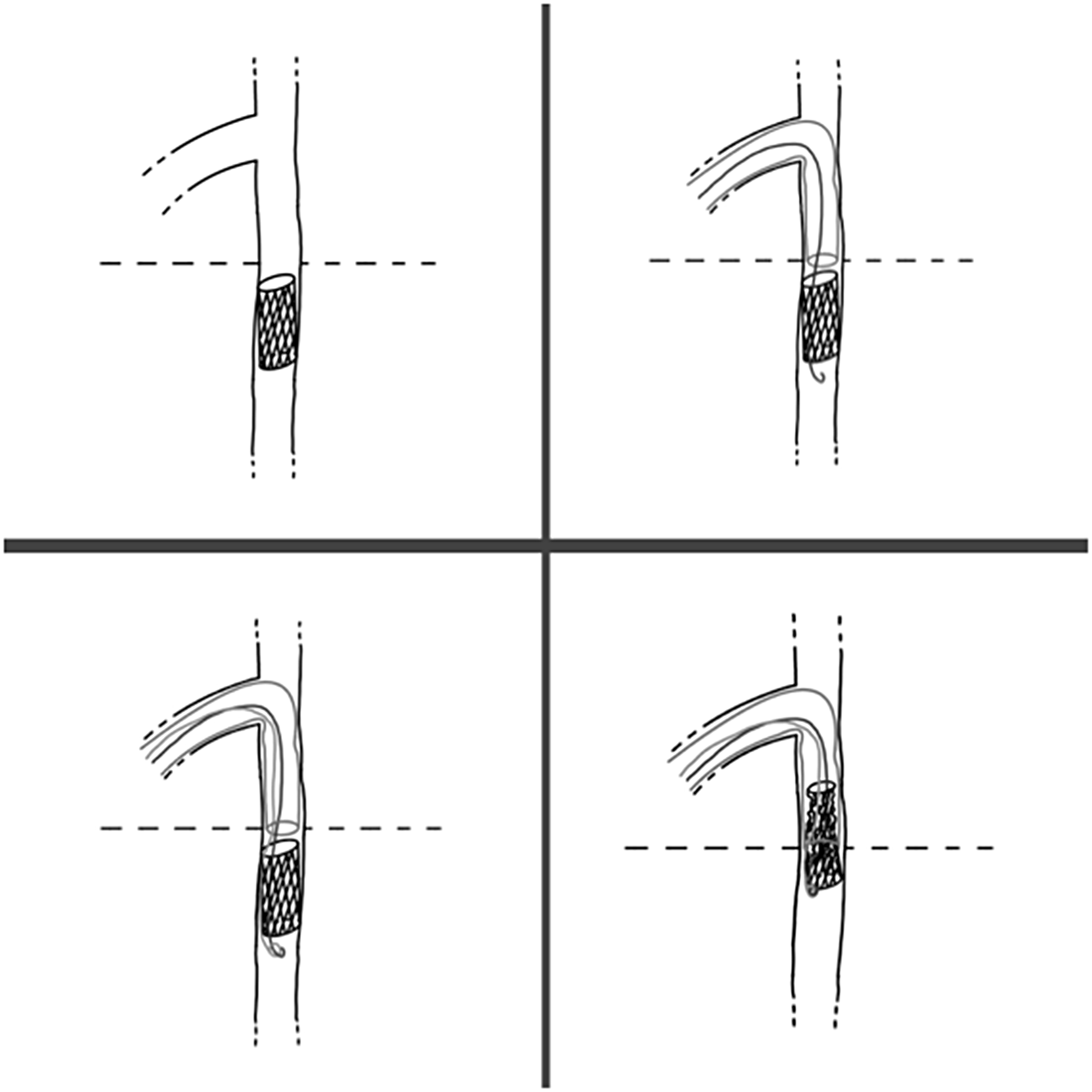
( a ) Duct angiography in lateral view. ( b ) Stent embolized in abdominal aorta, a guide-wire crossed the stent through the struts. ( c ) The loop made with the guide-wire and the microsnare engaged the stent. ( d ) Retrieval of the stent inside the Flexor catheter.

The left femoral artery was immediately accessed and angiography with a Pigtail 3.3 Fr was performed. We decided to retrieve the stent into the Flexor 4 Fr, anterogradely. During the advancement of the long sheath into the thoracic aorta, the stent migrated more distally in the abdominal aorta at the level of renal arteries (Figure 1B, Figure 2A). With a Pigtail positioned distally from the stent from the arterial access, we stopped stent movements and blocked it into the abdominal aorta. Numerous attempts were made to capture the stent with a Microsnare 4/6 (PFM Medical) without any success. After that, from the venous side through the ductus, a Galeo guide-wire of 0.014 inch was inserted into the struts of the stent (Figure 2B); with a Microsnare 4/6, from the same side, the distal part of this guide-wire was captured (Figure 1C, Figure 2C). Thanks to the support of the Microsnare/guide-wire loop, the stent was retrieved almost completely into the Flexor catheter, and the entire system was removed through the ductus into the right ventricle (Figure 1D, Figure 2D). During the procedure, the baby remained hemodynamically stable. The procedure was uncomplicated. A new attempt was made to stent the arterial duct with a drug-eluting stent (Ultimaster Tansei, Terumo), 3 x 15 mm, inflated at 16 atm until reaching a diameter of 3.2 mm, resulting in a good final result.
( a ) Embolized stent in the abdominal aorta. ( b ) Guide-wire inside the stent struts. ( c ) Loop made of guide-wire and microsnare. ( d ) Retrieval of the stent inside a Flexor.

The baby was discharged 3 weeks later in good clinical condition. At the last follow-up, a low gradient was detected echocardiographically at pulmonary level, and the stent was patent.
Discussion
Percutaneous embolization of stent into small vessels like those of a premature baby is an extremely challenging complication. In some cases, it is necessary lo leave the stent in a distal vascular branch or even to remove it surgically with subsequent morbidity. Reference Alwi and Mood2 Aortic flow obstruction caused by an embolized stent in newborn of such low weight can cause dramatic consequences such as mesenteric infarction, kidney failure, aortic dissection and even patient’s death. Reference Scott Hittinger, Varsha Bandisode and Shahryar Chowdhury3
In our case, albeit the very low weight of the patient, the stent was successfully retrieved thanks to a loop made with a guide-wire and a microsnare. Since it was impossible to recapture directly the stent, we retrieved it indirectly. In fact, we passed a coronary guide-wire through the stent struts and snared the distal end of the wire. In this way, the stent was pulled back inside a Flexor catheter. The wire was strong enough and the stent soft enough to be successfully retrieved.
To our knowledge, no reports about stent retrieval in premature babies were published. There are some reports about catheter fragment retrieval in small babies, mainly with goose-neck snare. Reference Demirel, Guven, Okan and Saygili4,Reference Young and Shaddy5
Some case reports describe successful coronary stent retrieval from the coronary circulation with different techniques: with snare devices, balloon angioplasty catheter or twisting guide-wire. Reference Colón-Arias, Moronta-Franco and Gutiérrez-Martínez6 The retrieval likelihood depends on the physical properties of the stent and its ability to lose the original shape and be captured inside a catheter. In our case, it was possible to retract the de-structured stent inside a Flexor sheath. Once removed, the stent appeared spiralized and elongated having conformed to the catheter shape.
Several risk factors concur in the occurrence of stent embolization, such as stent dimensions and ductal morphology. Reference Alwi and Mood2
Patient’s low weight and prematurity are important risk factors to be considered when performing ductal stenting. Backes et al Reference Backes, Cua and Kreutzer7 described the incidence of adverse events in relation to the weight of patients during cardiac catheterization, showing the highest incidence of adverse events in the group of children weighing less than 2 kg.
Aggarwal, Reference Aggarwal, Petit, Glatz, Goldstein and Qureshi8 describing ductal stenting technique, suggested this rule for stent size selection: for infants weighing > 3.0 kg, a 3.5–4 mm diameter stent is chosen; for those weighing 2.0–3.0 kg, a 3.5 mm diameter and for those < 2 kg, a 3 mm stent is chosen for deployment.
In our case, during the first attempt of stent implantation, we decided to use a stent of 2.5 mm in order to avoid pulmonary overcirculation. The discrepancy of stent size and ductal diameter may have contributed to instability and subsequent embolization in the abdominal aorta.
Other risk factor for stent embolization is the conical shape of the ductus Reference Aggarwal, Petit, Glatz, Goldstein and Qureshi8 that does not permit complete adherence of the stent to the ductal wall. This was also our case in which the ductus had a diameter of 4.5 mm at the aortic ampulla and 1.8 mm at the pulmonary end.
In conclusion, ductal stenting in congenital heart disease is a challenging procedure particularly in small babies. Presently reported was the successful retrieval of a coronary stent using a loop of a guide-wire and a microsnare via percutaneous approach without complications.