Case report
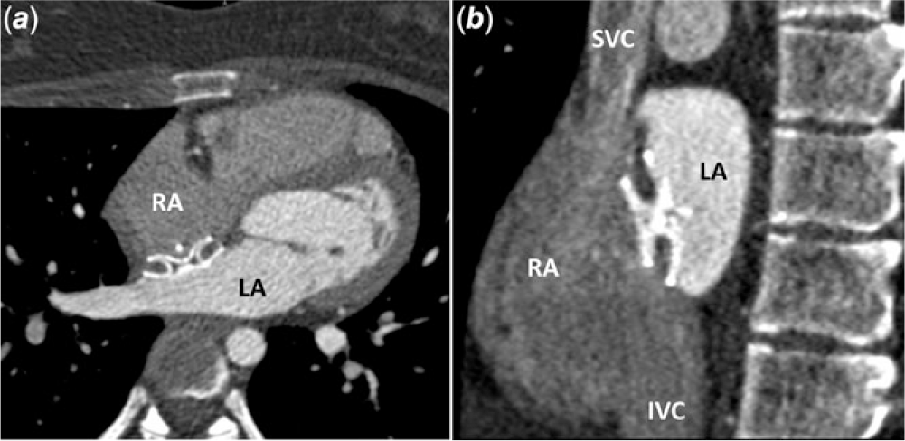
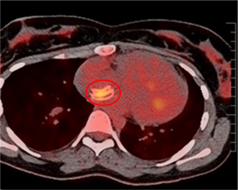
A 2-year-old female was diagnosed with atrial septal defect ostium secundum type because of a heart murmur. During the follow-up, a progressive dilatation of right heart cavities was gradually observed and an elective percutaneous atrial septal defect closure was performed when she was 14. The procedure was carried out under general anaesthesia and transesophageal echocardiographic monitoring. A 13 mm Amplatzer Septal Occluder device was deployed successfully. Aspirin was given for 6 months. Eleven months after the atrial septal defect closure, and a week following a viral upper airway infection, the patient came to the emergency department because of an oppressive chest pain, which increased with the decubitus position. The patient had no fever and the blood tests were not suggestive of active infection. Therefore, no microbiological testing was performed. Cardiac enzymes were negative. The electrocardiogram showed a very subtle ST elevation, and the transthoracic echocardiogram showed a mild pericardial effusion. All these data were compatible with the diagnosis of acute pericarditis. Since the patient presented a remarkable chest pain, the interventional team suggested steroids therapy as the first choice in order to achieve a prompt clinical remission. This treatment was maintained for 3 weeks. Unfortunately, the pericardial effusion decreased but the chest pain remained. A transesophageal echocardiogram and angio- CT demonstrated a norm-positioned device (Fig 1a and b) with no cardiac erosion. In a multidisciplinary meeting, an idiopathic pericarditis was regarded as the most probable diagnosis and empirical therapy with aspirin and colchicine was administered for 8 weeks. Nevertheless, the chest pain persisted. During the next following months, the patient underwent gynaecological assessment to discard breast pathology. An allergy and a rheumatologic evaluation searching for nickel and titanium allergy and autoimmune diseases, respectively, were also carried out. All these evaluations were negative. Five months after the pain started, a Positron Emission Tomography evidenced device radiotracer 18F-Fludeoxyglucose (FDG) captation of the tissue in contact with the discs, suggesting a low-grade inflammatory reaction (Fig 2). As the pain persisted, the possibility of a surgical device explantation was offered to the family but it was rejected. The patient also received psychological support. After 18 months of pain, a new Positron Emission Tomography scan continued to show a mild inflammatory reaction. Two and a half years later, the patient referred to a significant improvement of the pain, but it was not until 5 years after its onset that she became completely asymptomatic. No clear reason for the gradual improvement could be identified.
CT. (a) Axial plane. (b) Sagittal plane. The images show a norm-positioned device. IVC = inferior vena cava; LA = left atrium; RA = right atrium; SVC = superior vena cava.

Positron Emission Tomography scan. Axial plane showing radiotracer captation by the atrial septum tissue in contact with the discs of the device (yellow area, inside the red circle).

Discussion
Transcatheter closure of atrial septal defect is considered a safe and effective procedure. Reference Jalal, Hascoet and Baruteau1–Reference Tanghöj, Liuba, Sjöberg, Rydberg and Naumburg5 Most of the devices used, including the Amplatzer Septal Occluder, are made from nitinol, an alloy of nickel and titanium that confers not only special properties like superelasticity and shape memory, but also corrosion resistance and tissue biocompatibility. Reference Jalal, Hascoet and Baruteau1,Reference Sigler and Jux6,Reference Jalal, Hascoët and Gronier7 In some occasions, however, complications may occur either during the procedure or several years later. Reference Jalal, Hascoet and Baruteau1,Reference Jalal, Hascoët and Gronier7,Reference Mendirichaga, Smairat and Sancassani8 Thrombus formation, device embolisation, cardiac perforation, aortic erosion, and arrhythmias have been described after the procedure. Reference Jalal, Hascoet and Baruteau1,Reference Knepp, Rocchini, Lloyd and Aiyagari3,Reference Tanghöj, Liuba, Sjöberg, Rydberg and Naumburg5,Reference Jalal, Hascoët and Gronier7,Reference Divekar, Gaamangwe, Shaikh, Raabe and Ducas9 Although chest pain has been reported, its potential causes have been poorly investigated. Reference Knepp, Rocchini, Lloyd and Aiyagari3,Reference Alizadehasl, Neshati Pir Borj, Sadeghpour, Firouzi, Sanati and Movassaghi4,Reference Jalal, Hascoët and Gronier7 Knepp et al reported 94 case series of transcatheter atrial septal defect closure with an Amplatzer Septal Occluder 18 of whom presented chest pain during the follow-up. A possible cardiac cause, however, was identified in only two cases, a haemorrhagic pericarditis and a supraventricular tachycardia. Reference Knepp, Rocchini, Lloyd and Aiyagari3 Alizadehasl et al described 23 cases of chest pain over a total of 313 case series who underwent percutaneous atrial septal defect (n = 289) or PFO (n = 24) closure. Again, only two cases appeared to have a cardiac cause, myocarditis in one, and pericarditis with pericardial effusion in the other. Reference Alizadehasl, Neshati Pir Borj, Sadeghpour, Firouzi, Sanati and Movassaghi4 Thus, in both studies, the vast majority of patients presented a chest pain of unclear cause. In contrast, Jalal referred to a lower frequency, only 3 cases of atypical chest pain amongst 1326 children, who underwent percutaneous atrial septal defect closure with an Amplatzer Septal Occluder, but also without an apparent cardiac cause. Reference Jalal, Hascoët and Gronier7
When dealing with a sudden and intense chest pain after transcatheter atrial septal defect closure, it is essential to discard device erosion, which can occur from a few hours to several years after the procedure. Reference Jalal, Hascoet and Baruteau1,Reference Mendirichaga, Smairat and Sancassani8,Reference Divekar, Gaamangwe, Shaikh, Raabe and Ducas9 In the case, herein, described the transesophageal echocardiogram and the angio-CT showed a norm-positioned device with no erosion.
Other symptoms such as chest discomfort, palpitations, urticarial rash, pericardial effusion, or migraine headache have also been documented after an atrial septal defect closure, which in part have been ascribed to nickel hypersensitivity. Reference Jalal, Hascoet and Baruteau1,Reference Spina, Muller, Jansz and Gunalingam10 In our case, the chest pain started 11 months after the procedure and allergy to nickel and titanium were discarded with a skin patch test. We first hypothesised that the cause of her chronic chest pain was a pericardial inflammation associated with a respiratory infection that evolved chronically. Later on, however, the presence and persistence of radiotracer in the atrial septum tissue in the Positron Emission Tomography led us to suspect that it was a local inflammatory response rather than a chronic pericarditis, which is the most likely cause of the protracted chest discomfort. This local and chronic inflammatory reaction may be due to the mechanical friction between the device discs and the septal tissue and/or to a foreign body inflammatory response. Reference Sigler, Jux and Ewert2,Reference Suda, Raboisson, Piette, Dahdah and Miró11 In this regard, inflammatory reactions with lymphocytic infiltration and multinucleated foreign body giant cells have already been demonstrated in animal studies and also in explanted human devices. Reference Jalal, Hascoet and Baruteau1,Reference Sigler, Jux and Ewert2,Reference Sigler and Jux6 Why the chest pain appeared 11 months after the procedure and not earlier remains to be clarified. It is conceivable that the patient could have been suffering an asymptomatic and latent device-related inflammation, which was exacerbated by the acute respiratory infection.
The chronic chest pain after transcatheter atrial septal defect closure is an unusual complication and a multidisciplinary approach seems to be useful to rule out its principal causes. A chronic device-related mild inflammatory response could be the main cause of the pain.
Acknowledgements
None.
Financial support
This research received no specific grant from any funding agency, commercial, or not-for-profit sectors.
Conflicts of interest
None.
Ethical standards
The authors assert that all procedures contributing to this work comply with the ethical standards and with the Helsinki Declaration of 1975, as revised in 2008, and has been approved by the Institutional Ethics Committee of Clinical Investigation (Comité Ètic d’Investigació Clínica, Hospital Universitari Vall d’Hebron). A written informed consent was obtained from the girls’ parents.