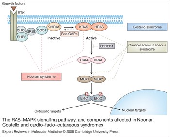
The Ras-induced mitogen-activated protein kinase (MAPK) cascade (the Ras–MAPK pathway) plays a central role in the signal transduction of extracellular stimuli to the intracellular environment (for review see Ref. Reference Hancock1). The small GTPase Ras is a critical signalling hub in the cell that is activated by receptor tyrosine kinases, G-protein-coupled receptors, cytokine receptors and extracellular matrix receptors. This pathway has been studied extensively in the context of oncogenesis since its dysregulation is one of the primary causes of cancer, with Ras found to be somatically mutated in ~20% of malignancies (Ref. Reference Bos2). Ras genes exist as a multigene family, with HRAS, NRAS and KRAS being the most commonly studied (for review see Ref. Reference Midgley and Kerr3). One signalling pathway activated by Ras is the MAPK cascade (Fig. 1). Activation of Ras leads to the activation of Raf (ARAF, BRAF and/or CRAF/RAF1), the first kinase of the cascade (‘MAPK kinase kinase’). Raf phosphorylates and activates MEK1 and/or MEK2 (‘MAPK kinase’, or MAP2K), which in turn phosphorylates and activates ERK1 and/or ERK2 (MAPK). ERK1/2 are the ultimate effectors of the MAPK cascade, and exert their function on a large number of downstream molecules that vary depending upon cell type and mechanism of pathway activation; activated ERK1/2 phosphorylates both nuclear and cytosolic substrates, including transcription factors, membrane proteins and protein kinases, which in turn control vital cellular functions including cell cycle progression and differentiation (Ref. Reference Yoon and Seger4).
The Ras–MAPK signalling pathway, and components affected in Noonan, Costello and cardio–facio–cutaneous syndrome. The Ras-induced mitogen-activated protein kinase (MAPK) casade (RAS–MAPK pathway) is highly conserved among eukaryotic organisms and is critically involved in cell proliferation, differentiation, motility, apoptosis and senescence. Ras (KRAS/HRAS in the figure) cycles between a GDP-bound, inactive state and a GTP-bound active state. The transition between these two states is controlled by GTPase-stimulating proteins (GAPs) and guanine-nucleotide-exchange factors (GEFs). The Ras–MAPK pathway is activated by multiple extracellular stimuli. In the figure, Ras activation is shown following growth factor binding to a receptor tyrosine kinase (RTK), which, through the docking proteins GRB2 and SHC, activates the GEF SOS1. Activated Ras recruits Raf (CRAF/BRAF) to the plasma membrane, which results in its activation. Active Raf, the first kinase of the cascade, phosphorylates MEK1 and/or MEK2 (MAP2Ks), which then phosphorylates ERK1 and/or ERK2 (MAPKs), the terminal effectors of the pathway. Activated ERK1 and ERK2 phosphorylate both nuclear and cytosolic substrates. The Ras–MAPK pathway proteins with germline mutations in their respective genes that are associated with Noonan syndrome, Costello syndrome and cardio–facio–cutaneous (CFC) syndrome are indicated by dashed lines. Noonan syndrome has been associated with mutations in PTPN11 (protein product SHP2, a nonreceptor tyrosine phosphatase), SOS1, RAF1 (encoding CRAF) and KRAS. The causal gene for Costello syndrome is HRAS. CFC is caused by mutations in BRAF, MAP2K1 and MAP2K2. The role of KRAS in Noonan syndrome and CFC has yet to be defined. SPRED1 is a member of the Sprouty/Spred family of proteins that act as a negative regulator of the Ras–Raf interaction. Abbreviations: BRAF, v-raf murine sarcoma viral oncogene homologue B1; HRAS/KRAS, v-Ha-ras Harvey or Ki-ras2 Kirsten rat sarcoma viral oncogene homologue; MAP2K1/MAP2K2, mitogen-activated protein kinase kinase 1 or 2 (also known as MEKs); PTPN11, protein tyrosine phosphatase, nonreceptor type 11 (encoding SHP2); RAF1, v-raf-1 murine leukaemia viral oncogene homologue 1 (encoding CRAF); RASA1, RAS p21 protein activator; SOS1, son of sevenless homologue 1; SPRED1, sprouty-related, EVH1-domain-containing 1.

Since the Ras–MAPK pathway has such an important function in all cell types, proper signalling of the pathway is critical for normal embryonic development. In Xenopus, elevated MAPK activity initiates oocyte meiotic maturation (Ref. Reference Haccard5) and metaphase arrest in cleaving embryos (Refs Reference Haccard6, Reference Haccard7). In addition, activation of the MAPK pathway in Xenopus embryos is important in mesoderm induction and patterning (Refs Reference Curran and Grainger8, Reference Gotoh9, Reference Gotoh and Nishida10). Highly regulated control of MAPK is also observed during Drosophila development (Ref. Reference Gabay, Seger and Shilo11). In zebrafish development, the Fgf–Ras–Mapk pathway inhibits bone morphogenic protein expression and controls the dorsoventral patterning of the embryo (Ref. Reference Furthauer12). In mice, sustained Erk activation is seen in both extraembryonic structures and the embryo proper including the ectoplacental cone, extraembryonic ectoderm, limb buds, branchial arches, frontonasal process, brain, tail bud, foregut and liver. Transient Erk phosphorylation is found within the neural crest, peripheral nervous system, nascent blood vessels and the early-forming structures of the ear, eye and heart (Ref. Reference Corson13).
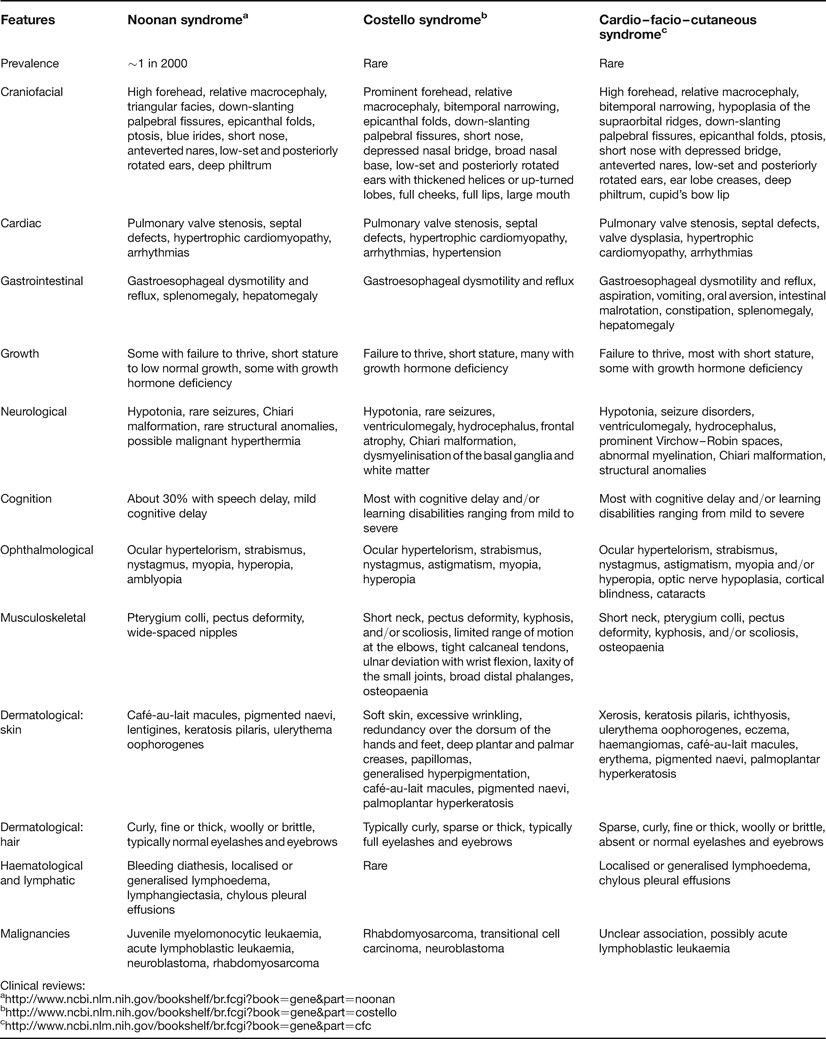
Dysregulation of the Ras–MAPK pathway in human development also has phenotypic consequences: over the past few years a class of genetic syndromes has emerged that are caused by germline mutations in genes that encode components of this pathway, as summarised in Table 1. Many of these syndromes share characteristic craniofacial dysmorphology, cardiac malformations and cutaneous, musculoskeletal and ocular abnormalities. In addition, mutations in this pathway resulting in dysregulation may cause varying degrees of neurocognitive impairment and, in some syndromes, an increased risk of developing cancer. While each syndrome has a distinct phenotype and clinical course, Noonan syndrome, Costello syndrome and cardio–facio–cutaneous (CFC) syndrome in particular often share numerous similarities of phenotypic features, making clinical diagnosis very challenging, especially in newborns and infants in whom distinguishing phenotypic features may not yet be evident. Because of this phenotypic overlap, a great deal of debate existed prior to our understanding of the molecular aetiology of these syndromes as to whether Noonan syndrome, CFC syndrome and Costello syndrome were distinct syndromes, a contiguous gene disorder, or allelic variants with variable expressivity (Refs Reference Leichtman14, Reference Fryer, Holt and Hughes15, Reference Neri and Zollino16, Reference Neri and Opitz17, Reference Tartaglia18). It is now established that each of these syndromes is genetically distinct, resulting from mutations in different genes of the Ras–MAPK pathway. Because of the developmental and phenotypic similarities between Noonan syndrome, Costello syndrome and CFC syndrome, this review focuses on the molecular aetiology and phenotypic features of these three disorders.
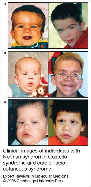
Clinical images of individuals with Noonan syndrome, Costello syndrome and cardio–facio–cutaneous syndrome. (a) This individual with Noonan syndrome, shown at 11 months and at 10 years of age, has a M504V SHP2 missense mutation. (b) This individual with Costello syndrome is pictured at 11 months of age and at 23 years. He has the common G12S HRAS missense mutation, seen in over 80% of individuals with this syndrome. (c) This child, who has typical craniofacial features of cardio–facio–cutaneous syndrome, has a rare small in-frame deletion in exon 11 of the BRAF gene; she is shown at 1 year and 10 years of age. Pictures were obtained and are reproduced with informed consent. Abbreviations: BRAF, v-raf murine sarcoma viral oncogene homologue B; HRAS, v-Ha-ras Harvey rat sarcoma viral oncogene homologue; SHP2, Src-homology-2-containing tyrosine phosphatase (encoded by PTPN11).

Genetic syndromes caused by germline mutations in genes encoding Ras–MAPK pathway components

aFunctions of most of these gene products in the Ras–MAPK pathway are shown in Figure 1.
Abbreviations: BRAF, v-raf murine sarcoma viral oncogene homologue B1; HRAS/KRAS, v-Ha-ras Harvey or Ki-ras2 Kirsten rat sarcoma viral oncogene homologue; MAP2K1/MAP2K2, mitogen-activated protein kinase kinase 1 or 2 (proteins also known as MEKs); PTPN11, protein tyrosine phosphatase, nonreceptor type 11 (encoding SHP2); RAF1, v-raf-1 murine leukaemia viral oncogene homologue 1 (encoding CRAF); RASA1, Ras p21 protein activator; SOS1, son of sevenless homologue 1; SPRED1, sprouty-related, EVH1-domain-containing 1.
Noonan syndrome
Clinical phenotype
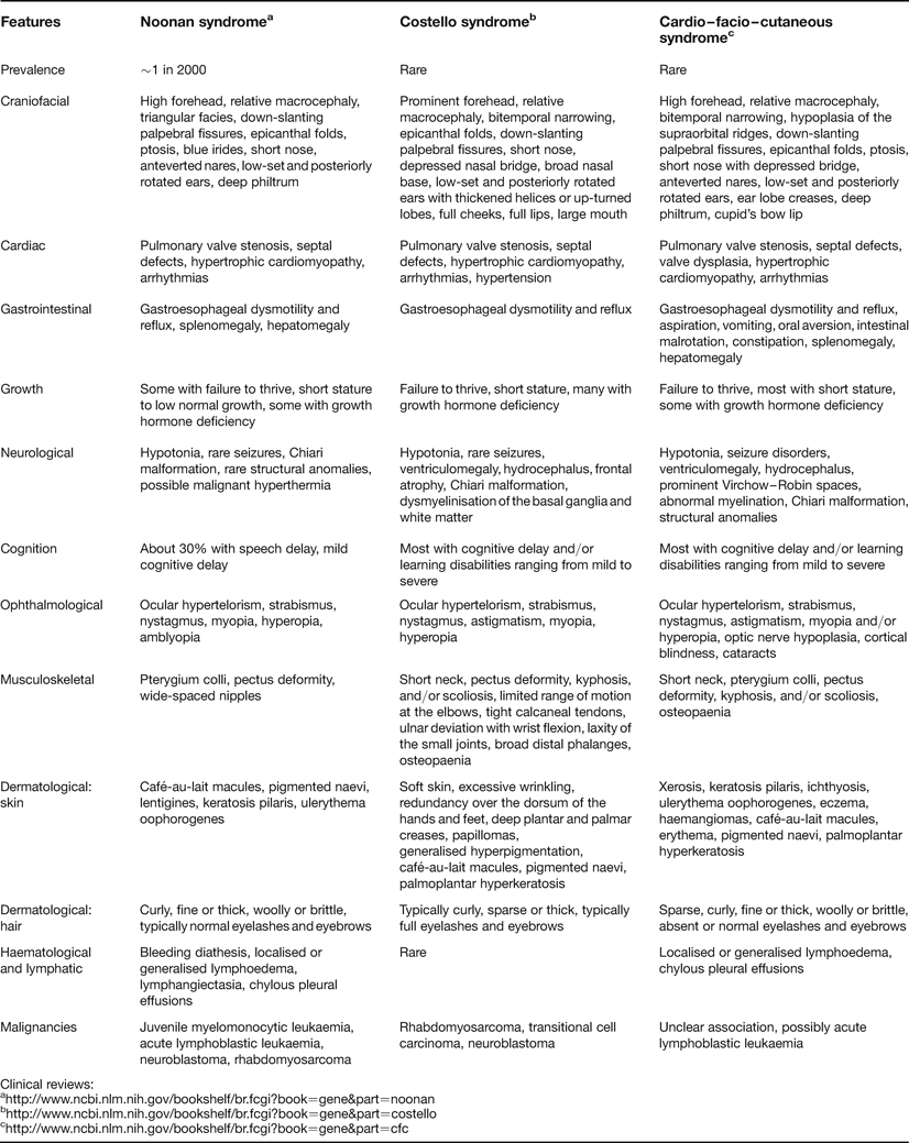
Noonan syndrome was first described by Noonan, a paediatric cardiologist, as an autosomal dominant genetic syndrome associated with congenital cardiac disease (Ref. Reference Noonan19; for review see Ref. Reference Shaw20). Noonan syndrome is characterised by distinctive craniofacial features (although many features overlap with CFC syndrome), including a broad forehead, hypertelorism, down-slanting palpebral fissures, ptosis, posteriorly rotated low-set ears and a high-arched-palate (Fig. 2a; Table 2). Congenital heart defects occur in the majority of individuals, including pulmonary valve stenosis, which is the most common, followed by hypertrophic cardiomyopathy (Ref. Reference Lin21). Other anomalies such as septal defects, tetralogy of Fallot and coarctation of the aorta may occur, but are less frequent. Individuals with Noonan syndrome are usually short in stature, and many have feeding difficulties and show failure to thrive in infancy. Undescended testicles are common in male infants. Ophthalmological abnormalities affect most and may include strabismus, myopia, hyperopia and nystagmus, as well as anterior segment and fundal abnormalities (Ref. Reference Reynolds22). Other phenotypic features of Noonan syndrome may include musculoskeletal and cutaneous anomalies, endocrine dysfunction and lymphoedema. Bleeding disorders may be seen in up to half of individuals with Noonan syndrome (Ref. Reference Massarano23). Neurological findings appear to be not as common in Noonan syndrome as in Costello syndrome and CFC syndrome; neurocognitive delay of variable degree is present in 25–30% of affected individuals (Ref. Reference Lee24). Chiari malformation has been reported in Noonan syndrome (Ref. Reference Holder-Espinasse and Winter25), a finding which is also seen in Costello syndrome and CFC syndrome. Individuals with Noonan syndrome may have an increased risk of developing cancer. The predisposition to cancer associated with Noonan syndrome includes acute lymphoblastic leukaemia, juvenile myelomonocytic leukaemia, neuroblastoma and rhabdomyosarcoma (Refs Reference Choong26, Reference Attard-Montalto, Kingston and Eden27, Reference Bader-Meunier28, Reference Moschovi29, Reference Roti30, Reference Piombo31).
General phenotypic characteristics of Noonan, Costello and cardio-facio-cutaneous syndromes
Molecular pathology of Noonan syndrome
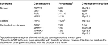
The inheritance of Noonan syndrome is autosomal dominant; however, many affected individuals have de novo mutations. To date, four genes have been associated with Noonan syndrome (Tables 1 and 3). The first gene mutation identified as causing Noonan syndrome, reported in 2001, was in PTPN11 (Ref. Reference Tartaglia32). Mutations in PTPN11 were identified in ~40–50% of individuals with clinically diagnosed Noonan syndrome. Subsequently, SOS1 mutations have been reported in ~13% of individuals with Noonan syndrome (Refs Reference Roberts33, Reference Tartaglia34). Mutations in RAF1 have also been reported to cause Noonan syndrome, with the percentage of mutation-positive individuals ranging from 3% to 17% (Refs Reference Pandit35, Reference Razzaque36). In addition, KRAS mutations have been reported to be responsible for <2% of individuals with Noonan syndrome (Refs Reference Schubbert37, Reference Zenker38).
Genetic characterisation of Noonan, Costello and cardio–facio–cutaneous syndromes

aApproximate percentage of affected individuals carrying mutations in each gene.
bPresently, HRAS is the only gene associated with Costello syndrome; however, this does not preclude the discovery of other genes associated with this disorder in the future.
PTPN11 mutations
PTPN11, the predominant gene associated with Noonan syndrome, encodes a nonreceptor protein tyrosine phosphatase known as SHP2; missense mutations are found in approximately half of all clinically diagnosed Noonan syndrome patients examined (Ref. Reference Tartaglia32). The locus of PTPN11 is in chromosome band 12q24.1. The PTPN11 gene spans 90 kb and comprises 16 exons containing a coding sequence of 1779 bases (Ref. Reference Tartaglia39). The SHP2 protein consists of N-terminal and C-terminal SH2 domains and a catalytic domain (PTP). The catalytic function of the protein is autoinhibited through a blocking interaction of the N-SH2 domain and the catalytic PTP domain (Ref. Reference Hof40). The majority of Noonan-syndrome-causing mutations in PTPN11 are missense, and are restricted to specific regions within the gene. The mutations cluster in exons 3, 7, 8 and 13, which mainly affect residues involved in the interface between the N-SH2 and PTP domains, with the most common mutation being an A to G change at nucleotide 922 that results in an N308D substitution. This mutation, and others in this region, affect the stability of the catalytically inactive form, and therefore interfere with the protein's ability to transition from the active to the inactive conformation (Refs Reference Keilhack41, Reference Tartaglia42). Thermodynamic modelling (Ref. Reference Tartaglia32), and in vitro (Refs Reference Tartaglia39, Reference Fragale43, Reference Niihori44) and in vivo (Refs Reference Araki45, Reference O'Reilly46) studies, have demonstrated that these SHP2 mutants produce a gain of function, resulting in increased signalling down the Ras–MAPK pathway. Additionally, mutations that affect regions outside the N-SH2–PTP interaction region have also been shown to cause a gain of function of SHP2 through other mechanisms (Ref. Reference Martinelli47).
Individuals with Noonan syndrome are predisposed to juvenile myelomonocytic leukaemia. One Noonan-syndrome-causing mutation, which results in a T73I residue substitution, has been specifically associated with this predisposition (Ref. Reference Choong26). This same mutation is somatically present in individuals with juvenile myelomonocytic leukaemia who do not have Noonan syndrome (Ref. Reference Kratz48). Other PTPN11 somatic mutations are also present in leukaemia and in some tumours (Refs Reference Kratz48, Reference Tartaglia49, Reference Bentires-Alj50, Reference Chen51, Reference Loh52, Reference Miyamoto53, Reference Schubbert54, Reference Yoshida55), but the somatic PTPN11 mutations associated with cancer are typically distinct from those associated with Noonan syndrome, and usually confer a higher activation of SHP2 function (Refs Reference Tartaglia42, Reference Schubbert54). Not only do PTPN11 mutations demonstrate a paternal bias, but Noonan syndrome affects males approximately twice as often as it does females. This sex ratio bias may be due to sex-specific developmental effects of PTPN11 mutations that result in a higher mortality of female embryos (Ref. Reference Tartaglia56).
KRAS mutations
KRAS mutations have been implicated in a small percentage of individuals with Noonan syndrome (Table 3). The KRAS gene is located on 12p12.1 and consists of four coding exons. Two splice variants exist: KRASA and KRASB (Ref. Reference Friday and Adjei57). KRASA is expressed in a tissue-specific and developmentally restricted fashion, whereas KRASB is ubiquitously expressed. KRAS mutations were independently identified in a small number of individuals clinically diagnosed with Noonan and CFC syndromes (Refs Reference Schubbert37, Reference Niihori58).
When 175 PTPN11 mutation-negative individuals with the clinical diagnosis of Noonan syndrome and 12 individuals with the clinical diagnosis of CFC syndrome were examined for KRAS mutations (Ref. Reference Schubbert37), five Noonan syndrome and one of the CFC syndrome individuals were found to have novel missense mutations in KRAS. The mutations in Noonan syndrome individuals included T58I, V14I and D153V. The phenotype associated with the T58I substitution was reported to be more severe than with the V14I substitution. Functional studies revealed that these mutations activate the MAPK pathway (Refs Reference Schubbert37, Reference Schubbert59). Ras cycles between a GDP-bound inactive state and a GTP-bound active state. The transition between these two states is controlled by GTPase-stimulating proteins (GAPs) and guanine-nucleotide-exchange factors (GEFs). Biochemical analyses of mutations T58I and V14I showed a reduced intrinsic GTPase activity of Ras compared with the wild-type protein, resulting in a reduction in Ras inactivation and therefore increased signalling down the MAPK pathway (Ref. Reference Schubbert37). The one CFC syndrome individual in the study had a missense KRAS mutation resulting in a P34R residue substitution.
In a separate study, 43 individuals with the clinical diagnosis of CFC syndrome were also screened for KRAS mutations (Ref. Reference Niihori58). Three individuals had missense KRAS mutations that resulted in either a G60R or D153V residue substitution, with the latter mutation also being reported in clinically diagnosed Noonan syndrome individuals (Ref. Reference Schubbert37). Subsequently, KRAS mutations have been identified in cohorts of individuals with severe Noonan syndrome (Refs Reference Zenker38, Reference Carta60).
These studies demonstrate that a variable phenotype is associated with KRAS mutations, and reinforce the difficulty of clinically distinguishing between Noonan syndrome and CFC syndrome, even given knowledge of underlying molecular defects. Further evaluation of the patient populations and functional studies will resolve these issues.
SOS1 mutations
SOS1 missense mutations were reported independently by two groups in individuals with Noonan syndrome and without mutations in either PTPN11 or KRAS (Refs Reference Roberts33, Reference Tartaglia34). At present, SOS1 mutations are the second most common cause of Noonan syndrome after PTPN11 mutations, and account for ~13% of individuals with Noonan syndrome (Table 3). SOS1, located on chromosome 2p22.1, consists of 23 exons and encodes a Ras-GEF. Ras-GEF proteins are responsible for stimulating the conversion of Ras from the inactive GDP-bound form to the GTP-bound active form. The majority of SOS1 missense mutations reported were located in codons encoding residues that are responsible for stabilising the SOS1 protein in an inhibited conformation. Therefore, alteration of these residues disrupts the autoinhibition of SOS1 Ras-GEF activity, resulting in a gain of function of SOS1 and a subsequent increase in the active form of Ras and increased Ras–MAPK pathway signalling (Refs Reference Roberts33, Reference Tartaglia34).
The phenotype resulting from SOS1 mutations, although it falls within the spectrum of characteristics encompassed by Noonan syndrome, appears to be distinctive. There is a higher prevalence of ectodermal abnormalities and a lower prevalence of cardiac abnormalities than observed with PTPN11 mutations (atrial septal defects were present in individuals with SOS1 mutations that produced the highest degree of SOS1 protein activation). Although Noonan and CFC syndromes may phenotypically overlap, one initial study did not identify SOS1 mutations in a cohort of individuals clinically diagnosed with CFC syndrome (Ref. Reference Zenker61). It is unclear if individuals with germline mutations in SOS1 are predisposed to cancer. In a survey of 810 primary human malignancies, somatic mutations in SOS1 were very rare, which may hold significant implications as to the predisposition for developing cancer in Noonan syndrome individuals who have SOS1 mutations (Ref. Reference Swanson62).
RAF1 mutations
Two groups simultaneously reported mutations in RAF1 in Noonan syndrome individuals who did not have mutations in PTPN11, SOS1 or KRAS (Refs Reference Pandit35, Reference Razzaque36). The majority of missense RAF1 mutations associated with Noonan syndrome were clustered in two regions: in conserved region 2 (CR2) flanking S259; and in conserved region 3 (CR3), in and surrounding the activation segment (Refs Reference Pandit35, Reference Razzaque36). In addition, two mutations in the C-terminus were also reported (Ref. Reference Pandit35). Mutations flanking S259 all resulted in a gain of function, since this region is responsible for autoinhibition of kinase activity (Refs Reference Pandit35, Reference Razzaque36). Mutations in the CR3 and the C-terminus also resulted in increased kinase activation (Refs Reference Pandit35, Reference Razzaque36). By contrast, mutations within the activation segment were shown to have lower kinase activity than wild-type CRAF (the protein product of RAF1) (Ref. Reference Pandit35). All the gain-of-function mutations in RAF1 were associated with a much higher prevalence of hypertrophic cardiomyopathy compared with other Noonan syndrome individuals who have other known gene mutations (~95% compared with ~18%, respectively) (Ref. Reference Pandit35).
The mammalian genome contains three Raf genes: ARAF, BRAF and RAF1. RAF1 is located on chromosome 3p25, contains 17 exons and spans 80 kb. Its protein product CRAF is a serine/threonine kinase that is one of the direct downstream effectors of Ras and is, in turn, responsible for the activation of MEK1 and MEK2. CRAF has a much lower basal kinase activity than BRAF, with somatic RAF1 mutations being very rare in cancer (Ref. Reference Emuss63). The few RAF1 mutations associated with cancer are not the same as those associated with Noonan syndrome.
Costello syndrome
Clinical phenotype
Two patients having characteristic craniofacial dysmorphology, high birth weight with subsequent failure to thrive, ectodermal anomalies including nasal papillomas and neurocognitive delay were initially reported by Costello at a genetics meeting in 1971 (Ref. Reference Costello64) and later described in more detail in 1977 (Ref. Reference Costello65). Subsequently, over 100 Costello syndrome patients have been reported in the literature. Costello syndrome is a rare congenital disorder with multiple anomalies that include characteristic dysmorphic craniofacial features, cardiac, musculoskeletal and ectodermal abnormalities, failure to thrive, and neurocognitive delay (for clinical and molecular reviews see Refs Reference Hennekam66, Reference Rauen67). Phenotypic features become apparent in the perinatal period, with polyhydramnios in utero and with many affected children born prematurely, albeit with increased birth weight. Newborns have many clinical features overlapping with Noonan syndrome and CFC syndrome, making the clinical diagnosis challenging (Fig. 2b). As the child matures, phenotypic features become more distinctive. Facial features become coarse and typically include macrocephaly with a prominent forehead, epicanthal folds, down-slanting palpebral fissures, and a short nose with a depressed nasal bridge and a broad tip. Additionally, the ears are low-set and posteriorly rotated, with thickened helices and lobes. The cheeks may be full and the mouth large with full lips (Table 2).
A more detailed examination of the phenotype reveals dermatological manifestations: soft skin, excessive wrinkling and redundancy over the dorsum of the hands and the feet, along with deep plantar and palmar creases. Other ectodermal features may include generalised hyperpigmentation, papillomas and curly hair. Musculoskeletal manifestations include limited range of motion at the elbows, tight calcaneal tendons, ulnar deviation with wrist flexion, laxity of the small joints and broad distal phalanges (Ref. Reference Yassir, Grottkau and Goldberg68). Cardiac abnormalities, which affect ~80% of individuals, vary and include structural and valve anomalies, hypertrophic cardiomyopathy and arrhythmias (Ref. Reference Lin69). Neurocognitive development is impaired, with impairment ranging from mild to severe (Ref. Reference Axelrad70). Structural abnormalities of the brain include enlarged ventricles, frontal atrophy, Chiari malformation, and dysmyelinisation of the basal ganglia and white matter (Ref. Reference Delrue71). In addition, individuals with Costello syndrome may have endocrine (Refs Reference Gregersen and Viljoen72, Reference Stein73) and ophthalmological abnormalities (Ref. Reference Estep74), as well as unique behavioural characteristics (Refs Reference Kawame75, Reference Galera76).
As with other syndromes linked to germline mutation within the Ras–MAPK pathway, individuals with Costello syndrome also are at increased risk of developing neoplasms, including benign papillomas and, of more concern, malignancies. More than half of the patients with Costello syndrome will develop cutaneous papilloma, which may arise as early as one year of age or may occur later, in the teenage years. Malignancies may develop in up to 20% of Costello syndrome individuals. The most common malignancies are rhabdomyosarcoma, transitional-cell carcinoma and neuroblastoma (Ref. Reference Gripp77). Embyronal rhabdomyosarcoma, a subtype of rhabdomyosarcoma, is most frequently reported; this is a small-cell tumour that typically occurs in the orbit or the genitourinary tract in children (Refs Reference Gordon78, Reference Tobar79). Because of the increased frequency of malignancies, a tumour-screening protocol is recommended (Ref. Reference Gripp80).
Molecular pathology of Costello syndrome
HRAS mutations
Costello syndrome was hypothesised to be caused by dysregulation of the Ras–MAPK pathway because of the high prevalence of cancer seen in this syndrome and phenotypic overlap with Noonan syndrome and neurofibromatosis 1 (Ref. Reference Tartaglia18). In the first report of mutations in individuals with a clinical diagnosis of Costello syndrome, heterogeneous germline mutations in HRAS were identified in 12 of 13 patients (Ref. Reference Aoki81). In this initial cohort, over half of the patients (7/13) had a nucleotide c.34G > A transition in codon 12 of coding exon 1; other mutations found at a lesser frequency included c.35G > C and c.35G > T in codon 12 and a c.38G > A substitution in codon 13. Subsequent confirmatory reports on larger cohorts of patients with the clinical diagnosis of Costello syndrome have shown that the c.34G > A substitution in codon 12 – resulting in a G12S missense substitution in HRAS – accounts for the vast majority of HRAS mutations associated with Costello syndrome (Refs Reference Estep74, Reference Gripp82, Reference Kerr83, Reference Lo84, Reference Schulz85, Reference van der Burgt86, Reference van Steensel87, Reference Zampino88); the distribution frequency of mutations reveals that more than 80% of individuals have a G12S (c.34G > A) substitution, with the second most common substitution being G12A. Rare mutations, which may result in an atypical phenotype, have also been reported that result in amino acid substitutions at residues T58, L117 and A146 (Refs Reference Kerr83, Reference Zampino88, Reference Denayer89, Reference Gripp90). Because phenotypic features in Costello syndrome are heterogeneous and most mutations affect codon 12, additional investigation will be needed on larger patient cohorts to establish if a genotype–phenotype correlation exists. In addition, the phenotypic overlap between Costello and CFC syndromes may make a clinical diagnosis challenging. However, individuals with HRAS mutations are considered to have Costello syndrome (Refs Reference Rauen91, Reference Kerr92).
The origin of constitutional germline mutations causing Costello syndrome reflects a paternal bias. One study examined 42 Costello syndrome mutation-positive individuals. Sixteen families were informative when four polymorphic markers were used to identify the parental origin (Ref. Reference Sol-Church93). Of these, 14 Costello syndrome patients had the causal mutation on the paternal chromosome, and in two the mutation was of maternal origin. Further support for the paternal bias of de novo mutations has been demonstrated (Refs Reference Schulz85, Reference Zampino88). In addition to paternal bias, somatic mosaicism for an HRAS G12S substitution has been identified in an individual with Costello syndrome (Ref. Reference Gripp94).
HRAS is a highly conserved gene located on 11p15.5 (Ref. Reference Midgley and Kerr3). Substitutions of glycine at codon 12 and 13 are gain-of-function somatic mutations that occur in cancer. These substitutions disrupt guanine-nucleotide binding, which results in a reduction in intrinsic and GAP-induced GTPase activity. This causes Ras to remain in the active state and leads to increased MAPK activity (Refs Reference Gibbs95, Reference McGrath96, Reference Sweet97). In fact, substitution of any amino acid except for proline at this location may cause activation with varying transformation efficiencies (Refs Reference Seeburg98, Reference Fasano99). However, activated Ras alone does not cause transformation of primary cells in culture, but rather transformation requires additional co-operating oncogenes (Refs Reference Newbold and Overell100, Reference Land, Parada and Weinberg101, Reference Hahn102).
Biochemical investigation of a novel HRAS mutant protein carrying the substitution L117R that causes Costello syndrome has demonstrated normal intrinsic GTP hydrolysis and responsiveness to GTPase-activating proteins; however, the nucleotide dissociation rate is increased, resulting in an effective 80-fold increase in guanine-nucleotide exchange (Ref. Reference Denayer89). The crystal structure of this mutant showed alteration of the side chain, predicting unfavourable nucleotide-binding properties. Therefore, this novel L117R mutant protein produces a phenotype similar to that arising from mutations in codons 12 and 13 that impair GTP hydrolysis, albeit by a different mechanism.
Ras mutations in codons 12, 13 or 61 resulting in aberrant protein activation are present in ~20% of all tumours (Ref. Reference Bos2). Although germline mutations in codon 12 and 13 have been identified in Costello syndrome, no mutations in codon 61 have been reported to date. Somatic point mutations in HRAS account for only a small fraction (~1%) of Ras mutations observed in sporadic cancers, with this mutation appearing most commonly in bladder cancer (Ref. Reference Levesque103). Costello syndrome patients have a predisposition to neoplasms including rhabdomyosarcoma. Interestingly, HRAS mutations are rarely seen in sporadic rhabdomyosarcoma (Refs Reference Takahashi104, Reference Yoo and Robinson105), implicating a possible alternative mechanism of rhabdomyosarcogenesis in Costello syndrome. Loss of heterozygosity (LOH) in Costello syndrome malignancies (Ref. Reference Kerr106) is supported by the observation that mRNA isolated from a ganglioneuroblastoma obtained from a Costello syndrome individual with a G12A germline mutation had monoallelic expression of the mutated allele, although there was no LOH detected in the tumour genomic DNA (Ref. Reference Aoki81). In addition, the loss of wild-type HRAS with retention of the activated HRAS allele has been demonstrated in a patient with recurrent biphasic embyronal rhabdomyosarcoma/fibrosarcoma who had a germline c.34G > A mutation (G12S) (Ref. Reference Estep74).
Cardio–facio–cutaneous syndrome
Clinical phenotype
Individuals who had unique craniofacial dysmorphology, ectodermal anomalies and cardiac defects were originally reported at a conference in 1979 (Ref. Reference Blumberg107). These three patients, along with five others, were subsequently distinguished as a new syndrome – cardio–facio–cutaneous (CFC) syndrome – because of their common phenotypic features (Ref. Reference Reynolds108). Since then, well over a hundred patients have been reported in the literature.
CFC syndrome shares many overlapping phenotypic features with Noonan and Costello syndromes (Ref. Reference Rauen91). Additionally, similar to Noonan and Costello syndromes, individuals with CFC syndrome display phenotypic variability and so each affected individual may not possess all characteristic features (Fig. 2c; Table 2). Craniofacial findings in CFC syndrome are similar to those described in Noonan syndrome and include macrocephaly, broad forehead, bitemporal narrowing, hypoplasia of the supraorbital ridges, down-slanting palpebral fissures with ptosis, short nose with depressed nasal bridge and anteverted nares, a high-arched palate and low-set, posteriorly rotated ears with prominent helices. Ectodermal findings typically consist of sparse, curly hair with sparse eyebrows and eyelashes, hyperkeratosis, keratosis pilaris, haemangioma, naevi and ichthyosis (Refs Reference Borradori and Blanchet-Bardon109, Reference Weiss110). Musculoskeletal abnormalities are common, as well as cardiac anomalies, with the most prevalent being pulmonic stenosis, septal defects and hypertrophic cardiomyopathy (Ref. Reference Wieczorek, Majewski and Gillessen-Kaesbach111). Ocular abnormalities, including strabismus, nystagmus, myopia, hyperopia and astigmatism, are also common (Ref. Reference Young, Ziylan and Schaffer112). Failure to thrive is typical in infancy, as is gastrointestinal dysfunction such as reflux, vomiting, oral aversion and constipation (Ref. Reference Grebe and Clericuzio113, Reference Sabatino114). Intestinal malrotation and renal anomalies are also common (Ref. Reference Herman and McAlister115). In addition, some instances of chylothoraces and lymphoedema have been reported at birth (Ref. Reference Chan, Chiu and Hwu116).
Neurological abnormalities are universally present in CFC syndrome, which distinguishes it from Noonan syndrome. These include hypotonia, motor delay, speech delay and learning disability (Ref. Reference Yoon117). Macrocephaly, ptosis, strabismus and nystagmus are present in greater than 50% of individuals, and corticospinal tract findings present in 32%. Ventriculomegaly, or hydrocephalus, are present in 66% and seizures in 38% of individuals. Additional neurological findings on magnetic resonance imaging include Virchow–Robin (perivascular) spaces (19%), abnormal myelination (13%) and brain structural anomalies (16%).
Unlike individuals with Noonan and Costello syndromes, it is unclear if individuals with CFC syndrome are at an increased risk for malignancies. Neoplasms such as benign papillomas or malignancies observed in Costello and Noonan syndromes have not been reported in CFC syndrome. However, acute lymphoblastic leukaemia has been reported in two CFC syndrome individuals with BRAF mutations (Refs Reference Van Den Berg and Hennekam118, Reference Makita119). One individual with a MAP2K1 mutation developed hepatoblastoma after a heart transplant while on immunosuppressive therapy (Ref. Reference Al-Rahawan120).
Molecular pathology of CFC syndrome
At the present time, four genes that encode proteins of the Ras–MAPK pathway have been identified as causing CFC syndrome (Table 3): BRAF (Refs Reference Niihori58, Reference Rodriguez-Viciana121), MAP2K1 and MAP2K2 (Ref. Reference Rodriguez-Viciana121), and KRAS (Refs Reference Schubbert37, Reference Niihori58). The role of KRAS remains unclear because KRAS mutations were also identified in individuals with clinically diagnosed Noonan syndrome as well as CFC syndrome (Refs Reference Schubbert37, Reference Niihori58).
BRAF mutations
Two research groups independently identified mutations in BRAF as causing CFC syndrome (Refs Reference Niihori58, Reference Rodriguez-Viciana121). When 23 unrelated individuals with the clinical diagnosis of CFC syndrome who lacked mutations in HRAS (Ref. Reference Estep74) and PTPN11 were screened for BRAF mutations (Ref. Reference Rodriguez-Viciana121), 18 (78%) were found to have heterogeneous missense mutations in BRAF. In a separate study, 43 CFC-syndrome-diagnosed individuals were screened for mutations, which identified eight unique BRAF mutations among 16 patients (37%) (Ref. Reference Niihori58). BRAF is located on chromosome 7q34, contains 18 exons and spans ~190 kb. The protein product BRAF is a serine/threonine protein kinase and is one of the many direct downstream effectors of Ras (for review see Ref. Reference Wellbrock, Karasarides and Marais122). The only known downstream effectors of BRAF are MEK1 and MEK2. There are three conserved regions in BRAF: CR1, part of the regulatory N-terminal region contains the Ras-binding domain and the cysteine-rich domain, both of which are required for recruitment of BRAF to the cell membrane; CR2, also part of the N-terminal region, is the smallest of the conserved regions; and CR3 encompasses the kinase domain and contains a glycine-rich loop (exon 11) and the activation segment (exon 15) of the catalytic domain. Mutations identified in CFC syndrome cluster in two regions: in the cysteine-rich domain of CR1 and in the protein kinase domain.
Examination of published CFC syndrome cohorts reveals that mutations in BRAF account for ~73% of mutation-positive individuals (Refs Reference Niihori58, Reference Schulz85, Reference Rauen91, Reference Al-Rahawan120, Reference Rodriguez-Viciana121, Reference Narumi123, Reference Nava124, Reference Gripp125). There is a wide distribution of BRAF mutations; 32 novel CFC syndrome BRAF mutations in 23 different codons have been identified. Of the 18 exons in BRAF, CFC syndrome mutations have been found in exon 6 and exons 11–16, with the majority of missense mutations in exon 6 (41%) and exon 12 (21%). The most frequent amino acid substitutions include exon 6 Q257R (29%), exon 12 E501 (12%) and exon 11 G469E (6%).
Functional analysis of expressed proteins from BRAF genes harbouring mutations reveal that the BRAF mutations associated with CFC syndrome are similar to the mutations identified in cancer (Ref. Reference Wan126). The majority of CFC syndrome mutations result in proteins with an increased kinase activity; however, a few have kinase-impaired activities (Refs Reference Niihori58, Reference Rodriguez-Viciana121). Increased BRAF activity results in increased signalling down the MAPK pathway. Curiously, kinase impairment of BRAF has also been shown to increase signalling down the MAPK pathway, possibly through CRAF interaction (Refs Reference Wan126, Reference Garnett127).
Somatic mutations in BRAF have been reported at a high frequency in melanoma and thyroid cancer and at a lower frequency in colorectal and ovarian cancer. Approximately 70 missense mutations affecting 34 codons have been reported (http://www.sanger.ac.uk/genetics/CGP/cosmic). The majority of somatic BRAF mutations result in missense substitutions found in, but not limited to, exon 11 (the glycine-rich loop) and exon 15 (the activation segment) in the kinase domain (for review see (Ref. Reference Wellbrock, Karasarides and Marais122). By contrast, only 18% of CFC syndrome mutations are found in exon 11 and 7% in exon 15. Mutations in these two regions are believed to convert BRAF into its active conformation (Ref. Reference Wan126). One somatic mutation, V600EBRAF, results from a T → A transversion at c.1796, substituting glutamic acid for valine at position 600. V600EBRAF, which has increased kinase activity, accounts for over 90% of somatic BRAF mutations identified in human cancer (Ref. Reference Davies128). This mutant BRAF protein may exert its oncological effect in a similar way to the oncogenic effect of an activated Ras. However, somatic V600EBRAF mutations are also found in benign skin lesions and premalignant colon polyps as well as a majority of benign naevi (Refs Reference Yuen129, Reference Rajagopalan130, Reference Pollock131). This suggests that the V600EBRAF mutation activation of MAPK signalling is important in melanocytic neoplasia, but in isolation is insufficient for tumourigenesis (Ref. Reference Pollock131).
MAP2K1 and MAP2K2 mutations
Missense mutations in MAP2K1 and MAP2K2 also cause CFC syndrome (Ref. Reference Rodriguez-Viciana121). The MAP2K1 gene is located on chromosome 15q22.31 and spans ~104 kb; MAP2K2 is located on chromosome 19p13.3 and spans ~34 kb (Ref. Reference Pearson132). Each gene contains 11 exons with intervening sequences. The MAP2K1 gene encodes the mitogen-activated protein kinase kinase 1 (MEK1); likewise MAP2K2 encodes the protein MEK2. MEK1 and MEK2 are threonine/tyrosine kinases activated by Raf, with both isoforms having the ability in turn to activate ERK1 and ERK2. The MEK1/2 proteins have about 85% amino acid identity (Ref. Reference Wu133) but do not serve redundant purposes (Refs Reference Brott134, Reference Alessandrini, Brott and Erikson135).
Missense MEK mutations were initially identified in three of five CFC syndrome individuals who were BRAF-mutation negative (Ref. Reference Rodriguez-Viciana121). MAP2K1 and MAP2K2 mutations make up ~27% of mutations in CFC syndrome individuals in whom a gene mutation has been identified (Refs Reference Niihori58, Reference Schulz85, Reference Rauen91, Reference Al-Rahawan120, Reference Rodriguez-Viciana121, Reference Narumi123, Reference Nava124, Reference Gripp125). The vast majority of these are missense substitutions located in exons 2 and 3. The most common mutation is MEK1 Y130C (~41% of all the MEK mutations). Rare in-frame deletions have been identified (Refs Reference Yoon117, Reference Nava124). Functional studies of the proteins encoded by the novel CFC syndrome MEK mutations have shown that all the mutant proteins studied are more active than wild-type MEK in stimulating phosphorylation of ERK (Ref. Reference Rodriguez-Viciana121; K. Rauen, unpublished).
MEK, like Raf, has been well studied in the context of cancer. Cellular transformation due to activation of the MAPK cascade ultimately is the result of MEK activation (Ref. Reference Cowley136). Constitutively active MEK mutants, produced in the laboratory by deleting the N-terminus of the protein, or by altering key regulatory residues, promote transformation of mammalian cells both in vitro and in vivo (Ref. Reference Mansour137). Several primary human tumour cell lines have increased MEK1 and MEK2 activities (Refs Reference Sivaraman138, Reference Hoshino139), but until recently no somatic kinase domain MAP2K1 and MAP2K2 mutations had been reported in cancer (Refs Reference Bignell140, Reference Davies141, Reference Hunter142, Reference Stephens143; http://www.sanger.ac.uk/genetics/CGP/cosmic). The first functional MEK1 mutation, D67N, was identified in an ovarian cancer cell line, with functional studies determining that this mutant protein has increased activity as measured by an increase in ERK phosphorylation (Ref. Reference Estep144). Interestingly, this mutation was also identified as causal for CFC syndrome (Ref. Reference Nava124).
Future research on Ras–MAPK disorders
Animal models
The development of transgenic animal models will be necessary to understand the underlying pathogenetic dysregulation of the Ras–MAPK pathway in vivo, especially with respect to the developmental aspects of each syndrome. Within each of the Ras–MAPK syndromes, there exists a range of phenotypic features due to the variety of different mutations in the specific genes associated with a particular syndrome. Therefore, the creation of transgenic animal models that encompass this variety of mutations will be essential to understanding and correlating the phenotypic features in each syndrome with each specific mutation. A few transgenic mouse models have been developed thus far. A transgenic mouse model for Noonan syndrome with the Shp2 missense mutation D61G is lethal in the homozygous state, which underscores the vital role of the Ras–MAPK pathway in development. Heterozygotes, however, are viable and have phenotypic features characteristic of Noonan syndrome, including craniofacial abnormalities, cardiac defects, myeloproliferative disease and small size (Ref. Reference Araki45). In addition, examination of a transgenic mouse harbouring a different Shp2 missense mutation, Q79R, under the control of a cardiac-specific promoter, revealed that expression of the mutant Shp2 protein in cardiomyocytes during embryonic development resulted in cardiac defects; however, when expression was restricted to after birth, no structural cardiac defects were observed, exemplifying the developmental aspect resulting from the dysregulation of the Ras–MAPK pathway (Ref. Reference Nakamura145). A mouse model for Costello syndrome, containing the oncogenic HRAS G12V substitution (an extremely rare cause of Costello syndrome), has recently been generated (Ref. Reference Schuhmacher146). Germline expression under the endogenous promoter produced mice that have mild craniofacial abnormalities, cardiomyopathy and hypertension.
Thus far, no transgenic animal models harbouring the specific mutations that cause CFC syndrome have been generated. A transgenic mouse harbouring a conditionally expressed V600EBraf mutation, the most common Braf mutation found in cancer, has been produced and is lethal, with death occurring around 7.5 days of gestation (Ref. Reference Mercer147). Similarly, a Braf-deficient mouse line has been created, but embryos die in utero by day 12 of gestation from vascular defects (Refs Reference Wojnowski148, Reference Galabova-Kovacs149). Abnormal vascularisation is also seen in Mek1-knockout mice, which results in embryonic death by the 10th day of gestation (Ref. Reference Giroux150). Interestingly, Mek2-deficient mice are viable and fertile with no apparent abnormal phenotype, suggesting very distinct roles of Mek1 and Mek2 in mouse development (Ref. Reference Belanger151). Mice harbouring deletions in both Mek1 and Mek2 show profound phenotype effects upon the epidermis, including hypoproliferation, apoptosis and skin barrier defects, whereas loss of either one alone has no effect, demonstrating the redundant function of Mek1 and Mek2 in the epidermis (Ref. Reference Scholl152).
Although transgenic mouse models with mutations and knockouts in the same genes as seen in the Ras–MAPK developmental disorders are informative, understanding the causative role of the Ras–MAPK dysregulation in these syndromes will require generating transgenic mice with the same mutations that have been identified as causative. In addition, transgenic animal models for each of the Ras–MAPK pathway disorders will be essential in the development and evaluation of possible therapeutic treatments based on modulation of Ras–MAPK signalling.
Development of potential treatments
Hyperactivated ERK is found in ~30% of human cancers (Ref. Reference Hoshino139). Because of this finding, the Ras–MAPK pathway has been an attractive target for inhibition in the treatment of cancer with small-molecule based therapeutics that specifically inhibit the pathway. Many are in development and several are currently undergoing clinical trials (Refs Reference Sebolt-Leopold and Herrera153, Reference Sebolt-Leopold154). Therefore, the same molecular inhibitors of the Ras–MAPK pathway may offer the ability to treat the developmental disorders due to hyperactivation of the Ras–MAPK pathway. Because many of the phenotypic symptoms are progressive, the possible use of systemic therapies after birth to reduce MAPK activity holds the potential to ameliorate the progression of some of these symptoms.
Currently, Ras-targeting agents, such as farnesyltransferase inhibitors that prevent post-translational modification of Ras, are being evaluated for cancer treatment and may be of therapeutic use for syndromes in this pathway, especially Costello syndrome. Also, inhibitors of Raf and MEK are in clinical trials for cancer and so far appear to be well tolerated (Ref. Reference Sebolt-Leopold154). At the present time, the kinase inhibitor sorafenib (Bayer Pharmaceuticals) has been approved by the US Food and Drug Administration for treatment of renal cancer. One BRAF inhibitor, PLX4720, specifically inhibits kinase-active mutant BRAF in cancer (Ref. Reference Tsai155), and perhaps offers promise for possible treatment of CFC syndrome caused by activating BRAF mutations. In addition, recent in vitro analysis of select CFC syndrome MEK1 and MEK2 mutant proteins have shown that these CFC syndrome MEK variants are sensitive to the MEK inhibitor U0126 (Ref. Reference Senawong156). This finding, combined with the observation that activating somatic BRAF mutations identified in cancer appear to have enhanced sensitivity to MEK inhibitors (Ref. Reference Solit157), makes MEK inhibitors an attractive potential therapeutic for CFC syndrome as well. Finally, because of the critical role the Ras–MAPK pathway plays in development, animal studies to determine the effects of inhibition of this pathway on normal development will be essential, in addition to studies utilising the transgenic animal models described above to establish the efficacy of these potential treatments before clinical trials can proceed.
Acknowledgments and funding
The authors thank the families of CFC International, the Costello Syndrome Family Network, the International Costello Syndrome Support Group and the Noonan Syndrome Support Group for their ongoing support of research in genetic medicine, as well as the reviewers of this article for their comments. The authors apologise for not citing all relevant references as a result of space limitations. This work was supported in part by NIH grant HD048502 (K.A.R.).